PODIUM PRESENTATION
Presented at
The 7th Academic Congress of Asian Shoulder Association (ACASA 2011), 7th to 8th July 2011, Urayasu, Chiba, Japan
Title:
Old Unreduced Acromio-clavicular Joint Separation
by
Dr. Hermawan Nagar Rasyid, MD, PhD
Department of Orthopaedics and Traumatology
Faculty of Medicine UniversitasPadjadjaran
dr. Hasan Sadikin Hospital
Old Unreduced Acromio-clavicularJoint Separation
Hermawan Nagar Rasyid
Department of Orthopaedics and Traumatology
Faculty of Medicine UniversitasPadjadjaran
Dr.Hasan Sadikin Hospital,Bandung – INDONESIA
Abstract
Acromio-clavicularjoint(AC joint) injuries commonly result from a fall onto the point of the shoulderwith the arm in adduction that produce a downward force on the acromion,it can lead to rupture of the AC and coracoclavicular (CC) ligaments leading to complete separation. It comprises 3-5% of all shoulder girdle injuries. Along with clinical examinationfor tenderness and instability, radiographic examination iscritical in the evaluation of AC joint injuries.Recommended treatment for stabilization in cases of old unreduced AC dislocationincludedreconstruction of coracoacromio (CA) and CC ligaments.In this presentation, we choose the old unreduced dislocation (Allman classification Type-III) as the subject, and treated using Neviaser method. Nine patientstreated bythis technique and followed up weredone. All patients were male and the average age was 38.5 (20 to 57) years. The causes of separation were falls on the shoulder in five (50%), traffic accidents in three (30%)and direct trauma to the shoulder in one (11%). The average interval between the accident and the surgery was 12 weeks (range 10 to16 weeks). The cases were followed up with an average period of two and a half years. The results were evaluated by Imatani’s scoring systemincluding movement, relief of pain, functional ability.The results were goodin six (66%) patients,two patients (22%) had fair results with pain of the upper extremity and one (11%) patient had poor result.Sinceanatomic reduction and firm are imperative to maintain the integrity post-repaired, old unreduced are best treated surgically, in particular for young and active persons, severely displaced cases and overhead workers.We consider that Neviaser method by reconstruct some important ligamentis a good alternative in the treatment of old unreduced acromio-clavicular dislocation.
Introduction
The AC jointis a joint in the shoulder where the collar-bone (clavicle) meets the
shoulder blade (scapula). This is in contrast to the glenohumeral joint, the main “ball
and socket” shoulder join.Comprises 3-5% of all shoulder girdle injuries, 40% of
high-performance athletes’ shoulder injuries.1Trauma may tear AC ligaments as well as
result in apparent superior subluxation of the clavicle.The majority of injuries to the
AC joint, which lies between the outer end of the clavicle and part of the scapula
called the acromion are sprains. These normally occur after a fall onto the point of
the shoulder with the arm in adduction that produce a downward force on the
acromion or tackle where the structures that normally control the joints stability are
loaded and partially torn. The forces from the fall can lead to rupture of the CC
ligaments leading to complete separation of the joint, along with clinical
examinationfor tenderness and instability. Radiographic examination iscritical in the
evaluation of AC jointinjuries.
Most sprains although painful will settle with time, applied ice and physiotherapy to
rehabilitate the injured area. If these structures are completely disrupted and the joint
is rendered unstable early surgery may be occasionally required particularly in
younger patients, particularly those engaged in contact and overhead sports
(basketball, soccer, tennis). In some cases of longstanding instability of the joint
reconstructive surgery may be required to restore more normal relationships between
the scapula and clavicle.Common reconstruction techniques include either
coracoclavicular (CC) ligament reconstruction with or without clavicle resection such
as modified Weaver-Dunn or CC stabilization with Bosworth screw with repair or
reconstruction of the CC ligaments. In these cases stabilizing of old unreduced AC
dislocation with CA ligament transfer, CC ligament reconstruction and imbrication of
AC jointroof were performed.
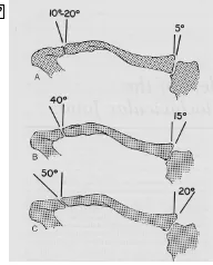
The acromioclavicular and sternoclavicular joints (anatomy and biomechanics)
The clavicle may be regarded as a link, jointed at each end, connecting the scapula
to the sternum (Fig. 1). Movement of the scapula must occur about a fulcrum at one
or both ends if this link. In the normal shoulder movement of the scapula, with
the arm above 90degrees; and 2) when the shoulders are braced backwards or
drawn forwards.2
Joint Stability:
Dynamic stability: deltoid and trapezius muscles inserts blended to the superior AC ligament and capsule. Anterior deltoid origin in the clavicle is just medial to the AC
joint, and superior trapezius fascia insertion blends to the posterior-superior AC
jointcapsule and dorsal clavicle.May add strength and stability when contracted but
dynamic stability in this joint is poorly understood.Static stability: AC and CC ligaments play an important role, wherein AC ligament works as a horizontal
stability and the CC ligaments for vertical stability.Conoidligament located in 46 mm
medial to AC jointand posterior, andtrapezoid ligamentlocated 26 mm medial to AC
joint, mid-portion of the inferior surface of the clavicle.4
Biomechanics:
Osteokinematics of the AC jointincludes clavicle rotation on its own axis and scapular
upward and downward rotations, sagital and horizontal plane adjustments.The
superior, inferior, and posterior AC ligaments insert an average of 16 to 20 mm
medial to the AC joint on the clavicle undersurface. The implication of this anatomic
observation is that aggressive distal clavicle excision (DCE) can destabilize the AC
jointand lead to symptomatic posterior impingement against the acromion.Motion in
the clavicle is necessary although when fused (AC or CC bone bar) still allows
normal elevation and abduction.Under physiological loads, AC ligaments and capsule
are the primary restraints for both horizontal and vertical forces. Under greater forces
and displacements the CC ligaments resist to vertical, horizontal and compressive
forces.3Trapezoid ligament resists axial compression.4
Clavicle rotates 40-50 degrees on its own axis in synchrony with scapular motion.
Most of the motion occurs on the sternoclavicular joint (SCJ). There are only 5 to 8
degrees of motion at the AC joint.3
Scapulothoracic elevation is an elevation on the SCJ and downward rotation at the
AC joint; wherein scapulothoracicprotaction is defined asprotaction of the SCJ with
slight horizontal slides and adjustments of the AC joint; scapulothoracic upward
rotation is defined as elevation of the SCJ and upward rotation at the AC joint.3
Clavicle motion is necessary to assist scapular movements on the thoracic wall. It
helps to keep the scapula in it functional position and motion,critical to the shoulder
normal function. It has a great role on maintaining scapulohumeralrhytm. Although
most of clavicle motion occurs on the SCJ, the AC joint contributes with an important
role in the shoulder biomechanics and it’s a very common source of traumatic,
inflammatory and degenerative problems.3
What structures are damaged in AC jointinstability?
Persistent upward displacement of the lateral end of the clavicle is a common sequel
to traumatic dislocation or subluxation of the AC joint. In most cases the
displacement is slight and causes no symptoms. Exceptionally there is pain, worse
during full elevation of the arm. On examination the lateral end of the clavicle is
unduly prominent, and a distinct step can be felt between it and the surface of the
acromion.
Classification
The most widely used classification is that of Rockwood et al.5 It is important to note
that this is a purely radiographic classification system. In a type I injury, there is
sprain of the AC ligament only. There is no radiographic abnormality. In type II injury,
the AC ligament and joint capsule are disrupted. The CC ligaments are intact but
sprained. There is 50% vertical subluxation of the distal clavicle. In Type III injury, the
AC ligaments and joint capsule as well as the CC ligaments are disrupted. The
deltotrapezial fascia is sprained. There is 100% superior displacement of the distal
clavicle. In Type IV injury, there is posterior subluxation of the clavicle into the
exaggeration of a type III because of additional complete rupture of the deltotrapezial
with 300% superior displacement of the clavicle. In the rare type VI injury, there is
subacromial or subcoracoid displacement of the clavicle.
Approach to patient evaluation
Symptoms of all injuries are local superior shoulder pain over the AC joint, deformity
occurs with types 2 and 3 injuries. Pain over AC jointwith active internal rotation can
be found. On inspection looks swelling, muscle wasting, deformity, outerclavicle
prominence, changing in scapular posture.Whereas failure to reduce indicates
violation of this fascia with the distal clavicle buttonholed through it (type
V).Distinguishing between type III and V injuries mayfacilitate by having the patient
shrug both shoulders, which in a type V injury exaggerates the degree of
displacement. Asking the patient to shrug his/her shoulder is a useful method to
determine if the deltotrapezial fascia is intact(clavicle reduces, type III injury) or if it
has been violated (clavicle remains dislocated, type V injury).Other symptoms are
localized tenderness to ACJ, piano-key sign range of motion (ROM) is usually
minimally affected
Special tests:
• Cross-arm adduction test: pain on forced adduction of elevated arm
• O’Brien’s active compression test: pain localized to the superior shoulder.6
(A recent analysis of these maneuvers found cross-arm adduction to be sensitive
(77%) and the active compression test of O’Brien to be 95% specific).6
• Scarf test: forced cross body adduction in 900flexion, pain at the extreme of
motion over the AC jointis indicative of AC jointpathology
Further Investigation Radiographs
a. Bilateral anteroposterior (AP) view of AC joint, the distance from the superior aspect of the coracoid process to the inferior aspect of the clavicle on both sides
is measured.
VI injury), which is rare and has not been seen in the authors’ practice.
c. The axillary view helps assess posterior displacement of the clavicle through a torn trapezial fascia (type IV injury),
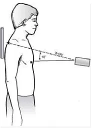
d. Bilateral Zanca view,7Zancaoriginally described a specialized view for the AC jointwhereby the x-ray beam is angled 10 cephalad to eliminate the overlap of the
clavicle with the spine of the scapula.Since the AC joint is more subcutaneous
than the glenohumeral joint, accurate visualization of the AC joint(Zanca view)
requires only a third to half of the radiographic penetration that is used for a
standard AP shoulder projection. The normal CC distance is between 1.1 and 1.3
cm, on average. Bearden and colleagues reported that complete CC ligament
disruption is indicated by an increase in the CC distance of 25% to 50%
compared with the contralateral shoulder.
Figure 2. Zanca view7
e. Basmania view defines AC joint injuries as either stable or unstable, and this is determined by the use of an AP radiograph of the affected shoulder with the arm
adducted across the chest.5
Management
Where treatment of types I, II, IV, V and VI are generally agreed on, treatment of type
III remains controversial. A new suggestion emerged for theRockwood classification
by further subdividing the type III AC joint injuries into IIIA (stable) and IIIB (unstable).
The basis for the sub-classification is mainly function rather than anatomic. Dr.
between stable and unstable AC joint. Patients who recover and regain function
within 6-8 weeks and whose X-rays show no override on Basmaniaview, are
considered stable (Type IIIA). If the clavicle overrides the acromion on the cross body
adduction AP view, it indicates instability of the CC ligaments in addition to the AC
joint disruption corresponding to a type IIIB. Normal positioning may be prognostically
positive and direct non-operative treatment. Superimposition of the acromion and
distal clavicle suggests instability and may indicate a role for surgery
Illustration
Author experience
According to Allman’s classification8, on plain radiographs all patients were evaluated
having old unreduced AC joint dislocation traumatic type III.
Since 2008, the authors have reconstructed in nine patients treated by transference
ligament of CA and CC ligaments, and followed in the Department of Orthopaedics
and Traumatology of Faculty of Medicine UniversitasPadjadjaran/ dr. Hasan Sadikin
Teaching Hospital, Bandung, Indonesia. The group comprised 9 men with a mean
age of 38.5 years (range, 20-57 years), the right side was affected in all patients. The
mechanism of injury was falls on the shoulder in five (50%) cases, traffic accidents in
three (30%) cases and direct trauma to the shoulder in one (11%) case. Mean time
from injury to surgery was 12 weeks (range, 10-16 weeks). Mean follow-up was 17
(range, 12-43 months). When evaluating the success or failure of the procedure, the
outcome measures utilized were variable with variable emphasis on pain, function,
movementability. Imatani scoring system was use for this evaluation.
The CA ligament has been used for the reconstruction of a dislocated AC joint. It lies
adjacent to the disrupted CC ligament. Primary fixation across the AC joint was done;
secondary stabilization of the joint by recreating the anatomic linkage between the
distal clavicle and the coracoid process; dynamic stabilization of the joint by creating
an inferiorly directed force on the distal clavicle; and imbrication of the superior roof
of the AC joint.The base of coracoid was excavated from scar tissue. Without
releasing the conjoined tendon from the tip of coracoid 3, 3-0 dexon sutures and a
piece of fascia lata (1.5 cm wide double layered) were passed under the bent portion
coracoid. Two Kirschner wire (1.8 mm) were used to fix the AC joint from acromion.
Results
According to the Imatani scoring system, mean therapeutic results was 66% yield
good result, 22% with fair and poor results in 11%.
The incidence of residual subluxation in the AC joint was evaluated at final
radiographic follow-up. In our cases subluxation that represented less than 5 mm of
superior translation of the clavicle occurred in two cases.
Meanwhile osteoarthritic changes occurred in the AC joint in 2 (22%) patients with
age 55 and 57-year old.
Table 1. Shoulder results after shoulder reconstruction
ofthe old AC joint separation (n=9)
Resuts Score
Good 6 (66%)
Fair 2 (22%)
Poor 1 (11%)
Discussion
AC joint separation are frequently treated in clinical practice. The degree or direction
of translation of the clavicle against the acromion depends on the injury states of the
AC and CC ligaments and detachment of deltoid or trapezius muscle from the
clavicle. Many surgical procedures exist for AC joint dislocation, including repair of
the AC ligament (Phemister9 or Neviaser procedures10,11), fixation between the
clavicle and the coracoid process (Bosworth procedure), reconstruction of the CC
ligament using the CA ligament (Weaver Dunn and Cadenat12 procedures), and
dynamic stabilization of the CC ligament by the transferred conjoined tendon (Dewar
procedure).
Since 2008, the author has reconstructed the anatomical structure of the CC
ligaments (trapezoid and conoid ligaments) with ipsilateral fascia lata used as
substitute ligaments, and reconstruction using coracoacromial(CAL) ligament.The
utilized for stabilization of AC joint. Neviaser published the technique of removing
CAL from its coracoid attachment to reconstruct superior acromioclavicular
ligament.10,11 The removal of CAL from its acromial end and subsequent transfer to
distal clavicle for AC joint dislocation was first describe by Cadenat10 in 1917. He
described anterior and posterior fascicle off acromion and sutured it to remnants of
the conoid ligament and periosteum superior aspect of clavicle.
The modified Neviaser procedure has some disadvantages, including a difficulty to
attach the CA ligament to posterior distal of the clavicle, a long period required for
range of motion recovery, incidence of residual subluxation, and postoperative
osteoarthritic changes on AC joint.
The mechanisms of stabilization for the AC joint established by the CA ligament
transferred from the acromion to the clavicle. This transferred CA ligament does not
anatomical reconstruct the trapezius and conoid ligaments that compose the CC
ligament. The conoid ligament attaches anatomically to the conoid tubercle, which is
located at the posterior edge of the clavicle, and the clavicle can make an axial
rotation during forward elevation of the shoulder joint. However, in the modified
Neviaser procedure it is possible that this axial rotation of the clavicle is restricted
because the transferred CA ligament is fixed to the anterior edge of the clavicle. For
this assumption, even if the dislocated AC joint is reduced in a normal position, it is
possible that osteoarthritic changes can occur to the AC joint.
Since anatomic reduction and firm are imperative to maintain the integrity
post-repaired, old unreduced are best treated surgically, in particular for young and active
persons, severely displaced cases and overhead workers.
Conclusion:
The modified Neviaser procedure can provide satisfactory therapeutic results and
avoid postoperative failure or loss of reduction of AC joint separations. Anatomic
reconstruction of both CC ligaments and AC ligaments can be best restore AC joint
function.
References:
posterior labral injury, Am JSports Med 2011;39(8):1687-96.
2. Adam JC. The shoulder region, in Adam JC (ed): Outline of orthopaedics. Churcill
Livingstone, Medical division of Longman Group UK Ltd. 1989, pp. 239-61.
3. DePalma AF: The role of the disks of the sternoclavicular and the
acromioclavicular joints. ClinOrthop 1959;13:7-12.
4. Fukuda K, Craig AV, An K, Cofield RH, Chao EYS: Biomechanical study of the
ligamentous systems of the acromioclavicular joint. J Bone Joint Surg
1986;68A:434-40.
5. Rockwood CA, Williams G, Yound DC: Injuries to the acromioclavicuar joint, in
Rockwood CA Jr, Green DP, Bucholz, Heckman JD (eds): Fractures in adults.
Philadelphia, Lippincott-Raven, 1996, pp 1341-413.
6. O'Brien SJ, Pagnani MJ, Fealy S, McGlynn SR, Wilson JB: The active
compression test: a new and effective test for diagnosing labral tears and
acromioclavicular joint abnormality. Am J Sports Med 1999;2:610-43.
7. Zanca P: Shoulder pain: Involvement of the acromioclavicular joint. Analysis of
1,000 cases. Am J Roentgenol 1971;112:493-500.
8. Allman FL., Jr: Fractures and ligamentous injuries of the clavicle and its
articulation. J Bone Joint Surg 1967;49A:774-84.
9. Phemister DB. The treatment of dislocation of the acromioclavicular joint by open
reduction and threaded-wire fixation. J Bone Joint Surg Am. 1942;24(1):166-8.
10. Neviaser JS. Acromioclavicular dislocation treated by transference of the
coraco-acromial ligament: A long-term follow-up in a series of 112 cases.
ClinOrthopRelat Res. 1968;58:57-68.
11. Neviaser JS. Acromioclavicular dislocation treated by transference of the
coracoacromial ligament. AMA Arch Surg. 1952;64:292-7.
12. Cadenat F. The treatment of dislocations and fractures of the outer end of the