Short communication
DNA vaccine-mediated immune responses in Coxsackie
virus B3-infected mice
Andreas Henke *, Roland Zell, Axel Stelzner
Institute of Virology,Medical Center,Friedrich Schiller Uni6ersity,Winzerlaer Str.10,D-07745Jena,Germany Received 29 March 2000; accepted 8 September 2000
Abstract
DNA immunizations with the major structural protein VP1 of Coxsackie virus B3 (CVB3) have been previously found to protect BALB/c mice from lethal challenge. Here we report that the other CVB3 capsid proteins, VP2, VP3, and VP4, were less effective at preventing CVB3-caused disease. The application of pCMV/VP1 as a vaccine caused decreased myocyte destruction, reduced viral load in the heart tissue, accelerated antibody induction, and an early cytokine expression in heart tissue. In summary, our results indicate that the induction of B cell and/or T cell memory in vaccinated mice prior to challenge is responsible for the protection observed. © 2001 Elsevier Science B.V. All rights reserved.
Keywords:DNA vaccine; Immune response; Coxsackie virus
www.elsevier.com/locate/antiviral
Infections with Coxsackie virus B3 (CVB3) are common causes of acute and chronic myocarditis in humans (Savoia and Oxman, 1995). In order to study virus-specific protection and CVB3-caused pathogenesis in more detail, several murine mod-els have been established (Chow et al., 1992 Hu-ber and Pfaeffle, 1994 Henke et al., 1995 Mena et al., 1999). Despite the well characterized molecu-lar structure of Coxsackie viruses (Natarajan and Johnson, 1998) and the successful use of common vaccinations in animal models (Fohlman et al., 1993 See and Tilles, 1997), no virus-specific
pre-ventive procedures against CVB3 are in clinical use today. A new method to prevent virus-caused disease is the administration of plasmid DNA encoding immunogenic viral epitops. The inocula-tion of DNA vaccines into muscle tissue or skin has been shown to induce humoral (Boyer et al., 1999 Konishi et al., 1999) as well as cellular (Schirmbeck et al., 1995; Qiu et al., 1999) immune responses and to confer protection against viral infections (Yokoyama et al., 1995 Operschall et al., 1999).
Using DNA plasmids encoding different se-quences of CVB3 structural proteins we demon-strated that the expression of the capsid protein VP1 was the most effective vaccine to protect mice against a lethal CVB3 infection. In order to analyze the putative protective effect of the other * Corresponding author. Tel.:+49-3641-657215; fax:+
49-3641-657202.
E-mail address:[email protected] (A. Henke).
the following sequences: VP2 (788 bp), VP3 (713 bp), and VP4 (206 bp), using the same method as described for pCMV/VP1 (Henke et al., 1998). Plasmid isolation from bacteria was performed using the endotoxin-free plasmid preparation kit (Qiagen, Hilden, Germany). Expression from these plasmids was determined in vitro by tran-sient transfection of HeLa cells. After RNA isola-tion, DNase digesisola-tion, and reverse transcriptase reaction, the transcriptional activity of all plas-mids was confirmed by PCR (data not shown).
After the expression from the DNA vaccines was analyzed in vitro, male BALB/c mice were inoculated intramuscularly (i.m.) twice in each quadriceps muscle separately with 100mg of plas-mid DNA at 4-week intervals. One group of mice remained untreated and one group received the parental vector pCMV as a negative control. Four weeks after the second immunization, mice were subjected to intraperitoneal (i.p.) challenge with a normally lethal dose of CVB3 (5 LD50). After this
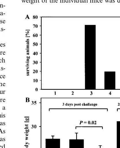
challenge, the number of surviving animals was monitored up to 4 weeks post infection (p.i.). As it is shown in 1A, the most effective vaccine was pCMV/VP1, confirming the result we obtained before (72.2% protection) (Henke et al., 1998). In this study, the percentage of surviving animals using this vaccine was 71.4% (10 of 14). Intramus-cular inoculations of all other plasmids were less effective, inducing incomplete protection of 21.4% (3 of 14 with pCMV/VP3) or 20% (2 of 10 with pCMV/VP2). Expression of the protein VP4 in-duced no protection (0 of 12 with pCMV/VP4). These results confirmed, that the major capsid protein VP1 — expressed as a single protein — is the best candidate among the capsid proteins to be used in plasmid DNA expression vectors reflecting the observation that several immuno-genic epitopes are located within this protein (Haarmann et al., 1994). In other experimental animal models using different picornaviruses like the encephalomyocarditis virus (Jun et al., 1995) or the foot and mouth disease virus (Huang et al., 1999), the VP1 protein expressed as a single
mice, the following approach was used: male BALB/c mice were inoculated with pCMV as a control or vaccinated with pCMV/VP1 in the same way as described above. Three days after the lethal CVB3 challenge with 5 LD50 doses, the
weight of the individual mice was determined. The
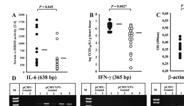
Fig. 2. Characterization of pCMV/VP1-mediated protection, 3 days after the lethal CVB3 challenge. The concentration of the cardiocyte-specific enzymea-HBDH (A) and the amount of CVB3-specific antibodies (C) — detected by ELISA (serum dilution 1:25) and demonstrated as the optical density at 490 nm (OD490) — were analyzed in sera of individual pCMV ( ) or pCMV/VP1 () inoculated mice. The viral load in the heart tissue of individual mice was determined using CCID50assays (B). The black bar in each figure indicates the mean value. and indicate the mean value of CVB3-specific antibody titers before the lethal challenge. The transcriptional activity of IL-6 and IFN-gwas analyzed using individual heart tissue obtained from two pCMV- and five pCMV/VP1-inoculated mice by RT-PCR (D). The statistical comparison was carried out with Microsoft Excel by using Students’t-test.
significant loss of body weight of pCMV-treated mice indicated that these mice already had an ongoing infection, which is common in CVB3-in-fected mice at this time (1B, III). Mice which received the pCMV/VP1 vaccine remained in the same body weight range as normal non-infected control mice (1B, II and I). Four weeks after the CVB3 challenge, the body weight of surviving pCVM/VP1-immunized mice was identical to that of non-infected mice, indicating that no ongoing pathogenic process was present in these mice (1B, V and IV). At this time, no pCMV-inoculated mouse survived the lethal CVB3 challenge.
Three days after the CVB3 challenge, serum as well as heart tissue were obtained from plasmid DNA-inoculated mice. In sera, the concentration of the cardiocyte-specific enzyme a -hydroxybu-tyrate-dehydrogenase (a-HBDH) was analyzed using a commercially available kit (Sigma Diag-nostics, St Louis, USA) according to the instruc-tions of the manufacturer. Heart muscle cells contain high levels of a-HBDH and myocardial damage results in an increased release of this
enzyme into the blood (Franken et al., 2000).
a-HBDH activity was compared to the whole LDH (lactate dehydrogenase) activity (Sigma Di-agnostics) demonstrating that the quotient LDH/
a-HBDH was always below 1.3, which indicates heart tissue destruction (data not shown). In addi-tion, no significant inflammation at the site of the DNA vaccine inoculation was observed at this time, indicating that only minor levels of a -HBDH might be contributed by skeletal myocyte damage. As is demonstrated in 2A, high levels of
a-HBDH-activity were detectable in sera of pCMV-treated control mice 3 days after the chal-lenge. In contrast, a-HBDH activity in the sera of pCVM/VP1-vaccinated mice was significantly de-creased. Furthermore, the infectious virus was isolated from the heart tissue of the same mice as described previously (Henke et al., 1995) and the virus titer was determined by cell culture infec-tious dose 50% assays (CCID50) on HeLa cell
animals did not show histopathological disorders, indicating that CVB3 was unable to induce tissue destruction, massive inflammation, or fibrosis 45 days p.i., as has been demonstrated before (Henke et al., 1998). Furthermore, levels of immunoglob-ulin M (IgM)- and IgG-specific antibodies were assessed together by enzyme-linked immunosor-bent assay (ELISA), using purified CVB3 as the target antigen. As is demonstrated in 2C, the vaccination procedure induced low levels of anti-CVB3 antibodies. However, 3 days after the chal-lenge, a significant increased concentration of CVB3-specific antibodies was detectable in the sera of pCMV/VP1-vaccinated mice, indicating that the vaccination procedure induced an im-munologic memory that was accompanied by a secondary antibody response. This may then lead to reduced viral replication in the heart tissue detected in the vaccinated mice. In addition, the activated immune response was also reflected by an early induction of the transcriptional activity of immunoregulatory cytokines in the myocardial tissue, indicating an early immune infiltration of the heart tissue. In general, cytokine transcription and translation were found to be highly increased in CVB3-caused myocarditis, as has been de-scribed by several different investigators (Huber et al., 1996 Gebhard et al., 1998 Opavsky et al., 1999). During our vaccination experiments we analyzed the activation of interleukin-6 (IL-6) and interferon-g(IFN-g) transcription in heart tissue 3 days after the lethal challenge. Briefly, cytokine expression was determined in individual tissue samples of pCMV- or pCMV/VP1-inoculated mice by RT-PCR. After RNA isolation, DNase digestion, and reverse transcriptase reaction, the transcriptional activity of IL-6, as well as that of IFN-g, was confirmed by PCR and compared to
b-actin expression (2D). At this time point, only in pCMV/VP1-vaccinated mice was cytokine ex-pression detectable, indicating an early activation of immunoreactivity against the viral infection.
Furthermore, the activation of the cellular im-mune response in pCMV/VP1-vaccinated mice
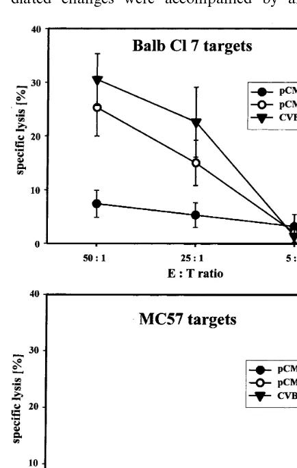
are normally not susceptible to infections with CVB3. Therefore, the recombinant VV/VP1 was established as described before (Henke et al., 1998 Wessely et al., 1998). BALB/c mice were plasmid-inoculated using the same schedule as described above. Four weeks after the second immuniza-tion, spleen cells were obtained from pCMV/ VP1-and pCMV-treated mice VP1-and a non-radioactive cytotoxic T lymphocyte (CTL) assay was per-formed detecting the release of LDH in cell cul-ture supernatants according to the instructions of the manufacturer (Boehringer, Mannheim, Ger-many). For this assay, murine Balb Cl 7 (H-2d)
target cells as well as MC57 (H-2b) target cells
(Klavinskis et al., 1990) were infected with VV/ VP1, but no CVB3-specific CTL activity was ob-served (data not shown). Therefore, pCMV/VP1-vaccinated as well as pCMV-treated control BALB/c mice were challenged with 5 LD50doses of CVB3 and 3 days later, spleen cells
were obtained and used as effector cells for CTL assays. Under these test conditions, only Balb Cl 7 target cells were lysed, indicating MHC-class I-restricted CTL activity of spleen cells obtained from pCMV/VP1-vaccinated BALB/c mice, whereas no lysis was detectable using MC57 target cells. The specific lysis observed was almost in the same range as the CTL activity detectable from spleen cells obtained from BALB/c mice infected twice with a 0.1 LD50dose of CVB3 (Fig.
3). The CTL activity of these spleen cells was analyzed 3 days after the second infection, whereas no CTL activity was observed using spleen cells from mice after a single infection (data not shown).
CVB3-caused myocyte destruction was less severe, (ii) the number of infectious virus particles was re-duced, and (iii) increased levels of CVB3-specific antibodies were detectable by ELISA, indicating induced immunologic memory in vaccinated mice in comparison to control mice. These vaccine-me-diated changes were accompanied by an early
expression of immunoregulatory cytokines in the heart tissue of vaccinated mice and increased virus-specific cytotoxic activity of spleen cells. Further experiments will be focused on suitable methods to increase the efficiency of the estab-lished DNA vaccine pCMV/VP1.
Acknowledgements
We thank Katrin Klement, Birgit Meißner, and Birgit Schikowski for excellent technical assis-tance. This work was supported by BMBF grant 0122960/01229104 ZP5.
References
Boyer, J.D., Kim, J., Ugen, K., Cohen, A.D., Ahn, L., Schu-mann, K., Lacy, K., Bagarazzi, M.L., Javadian, A., Cic-carelli, R.B., Ginsberg, R.S., MacGregor, R.R., Weiner, D.B., 1999. HIV-1 DNA vaccines and chemokines. Vac-cine 17 (suppl. 2), 53 – 64.
Chow, L.H., Beisel, K.W., McManus, B.M., 1992. Enteroviral infection of mice with severe combined immunodeficiency. Evidence for direct viral pathogenesis of myocardial injury. Lab. Invest. 66, 24 – 31.
Fohlman, J., Pauksen, K., Morein, B., Bjare, U., Ilback, N.G., Friman, G., 1993. High yield production of an inactivated Coxsackie B3 adjuvant vaccine with protective effect against experimental myocarditis. Scand. J. Infect. Dis. 88 (suppl.), 103 – 108.
Franken, N.A., Strootman, E., Hollaar, L., van der Laarse, A., Wondergem, J., 2000. Myocardial enzyme activities in plasma after whole-heart irradiation in rats. J. Cancer Res. Clin. 126, 27 – 32.
Gebhard, J.R., Perry, C.M., Harkins, S., Lane, T., Mena, I., Asensio, V.C., Campbell, I.L., Whitton, J.L., 1998. Cox-sackievirus B3-induced myocarditis: perforin exacerbates disease, but plays no detectable role in virus clearance. Am. J. Pathol. 153, 417 – 428.
Haarmann, C.M., Schwimmbeck, P.L., Mertens, T., Schultheiss, H.P., Strauer, B.E., 1994. Identification of serotype-specific and non-serotype-specific B-cell epitopes of Coxsackie B virus using synthetic peptides. Virology 200, 381 – 389.
Henke, A., Huber, S., Stelzner, A., Whitton, J.L., 1995. The role of CD8+ T lymphocytes in Coxsackie virus B3-in-duced myocarditis. J. Virol. 69, 6720 – 6728.
Henke, A., Wagner, E., Whitton, J.L., Zell, R., Stelzner, A., 1998. Protection of mice against lethal Coxsackie virus B3 infection by using DNA immunization. J. Virol. 72, 8327 – 8331.
Huang, H., Yang, Z., Xu, Q., Sheng, Z., Xie, Y., Yan, W., You, Y., Sun, L., Zheng, Z., 1999. Recombinant fusion Fig. 3. Characterization of the cellular immune response in
virus B3 infections of BALB/c mice initiated by cells expressing the gamma delta+T-cell receptor. J. Virol. 70, 3039 – 3044.
Huber, S.A., Pfaeffle, B., 1994. Differential Th1 and Th2 cell responses in male and female BALB/c mice infected with Coxsackie virus group B type 3. J. Virol. 68, 5126 – 5132. Jun, H.S., Yoon, S.W., Kang, Y., Pak, C.Y., Lee, M.C.,
Yoon, J.W., 1995. Cloning and expression of the VP1 major capsid protein of diabetogenic encephalomyocarditis (EMC) virus and prevention of EMC virus-induced dia-betes by immunization with the recombinant VP1 protein. J. Gen. Virol. 76, 2557 – 2566.
Klavinskis, L.S., Whitton, J.L., Joly, E., Oldstone, M.B.A., 1990. Vaccination and protection from a lethal viral infec-tion: identification, incorporation, and use of a cytotoxic T lymphocyte glycoprotein epitope. Virology 178, 393 – 400. Konishi, E., Yamaoka, M., Khin Sane, W., Kurane, I.,
Takada, K., Mason, P.W., 1999. The anamnestic neutraliz-ing antibody response is critical for protection of mice from challenge following vaccination with a plasmid en-coding the Japanese encephalitis virus premembrane and envelope genes. J. Virol. 73, 5527 – 5534.
Mena, I., Perry, C.M., Harkins, S., Rodriguez, F., Gebhard, J., Whitton, J.L., 1999. The role of B lymphocytes in Coxsackie virus B3 infection. Am. J. Pathol. 155, 1205 – 1215.
Natarajan, P., Johnson, J.E., 1998. Molecular packing in virus crystals: geometry, chemistry, and biology. J. Struct. Biol. 121, 295 – 305.
Operschall, E., Schuh, T., Heinzerling, L., Pavlovic, J., Moelling, K., 1999. Enhanced protection against viral in-fection by co-administration of plasmid DNA coding for viral antigen and cytokines in mice. J. Clin. Virol. 13, 17 – 27.
Qiu, J.T., Song, R., Dettenhofer, M., Tian, C., August, T., Felber, B.K., Pavlakis, G.N., Yu, X.F., 1999. Evaluation of novel human immunodeficiency virus type 1 gag DNA vaccines for protein expression in mammalian cells and induction of immune responses. J. Virol. 73, 9145 – 9152. Savoia, M.C., Oxman, M.N., 1995. Myocarditis and
pericardi-tis. In: Mandell, G.M., Benett, J.E., Dolin, R. (Eds.), Principles and Practice of Infectious Disease. Churchill Livingstone, New York, pp. 799 – 813.
Schirmbeck, R., Bohm, W., Ando, K., Chisari, F.V., Rei-mann, J., 1995. Nucleic acid vaccination primes hepatitis B virus surface antigen-specific cytotoxic T lymphocytes in nonresponder mice. J. Virol. 69, 5929 – 5934.
See, D.M., Tilles, J.G., 1997. Occurrence of Coxsackie virus hepatitis in baby rabbits and protection by a formalin-in-activated polyvalent vaccine. Proc. Soc. Exp. Biol. Med. 216, 52 – 56.
Wessely, R., Henke, A., Zell, R., Kandolf, R., Knowlton, K.U., 1998. Low-level expression of a mutant Coxsackie viral cDNA induces a myocytopathic effect in culture: an approach to the study of enteroviral persistence in cardiac myocytes. Circulation 98, 450 – 457.
Yokoyama, M., Zhang, J., Whitton, J.L., 1995. DNA immu-nization confers protection against lethal lymphocytic cho-riomeningitis virus infection. J. Virol. 69, 2684 – 2688.