The effects of fatty acids on apolipoprotein B secretion by human
hepatoma cells (HEP G2)
Sharon Arrol, Michael I. Mackness, Paul N. Durrington *
The Uni6ersity Department of Medicine,Manchester Royal Infirmary,Oxford Road,Manchester M13 9WL,UK
Received 5 February 1999; received in revised form 18 August 1999; accepted 8 September 1999
Abstract
We have investigated the effect of fatty acids on the rate of apolipoprotein B (apo B) secretion by human hepatoma cells (Hep G2). When Hep G2 cells were maintained in tissue culture flasks oleic acid up to 0.4 mM increased apo B secretion in a dose-dependent manner, whereas increases in triacylglycerol (TG) were smaller and dose dependency was less evident. In the absence of oleic acid, apo B accumulating in the tissue culture medium was predominantly in lipoproteins of higher density than very low density lipoproteins (VLDL). However, when the rate of secretion was stimulated with oleic acid the apo B-containing lipoproteins became lower in density. We postulated that there was a high rate of lipolysis of newly secreted VLDL by Hep G2 cells, which would account both for the relatively smaller effect of oleic acid on TG as opposed to apo B accumulating in the culture medium and the predominance of apo B in lipoproteins of a higher density than VLDL, which became less evident when VLDL secretory rates were stimulated by oleic acid. To test this hypothesis, cultured Hep G2 cells were transferred to columns containing Cytodex beads, permitting their continuous perfusion with culture medium so that newly secreted VLDL did not remain in contact with the cells. Apo B recovered from the perfusate was largely in VLDL range lipoproteins and the TG measured in the perfusate indicated that the true secretory rate of TG-rich lipoproteins was substantially higher than had been reflected by TG accumulating in culture medium left in contact with cells. Apo B measured in the culture medium of Hep G2 cells may thus be a better reflection of VLDL secretion, even though it is contained in higher density lipoproteins due to removal of TG by lipolysis. The effects of saturated fatty acids (SFA), monounsaturated fatty acids (MUFA) and polyunsaturated fatty acids (PUFA) on apo B (apo B) secretion by Hep G2 cells maintained in tissue culture flasks were next investigated. SFA (0.4 mM), with the exception of stearic acid (C18:0), increased apo B secretion. Lauric acid (C12:0) increased apo B secretion by 32%, myristic acid (C14:0) by 41% (PB0.005), palmitic acid (C16:0) by 154% (PB0.025), and arachidic acid (C20:0) by 186% (PB0.005). The effect of MUFA (0.4 mM) was to increase apo B secretion, oleic acid (C18:1) by 239% ((PB0.0005) and palmitoleic acid (C16: 1) by 125% (PB0.005). Of the PUFA investigated, linolenic acid (C18:3) (0.4 mM) did not have any significant effect on apo B secretion, whereas linoleic acid (C18:2) (0.4mM) arachidonic acid (C20:4) (0.1 mM) and eicosapen-taenoic acid (C20:5) (0.1 mM) caused significant increases of 164, 171 and 171%, respectively (PB0.005). The fatty acids studied increased intracellular TG and cholesteryl ester concentrations to varying extents. The increase in intracellular TG produced by the different fatty acids correlated with the rate of apo B secretion (r=0.6;PB0.05). In this human hepatoma cell line, with the exception of the saturated fatty acids, the rate of secretion of apo B-containing lipoproteins does not follow the same pattern as changes in circulating low density lipoprotein (LDL) concentrations reported with dietary manipulation in man. If our findings reflect the in vivo situation, we suggest that whilst the dietary effects of SFA on serum LDL may in part be determined by the hepatic apo B secretory rate, the effects of MUFA and PUFA must be largely mediated through a catabolic effect rather than an effect on hepatic secretion. The marked increase in apo B secretion with the more highly polyunsaturated fatty acids, such as eicosapentaenoic acid, may also explain why they do not lower circulating LDL, despite reports of their apparently favourable effect on LDL-receptor mediated clearance. © 2000 Elsevier Science Ireland Ltd. All rights reserved.
Keywords:Human hepatoma cells; Apolipoprotein B; Saturated fatty acids; Monounsaturated fatty acids; Polyunsaturated fatty acids www.elsevier.com/locate/atherosclerosis
* Corresponding author. Tel: +44-161-276-4226; fax:+44-161-274-4833.
1. Introduction
Dietary fatty acids play an important role in deter-mining plasma total cholesterol (TC) concentrations in humans. Saturated fatty acids (SFA) of chain length C12 – C16 have been demonstrated to be the most hy-percholesterolaemia-inducing fatty acids [1]. In con-trast, polyunsaturated fatty acids (PUFA), such as linoleic acid, and monounsaturated fatty acids (MUFA), such as oleic acid, when substituted for SFA in the diet, lower plasma TC concentrations [2,3] and this forms the basis of current dietary recommendations to reduce plasma TC and low density lipoprotein-cholesterol (LDL-C) concentrations [4]. Highly polyun-saturated fatty acids such as eicosapentaenoic acid and docosahexaenoic acid, although they do not decrease serum cholesterol, are effective in lowering serum tria-cylglycerol (TG) concentrations [5].
It has been well documented that Hep G2 cells synthesise and secrete lipoproteins which contain the apolipoproteins B-100, A-I, A-II, A-IV, B, C-I, C-II, C-III and E [6 – 10]. Apolipoprotein B-100 (apo B) is the sole apo B species secreted by the human liver [11], and is also the apo B species synthesised and secreted by the Hep G2 cell line [7]. Hep G2 cells maintain many of the morphological and biochemical characteristics of normal hepatocytes [7,10,12,13]. They have, however, been criticised as a model of the hepatic secretion of apo B-containing lipoproteins, because unlike, for ex-ample rat hepatocytes, the apo B which accumulates in the culture medium of Hep G2 cells is not principally in very low density lipoprotein (VLDL), but is also sub-stantially present in the low density lipoprotein (LDL) and even denser lipoprotein particles. We have previ-ously suggested, however, that the apo B-containing VLDL secreted by Hep G2 cells is rapidly converted to LDL, because of the high activity of extracellular hep-atic lipase produced by Hep G2 cells [14]. Thus the apo B accumulating in the medium reflects the rate of VLDL secretion, although the concentration of TG in the medium does not necessarily reflect its rate of secretion because of lipolysis. In the present investiga-tion we examined this hypothesis further in a cell culture system which allows the rapid removal of newly secreted lipoproteins from further contact with Hep G2 cells. This part of the investigation confirmed our hy-pothesis. We therefore progressed to study the effects of individual saturated, monounsaturated and polyunsatu-rated fatty acids on apo B secretion, using a conven-tional Hep G2 cell culture system, which may provide an in vitro model to assist in explaining the results of dietary experiments with foods of different fatty acid composition, which cannot readily distinguish effects on the secretion of apo B-containing lipoproteins from those on their catabolism.
2. Methods
2.1. Materials
Dulbecco’s Modified Eagle’s Medium (DMEM) con-taining 25 mM glucose, foetal calf serum (FCS), peni-cillin/streptomycin solution, trypsin/EDTA solution (10×) and nystatin were purchased from Life Tech-nologies Ltd (Paisley, UK).
Fatty acid free-fraction V bovine serum albumin (BSA), saturated fatty acids, monounsaturated and polyunsaturated fatty acids, beta-hydroxybutyrate kits and lactate dehydrogenase kits were all supplied by the Sigma Chemical Co. (London, UK).
Tissue culture flasks, 25 cm2, were purchased from
Bibby, (Nottingham, UK). Pierce BCA protein reagent was supplied by Pierce Chemical Co. (Pierce & War-riner, Chester, UK). Horse anti-sheep IgG decanting Solution 2 was from Pharmacia LKB (Milton Keynes, UK). Monoclonal antibody to apolipoprotein B was obtained from Biogenesis Ltd. (Bournemouth, UK). The non-esterified fatty acid kit was supplied by the WAKO Chemical Co. (Alpha Laboratories, Hants, UK).
TG and cholesterol reagent (GPO-PAP and CHOD-PAP) were purchased from Biostat Ltd. (Stockport, UK) and free cholesterol reagent was from Boehringer Mannheim BCL (Lewes, Sussex, UK).
Amicon centriflo membrane cones type CF25, conical supports CS1A, and centrifuge tubes CTI, were sup-plied by Amicon Ltd. (Storehouse, Gloucestershire, UK).
2.2. Cell culture
Human hepatoblastoma cells, Hep G2, were supplied from the European Collection of Animal Cell Culture [15]. Hep G2 cells were cultured in DMEM containing 25 mM glucose, penicillin (1000 U/ml), streptomycin (100 mg/ml) and 10% FCS (v/v) at 37°C in an atmo-sphere of 95% air/5% CO2, with one medium change,
for 7 days prior to an experiment, after which time they were confluent, as observed by light microscopy (4× 10⁶ cells per flask), viability was 9690.5%as assessed
ratio 10.8:1). After incubation, medium was removed for analysis and the cells washed with phosphate buffered saline (PBS) pH 7.2 and harvested using a rubber policeman. The cells in PBS containing 1% triton X-100 were then disrupted using a Kontes micro ultrasonic cell disrupter. The average cell protein per incubation was between 0.5 and 0.6 mg.
2.3. Cell culture on perfused beads
2.3.1. Preparation of Cytodex 2 microcarrier beads A total of l g of Cytodex 2 beads were added to 50 ml PBS in a glass bottle and incubated over-night at 37°C to hydrate the beads. They were then autoclaved, washed in PBS and allowed to settle for 10 min. The PBS was removed and to 5 ml of beads an equal volume of DMEM was added to make 50% slurry [16].
2.3.2. Adhering Hep G2 cells to the microcarrier beads Two confluent flasks of Hep G2 cells were trypsinised, centrifuged and resuspended in DMEM. These cells (4×10⁶
) and the slurry of Cytedex beads were added to an autoclaved 125 ml Techne stirrer bottle (Phillip-Harris, Hull, UK) and the volume made to 50 ml with DMEM. This was placed on a hot plate magnetic stirrer (35 – 40°C) to stir continuously at ap-proximately 40 rpm. After 3 h the volume was in-creased to 100 ml with DMEM and allowed to continue stirring over-night. Following this, the beads were al-lowed to settle and the medium removed and replaced with 10 ml of DMEM.
2.3.3. Cell perfusion
The beads were poured into a 10×0.5 cm2
chro-matography column. The column was perfused with fresh sterile medium, kept at 37°C in a sterilised con-tainer. Medium was passed over the Hep G2 cells at a constant rate of 6 ml/h and collected in 2 ml fractions over a period of 24 h. These fractions were used for lipoprotein fractionation by discontinuous gradient ul-tracentrifugation (DGU) and subsequent measurement of protein apo B and TG concentrations.
2.4. Lipid and protein measurements
Protein was determined in cell homogenates and culture media using the Pierce BCA protein reagent (Pierce and Warriher, Chester, UK). TG, free choles-terol (FC) and total cholescholes-terol (TC) concentrations were measured using enzymic methods (GPO-PAP and CHOD-PAP) adapted and standardised for use in cell culture experiments as previously described by us [14]. Recovery experiments were carried out to compare cell homogenates with chloroform/methanol (2:1 v/v) dried lipid extracts, and the recoveries for TG, FC, and TC
were 82.595.0%, 87.593.Q%, and 9094.5%, respec-tively. Cholesteryl ester (CE) was calculated by sub-tracting the FC from the TC measured in the cells.
2.5. Discontinuous density gradient ultracentrifugation (DGU)
A volume of 10 ml of medium in the flask experi-ments or a 2 ml fraction in the perfusion experiexperi-ments were concentrated to 1 ml in Amicon Cones and lipo-proteins separated by DGU as described previously [17] to produce fractions of Sf 60 – 400, 20 – 60, 12 – 20, 0 – 12 and B0.
2.6. Immunoradiometric assay (IRMA)of apolipoprotein B
Apolipoprotein B secreted into the culture medium from Hep G2 cells was measured using an immunora-diometric assay (IRMA) (sensitivity 6 ng/ml) as previ-ously described in detail [14] using 100ml of sample and standard in the assay.
2.7. Measurement of non-esterified fatty acids(NEFA)
To ensure that all the FA had been complexed to the BSA, serum-free medium containing 1% BSA and fatty acids at the concentration used in the experiments, but which had not been exposed to Hep G2 cells, was concentrated 10-fold using Amicon centriflo cones. The cones have a filter with a molecular weight cut-off of 30 000 Da, therefore the BSA is retained within the cone and the medium which has passed through the cone will contain any non-BSA bound FA. The NEFA concentrations in a 50 ml sample of medium which had passed through the cone was determined using the WAKO NEFA-C kit according to the manufacturer’s instructions. Three standards (0.5, 1.0 and 1.97 mEq/l) were assayed in parallel and used to construct a stan-dard curve.
2.8. Measurement of beta-hydroxybuyrate (b-HBA)
Beta-Hydroxybutyrate was measured in the medium from Hep G2 cells using a commercial method (Sigma Chemical Co) according to the manufacturer’s instructions.
2.9. Measurement of cytotoxicity
2.10. Statistical analysis
Differences between results were sought using a Stu-dent’s t-test and correlations between data sets were calculated using the Pearson’s correlation coefficient.
3. Results
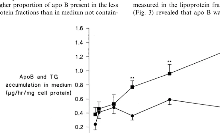
In initial experiments carried out to determine the optimal concentration of FA stimulating VLDL secre-tion apo B was measured in the medium after 24 h incubation with increasing concentrations of FA. With all FAs there was progressive increase in apo B until at least 0.4 mM. The example of oleic acid is shown in Fig. 1. FA concentration of 0.4 mM is within the range encountered in the hepatic circulation [18]. At concen-trations greater than 0.5 mM some of the FA became cytotoxic at the concentration of albumin used in our experiments. The exceptions were arachidonic acid and eicosapentaenoic acid, which caused Hep G2 cells to die at lower concentration. They were therefore studied at 0.1 mM. Medium TG concentration, unlike apo B, did not increase with increasing oleic acid and remained relatively constant over 24 h (Fig. 1).
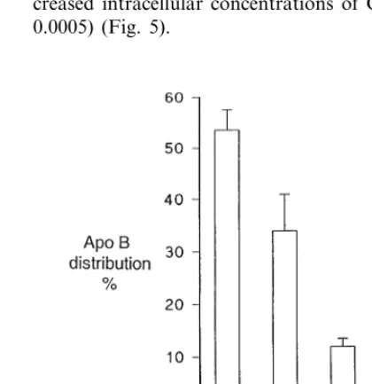
When the medium from Hep G2 cells cultured in the presence and absence of 0.4 mM oleic acid was sub-jected to discontinuous gradient ultracentrifugation, the distribution of apo B between the density subfractions showed a higher proportion of apo B present in the less dense lipoprotein fractions than in medium not
contain-ing oleic acid in which Hep G2 cells had been cultured (Fig. 2). This could be interpreted as indicating that larger apo B-containing lipoprotein particles are secreted when oleic acid is present as a substrate. However, given the much higher rate of apo B secretion by cells grown in oleic acid-containing media (Fig. 1), this seemed unlikely to be the whole explanation for the phenomenon. We postulated that apo B was initially secreted by Hep G2 cells in VLDL both when oleic acid was present and absent, but that when oleic acid was present VLDL production was higher and the VLDL substrate in the culture medium exceeded the capacity of hepatic lipase present on the surface of hepatocytes to remove its TG and phospholipid, so that the propor-tion of less dense lipoproteins remaining in the culture medium was greater than when VLDL secretory rates were lower in the absence of oleic acid. In order to test further this hypothesis, we cultured the same number of hepatocytes (4×10⁶) on Cytodex beads in a column
which was perfused with culture medium at a rate of 6 ml/h so that newly secreted lipoproteins were not left in contact with cells. (Hepatic lipase is bound to glycoso-aminoglycans adherent to the cell surface). Under these conditions, 216.7941.7 mmol/24 h of TG was recov-ered in the perfusate, whereas cells grown in flasks yielded only 20.094.2mmol/24 h. The concentration of apo B in the perfusate was below the detection limit of our assay. However, when the perfusate was subjected to DGU after concentration twofold apo B could be measured in the lipoprotein fractions. This experiment (Fig. 3) revealed that apo B was largely present in the
Fig. 2. Apolipoprotein B (apo B) distribution in lipoproteins isolated by discontinuous gradient ultracentrifugation from the culture medium from Hep G2 cells incubated for 24 h in the presence and absence of oleate 0.4 mM. Mean9SE of 12 experiments. Significantly different compared to no DMEM addition *PB0.01, **PB0.05.
VLDL density range (Sf60 – 400 and 20 – 60). We have previously demonstrated that high lipase activity is expressed by Hep G2 cells [14]. The heparin-releasable lipase activity in our experiments was 8.8291.42mmol/ min per mg cell protein (n=9) (mean9SD) and was not significantly affected by the presence of any of the FA’s studied (results not shown). The concentration of apo B accumulating in the culture medium over a given time period in non-perfusion experiments is thus a better indication of VLDL secretion than TG which is rapidly hydrolysed.
3.1. Effects of fatty acids on apo B secretion, and intracellular cholesteryl ester and triacylglycerol
Differences in response to different FA were proba-bly not due to differential binding of FA to albumin, because no detectable unbound FA was found in the medium with any of the FAs used. The different FAs did not cause ketoacidosis or cytotoxicity in the Hep G2 cells at the concentrations employed, as determined by beta-hydroxybutyrate and, LDH determinations. which were not significantly different for any fatty acid and were not different from control incubations (results not shown).
3.2. Saturated fatty acids
Lauric acid (C12:0) caused a small, non-significant increase in apo B secretion from Hep G2 cells (36%) (Fig. 4), but myristic acid (C14:0) caused apo B secre-tion to be significantly raised when compared to con-trols (46%, PB0.005), as did palmitic acid (C16:0)
(154%, PB0.025). Stearic acid (C18:0), however, did not change apo B secretion compared to control values. Arachidic acid (C20:0), was found to increase apo B secretion by Hep G2 cells more than any of the other SFAs (186%, PB0.005).
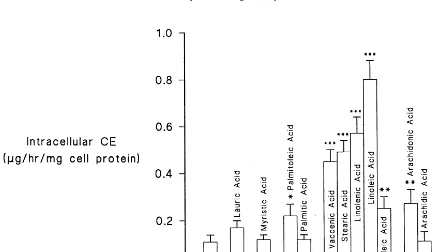
When cells were incubated with SFA, only those incubated with stearic acid showed significantly in-creased intracellular concentrations of CE (345%, PB 0.0005) (Fig. 5).
Fig. 4. Apolipoprotein B accumulation in the medium from Hep G2 cells after incubation with different fatty acids. Experimental details are described under Section 2. Values are Mean9SE of 15 experiments. Significantly different from 1% BSA, *PB0.05, **PB0.005, ***PB
0.0005.
Fig. 5. Intracellular cholesteryl ester (CE) accumulation after incubation with different fatty acids. Experimental details are described under Section 2. Values are Mean9SE of 15 experiments. Significantly different from 1% BSA, *PB0.05, **PB0.005, ***PB0.0005.
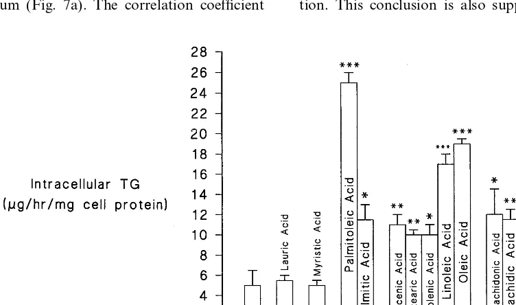
SFA chain length influenced the intracellular TG accumulation (Fig. 6). Thus, intracellular TG concen-tration in the presence of lauric acid and myristic acid was not significantly increased, whereas it was by palmitic acid (PB0.01), arachidic acid (PB0.005) (both by 130%) and stearic acid (PB0.005) by 100%.
3.3. Monounsaturated fatty acids
did not have any significant effect on apo B produc-tion.
All the MUFA studied increased the CE and TG content of Hep G2 cells (Figs. 5 and 6) to varying degrees.
3.4. Polyunsaturated fatty acids
Linoleic acid (C18:2) significantly increased apo B production by 164%, PB0.005) as did arachidonic (C20:4) and eicosapentaenoic (C20:5) acids, both by 171% (PB0.0005) (Fig. 4). Linolenic acid (C18:3), however, did not significantly affect apo B production. All of the PUFA investigated caused significant in-creases in the CE content of the cells (Fig. 5). The increases ranged from 627% (PB0.0005) for linolenic acid and 418% for linoleic acid (PB0.0005) to 127% for both eicosapentaenoic and arachidonic acid (PB 0.025). All the PUFA also increased intracellular TG (Fig. 6). Both arachidonic acid and eicosapentaenoic increased intracellular TG by 140% (PB0.005), while linolenic acid caused a 100% increase (PB0.01). The greatest increase was observed with linoleic acid (240%, PB0.0005).
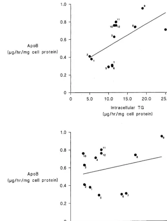
3.5. Relationship between intracellular triacylglycerol and cholesteryl ester content and the rate of apo B secretion
There was a significant (PB0.05) correlation (r= 0.6) between Hep G2 cell TG content after incubations with the various FAs and the rate of apo B secretion into the medium (Fig. 7a). The correlation coefficient
(r=0.32) between Hep G2 cell cholesteryl ester content and apo B secretion did not, however, achieve statisti-cal significance (Fig. 7b).
4. Discussion
Apo B accumulation in the medium of Hep G2 cells was chosen as a measure of VLDL secretion because of our finding, reported earlier [14] and confirmed in these experiments, that TG rapidly undergoes lipolysis after its secretion into the culture medium, presumably by hepatic lipase, which is more actively expressed by Hep G2 cells than, for example, by adult rat heptocytes in tissue culture [14,19].
Our results show that when VLDL secretion is as-sessed in terms of TG accumulation in the medium, no apparent increase is observed with increasing FA con-centration, whereas measurement of apo B accumula-tion clearly shows secreaccumula-tion of apo B-containing lipoproteins to be stimulated. Previous investigations are divided about whether oleic acid and eicosapen-taenoic acid stimulate VLDL secretion by Hep G2 cells, but any positive effects are not marked [8,20,21]. To explain the lack of apparent stimulation of TG secre-tion by FA in Hep G2 cells, it has been suggested that VLDL secretion by hepatocytes is defective and that the dense, TG deficient, apo B-containing lipoproteins present in the culture medium are secreted in that form by the Hep G2 cells [22,23]. Our results suggest that an alternative explanation for these observations is that there is rapid lipolysis of TG in VLDL after its secre-tion. This conclusion is also supported by our earlier
Fig. 7. (a) Correlation of Apo B with Intracellular TG Fatty Acids: (1) lauric acid; (2) myristic acid; (3) palmitoleic acid; (4) palmitic acid; (5) vaccenic acid; (6) stearic acid; (7) linolenic acid; (8) linoleic acid; (9) oleic acid; (10) arachidonic acid; (11) arachidic acid; and (12) eicosapentaenoic acid. (b) Correlation of Apo B with intracellular CE fatty acids: — as per Fig. 7a.
study in which there was rapid reuptake of radioac-tively labelled fatty acids in TG secreted by Hep G2 cells into the culture medium [14].
It is well accepted that dietary SFA with the excep-tion of stearate increase plasma TC and LDL-C con-centrations, whereas unsaturated fatty acids such as oleate and linoleate lower serum TC and LDL-C and the more unsaturated eicosapentanoate and docosahex-aenoate found in fish-oils lower TG and VLDL-C [5,20,24]. This forms the basis of current dietary recom-mendations to decrease serum cholesterol and TG [1]. In the experiments reported here, SFAs with the excep-tion of stearate increased apo B secreexcep-tion by Hep G2 cells. The effects of SFA on the secretion of apo
B-containing lipoproteins in our human hepatocyte model thus reflect the dietary effects of SFAs on serum concentrations of these lipoproteins, including the neu-tral effect of stearate [20]. However, the MUFAs and PUFAs investigated with the exception of linolenic acid and vaccenic acid, which is a trans-FA, also increased the secretion of apo B-containing lipoproteins.
some investigators have suggested that CE is the major lipid species stimulating VLDL secretion by Hep G2 cells [25]. It is possible that in the presence of abundant FA for TG synthesis, CE becomes less important. Furthermore it is well-known that in the human very little CE is secreted in hepatic VLDL, most of the cholesterol secreted by the liver being in the form of free cholesterol, esterification of which occurs in the circulation [18]. Thus the situation in human liver, unlike for example that in the rat in which most cholesterol is esterified before secretion, may mean that hepatic TG synthesis has a greater influence on apo B secretion. Our study thus supports the conclusion of Benoist and Grand-Perret [26] using inhibitors of lipid biosynthesis in Hep G2 cells.
Conclusions based on the Hep G2 cell model of lipoprotein secretion must necessarily be limited, be-cause it is an immortalised cell line derived from neo-plastic tissue. However, the present observations of the differential effects of different fatty acids on apo B secretion by Hep G2 cells are interesting, firstly because they indicate that TG synthesis is the dominant influ-ence on apo B secretion in human hepatocytes and secondly because the primary effect of FAs on apo B secretion does not reflect their dietary effects on serum VLDL and LDL levels in human experiments. The implications of this are that the nutritional effects of different fatty acid components in the diet on serum apo B-containing lipoproteins are likely to include indi-rect effects on hepatic lipoprotein secretion or post-secretory effects. The mechanism by which polyunsaturated fatty acids decrease hepatic TG secre-tion [27] may, for example, be due to decreased circu-lating NEFA levels [28] which are a major determinant of hepatic TG synthesis [29]. Recent reviews have also emphasised the importance of the decreased receptor-mediated catabolism of LDL during saturated fatty acid feeding [30] and the increase in cholesteryl ester transfer from HDL to apo B-containing lipoproteins [31]. The high rates of apo B secretion induced by highly polyunsaturated fatty acids in our experiments might also explain why, in human feeding experiments, eicosapentaenoic acid and docosahexaenoic acid do not lower cholesterol, despite their potentially favourable effect on LDL receptor-mediated clearance. On the other hand less highly unsaturated fatty acids, such as linoleate and oleate, do lower serum LDL cholesterol levels, perhaps because they stimulate VLDL and LDL production to a lesser extent, thus allowing their fa-vourable effect on LDL receptor mediated clearance to come to the fore.
Hep G2 cells cultured in flasks are not a suitable model for the investigation of the effects of different fatty acids on TG secretion, because of high rates of lipolysis when TG-containing lipoproteins remain in contact with the cells. However, our preliminary
exper-iments and those of others [16] suggest that perfused Hep G2 may provide a suitable method for studying TG metabolism and other aspects of lipoprotein metabolism in future studies.
Acknowledgements
We thank Dr D.E. Bowyer and Mrs C.F. Fitzsim-mons for advice about cell culture on microcarrier beads. Support for this work was gratefully accepted from the National Health Service Research and Devel-opment Levy. We are indebted to C. Price for expertly preparing the manuscript.
References
[1] Keys A, Anderson JT, Grande F. Serum cholesterol response to changes in the diet. IV. Particular saturated fatty acids in the diet. Metabolism 1965;14:776 – 87.
[2] Hegsted DM, McGandy RB, Myers ML, Stare FJ. Quantitative effects of dietary fat on serum cholesterol in man. Am J Clin Nutr 1965;17:281 – 95.
[3] Hegsted DM, Ausman LM, Johnson JA, Dallal GE. Dietary fat and serum lipids: an evaluation of the experimental data. Am J Clin Nutr 1993;57:875 – 83.
[4] Diet and Health Implications for reducing chronic disease risk. Washington, DC: National Academy Press, 1989. p. 159 – 258. [5] Connor SL, Connor WE. Are fish oils beneficial in the
preven-tion and treatment of coronary artery disease? Am J Clin Nutr 1997;66(Suppl):1020S – 31S.
[6] Zannis VI, Breslow JL, San Giacomo TR, Aden DP, Knowles BB. Characterisation of the major apolipoproteins secreted by two human hepatoma cell lines. Biochemistry 1981;20:7089 – 96. [7] Rash JM, Rothblat GH, Sparks CE. Lipoprotein apolipoprotein synthesis by human hepatoma cells in culture. Biochim Biophys Acta 1981;666:294 – 8.
[8] Dashti N, Wolfbauer G. Secretion of lipids, apolipoproteins, and lipoproteins by human hepatoma cell line, Hep G2: effects of oleic acid and insulin. J Lipid Res 1987;28:423 – 36.
[9] Pullinger CR, North JD, Teng B-B, Rifici VA, Ronhild de Brito AE, Scott J. The apolipoprotein B gene is constitutively ex-pressed in Hep G2 cells: regulation of secretion by oleic acid, albumin, and insulin, and measurement of the mRNA half-life. J Lipid Res 1989;30:1065 – 77.
[10] Ellsworth JL, Erickson SK, Cooper AD. Very low and low density lipoprotein synthesis and secretion by the human hep-atoma cell line Hep G2: effects of free fatty acid. J Lipid Res 1986;27:58 – 874.
[11] Kane JP. Apolipoprotein B: structural and metabolic hetero-geneity. Annul Rev Physiol 1983;43:637 – 50.
[12] Erickson SK, Fielding PE. Parameters of cholesterol metabolism in the human hepatoma cell line, Hep G2. J Lipid Res 1986;27:875 – 83.
[13] Wang SR, Pessah M, Infante J, Catala D, Salvat C, Infante R. Lipid and lipoprotein metabolism in Hep G2 cells. Biochim Biophys Acta 1988;961:351 – 63.
[15] Knowles BB, Howe CC, Aden DP. Human hepatocellular car-cinoma cell lines secrete the major plasma proteins and hepatitis B surface antigen. Science 1980;209:497 – 9.
[16] Dovey LA. Measurement of the LDL receptor activity in Hep G2 cells grown on microcarrier beads. MPhil Thesis, Open University, 1994.
[17] Mackness MI, Durrington PN. In: Converse CA, Skinner ER, editors. Lipoprotein Analysis: a Practical Approach. Oxford: IRL Press, 1992:1 – 42.
[18] Durrington PN. Hyperlipidaemia. Diagnosis and Management, 2nd Edition. Oxford: Butterworth Heinemann, 1995.
[19] Durrington PN, Newton RS, Weinstein DB, Steinberg D. Effects of glucose and insulin on the secretion of very low density lipoprotein triglyceride by cultured adult rat hepatocytes. J Clin Invest 1982;70:63 – 73.
[20] Wong S-H, Fisher EA, Marsh JB. Effects of eicosapentaenoic and docosahexaenoic acids on apoprotein B mRNA and secre-tion of very low density lipoprotein in Hep G2 cells. Arterioscler Thromb Vasc Biol 1989;9:836 – 41.
[21] Kurokawa M, Hirano T, Furakawa S, Nagano S, Adachi M. Similar to oleic acid, eicosapentaeoic acid stimulates apolipo-protein B secretion by inhibiting its intracellular degradation in Hep G2 cells. Atherosclerosis 1995;112:59 – 68.
[22] Wu X, Shang A, Jiang H, Ginsberg HN. Low rates of apo B secretion from Hep G2 cells result from reduced delivery of newly synthesised triglyceride to a ‘secretion-coupled’ pool. J Lipid Res 1996;37:1198 – 206.
[23] Gibbons GF, Khurana R, Odwell A, Seelaender MC. Lipid
balance in Hep G2 cells. Active synthesis and impaired mobilisa-tion. J Lipid Res 1994;35:1801 – 8.
[24] Grundy MS, Denke MA. Dietary influences on serum lipids and lipoproteins. J Lipid Res 1990;31:1149 – 72.
[25] Avramoglu RK, Cianflone K, Sniderman AD. Role of the neutral lipid accessible pool in the regulation of secretion of apo B-100 lipoprotein particles by Hep G2 cells. J Lipid Res 1995;36:2513 – 28.
[26] Benoist F, Grand-Perret T. Apo B-100 secretion by Hep G2 cells is regulated by the rate of triglyceride biosynthesis but not by intracellular lipid pools. Arterioscler Thromb Vasc Biol 1996;16:1229 – 35.
[27] Sanders TAB, Sullivan DR, Reeve J, Thompson GR. Triglyce-ride-lowering effect of marine polyunsaturates in patients with hypertriglyceridaemia. Arteriosclerosis 1985;5:459 – 65.
[28] Singer P, Wirth M, Berger I. A possible contribution of decrease in free fatty acids to low serum triglyceride levels after diets supplemented with n-6 and n-3 polyunsaturated fatty acids. Atherosclerosis 1990;83:167 – 75.
[29] Havel RJ, Kane JP, Balasse EO, Segal N, Basso LV. Splanchnic metabolism of free fatty acids and production of triglycerides of very low density lipoproteins in normotriglyceridaemic and hy-pertriglyceridaemic humans. J Clin Invest 1970;49:2017 – 35. [30] Spady DK, Woollett LA, Dietschy JM. Regulation of plasma
LDL-cholesterol levels by dietary cholesterol and fatty acids. Ann Rev Nutr 1993;13:355 – 82.
[31] Harris WS, Connor WE, Illingworth DR, Rothrock DW, Foster DM. Effect of fish oil on VLDL triglyceride kinetics in man. J Lipid Res 1990;31:1549 – 58.
.