Open access:
www.balimedicaljournal.org
and www.ojs.unud.ac.id 76
SPILANTHES ACMELLA AND PHYSICAL EXERCISE
INCREASED TESTOSTERONE LEVELS AND OSTEOBLAST
CELLS IN GLUCOCORTICOID-INDUCED OSTEOPOROSIS
MALE MICE
1
Hening Laswati,
1Imam Subadi,
2Retno Widyowati,
2Mangestuti Agil,
3
Jahya Alex Pangkahila
31
Department of Physical Medicine and Rehabilitation, Faculty of Medicine,
Airlangga University, Surabaya-Indonesia
2
Department pf Pharmacognosy and Phytochemistry, Faculty of Pharmacy,
Airlangga University, Surabaya-Indonesia
3
Department of Andrology and Sexology, Faculty of Medicine, Udayana
University, Bali-Indonesia
Background:
Glucocorticoid-induced osteoporosis is leading cause of secondary osteoporosis by
decreasing formation activity and increasing resorption activity. Spilanthes acmella, is one of
Indonesia medicinal plants that contain of polyphenol and flavonoids. Previously in vitro study
showed that buthanol and water fraction from this plant have increased alkaline phosphatase that
known as marker of bone formation.
The objective of this study to analyze the effect of
Spilanthes
acmella
and physical exercise in increasing testosterone and osteoblast cells of femoral’s
trabecular
glucocorticoid-induced osteoporosis male mice.
Method:
This study using a posttest control group
design, 36 male healthy mice (5 months old) were randomizely devided into 6 groups, there are :
1.Healthy control group (without induction dexamethaxone), 2.Osteoporosis groups (induction with
dexamethaxone without treatment), 3.Positive control receive suspension alendronat, 4.70% Ethanol
extract of Spilanthes acmella group, 5.Combination group of 70% extract ethanol of Spilanthes
acmella and exercise, and 6.Exercise group (walking using mice treadmill 10m/minute, 5-12 minutes
3 times a week). All of the intervention were given for 4 weeks. The serum levels of testosterone were
determined using immunoserology (ELISA) and osteoblast cells were determined histomorphometry
by light microscopy. All statistical test were carried out using SPSS 23 and statistical significance was
set at
p
<0.05 for all analysis. The testosterone levels between group were compared using
Mann-Whitney test and osteoblast cells between group were compared with multiple comparison.
Results:
It
showed that the alendronate group, combination group and the exercise group increasing testosterone
level (
p
<0.05) from that osteoporotic group. There were also increasing osteoblast cells (p<0.05) in the
alendronate group and combination group. There was no correlation between testosterone level and
osteoblast cells (
p
>0.05).
Conclusion:
It proved that 70% ethanol extract of Spilanthes acmella have
an additive effect to weight bearing exercise in glucocorticoid-induced osteoporosis male mice.
Keywords: Glucocorticoid; Induced; Osteoporosis; Spilanthes acmella; Testosterone
INTRODUCTION
Osteoporosis is defined as a skeletal disorder of compromised bone strength predisposing to an increased risk of fracture.1 Recent epidemiological studies have shown that osteoporosis become as a
Address for correspondence: Hening Laswati
Department of Physical Medicine and Rehabilitation, Faculty of Medicine, Airlangga University, Surabaya-Indonesia
Email: [email protected]
Open access:
www.balimedicaljournal.org
and www.ojs.unud.ac.id 77
States have osteoporosis (T-score <-2.5) and 8 to 13 million have osteopenia (Tscore between 1.0 and -2.5) or the prevalence are 6% for osteoporosis and 47% for osteopenia.2 In aging population, morbidity and mortality from hip fractures are higher in men than in women with fatality rates among over 75 years is 20.7% in men versus 7.5% in women.1 The causes osteoporosis in men are related to genetics, environmental, hormonal and disease-specific factors, and approximately 50% of men with osteoporosis are secondary osteoporosis.2 The three major causes of secondary osteoporosis in men are alcohol abuse, glucocorticoid excess (Cushing’s syndrome or long-term glucocorticoid therapy) and hypogonadism. The prevention and treatment according Recommendation of American College of Rheumatology Ad Hoc Committe including supplementation with calcium and vitamin D, antiresorptive agents (bisphosphonates),calcitonin, replacement of gonadal sex hormone (testosterone replacement therapy), and modify lifestyle risk factors.4 Clinical evidence suggests a role for phytoestrogen in the treatment of post-menopausal osteoporosis.5,6 Based on screening of 32 Indonesian tradisional medicinal plants Spilanthes acmella aerial parts stimulated ALP activity. Previously in vitro study showed that buthanol and water fraction from this plant have increased alkaline phosphatase (ALP) that known as marker of osteoblast differentiation.7 Phytochemistry study showed the major constituent in this plant was spilanthol (N-isobutylamide) and there also triterpenoid.8 Suthikrai et al.,(2010) reported that Spilanthes acmella contains 0.59-1.39 ng/g of phytotestosterone.9 There have been many reports the effect of phytoestrogen and exercise in invivo studies, but still few invivo studies which reports the effect of phytotestosterone and exercise. The objective of this study to analyze the effects of
Spilanthes acmella and physical exercise in increasing testosterone and osteoblast cells of femoral’s trabecular in glucocorticoid–induced osteoporosis male mice. group, alendronate group: osteoporosis received alendronate suspension (0.026 mg/20 g BW/day), Spilanthes acmella group : osteoporosis received 70% ethanol extract of Spilanthes acmella (4.14 mg/20 g BW/day), exercise group: osteoporosis with intervention walking with velocity 7-10m/min for 5-12 minutes, 3 times/week, and combination group: osteoporosis received 70% ethanol extract of Spilanthes acmella and exercise. To determined the
effect of dexamethasone (0.002 mg/20g BW/day for 4 weeks) the trabecular area of proximal femur from six normal male rats and six male rats who received 4 weeks dexamethasone were determined histomorphometry. Four weeks after intervention the serum testosterone levels were determined with immunoserology (ELISA). After femurs were removed, immediately fixed in 10% neutral-buffered formalin, and placed in decalcifying solution for 24 h at 37°C, continuous with dehydrated and embedded in paraffin. The proximal femur section dyed with a haematoxylin-eosin (HE)
stain. Osteoblast cells were determined
histomorphometry by light microscopy, magnifying 2000 times. The amount of osteoblast cells can be counted as a total osteoblast per five field area. Because the test of normality in osteoporosis and spilanthes acmella groups significant (p<0.05), the testosterone levels between the groups were compared using Mann-Whitney test. Multiple comparison test was applied to determine the specific difference between the groups of osteoblast cells. All statistical test were carried out using SPSS 23 and statistical significance was set at p<0.05 for all analysis.
RESULTS
Histomorphometry were determined based on the effect of dexamethasone (0.002 mg/20g BW/day for 4 weeks) the trabecular area of proximal femur from six normal male rats and six male rats who received 4 weeks. The result was presented in Figure1.
Figure 1
Histomorphometry trabecular area of the proximal femur of the normal control male rat (A) and after
received dexamethasone for 4 weeks (B). The section dyed with a haematoxylin-eosin, magnifying 100 times. There was decrease in the thickness of trabeculae (T) in the dexamethasone group (B) compared to the normal control groups
Open access:
www.balimedicaljournal.org
and www.ojs.unud.ac.id 78
There is a significant increase of testosterone levels after intervention compared to osteoporosis and Spilanthes acmella groups (p<0.05). The result can be seen in Figure 2.
Figure 2
Testosterone levels after intervention. Note there is a significant increase (p<0.05) the testosterone levels in the alendronate group, combination group
and exercise group, compared to osteoporosis and Spilanthes acmella groups.
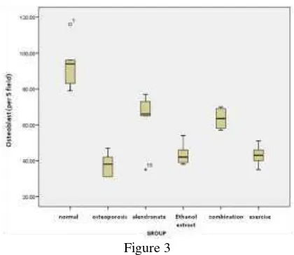
In this study we obtained that osteoblast cells after interventions is a significant increase osteoblast cells in the alendronate and combination groups (p<0.05), compared to the osteoporosis, spilanthes acmella and exercise groups (Figure 3)
Figure 3
Osteoblast cells after interventions. Note there is a significant increase osteoblast cells in the alendronate and combination groups (p<0.05), compared to the osteoporosis, spilanthes acmella
and exercise groups.
Histomorphometry trabecular area of the proximal femur can be seen in Figure 4.
DISCUSSION
There were significancy difference of testosterone levels on alendronate group (p=0.016), combination group (p=0.048) and exercise group (p=0.016) from osteoporotic group but not significance difference in ethanol extract group (p=0.112). The effect of combination group same with the effect of alendronate group (p=0.789) and exercise group (p=0.895).
Figure 4
Histomorphometry trabecular area of the proximal femur. The section dyed with a haematoxylin-eosin,
osteoblast cells was counted by light microscopy, magnifying 2000 times. Note there is an increase osteoblast cell (arrow) in the alendronate (B) and
combination group (D) compared to the osteoporosis (A), Spilanthes acmella (C) and
exercise (E) groups.
This study showed that in the osteoporotic group (after intervention with dexamethasone for 4 weeks), the testosterone level is lower than healthy group and ethanol extract group but not significant.
In human, after 3 months glucocorticoid administration will decline sex steroid production by supression of gonadal function and pituitary gonadotropin secretion.3 Low serum total testosterone (below 3 ng/ml) was the cause of osteoporosis. There is less information regarding
hypogonadism secondary to glucocorticoid
treatment.
androgen-Open access:
www.balimedicaljournal.org
and www.ojs.unud.ac.id 79
replaced men with long-term hypogonadism after 12 months of alendronate treatment. The urinary
marker of bone resorption (urinary
deoxypyridinoline) decreased significantly after 6 months of therapy with alendronate.10 This condition will result balance of bone remodeling and increasing of serum marker of bone formation (osteocalcin). Osteocalcin promotes testosterone production in the Leydig cell by activating steroidogenesis enzymes.11,12
In this study 4.14 mg/20g BW/day ethanol extract of Spilanthes acmella treatment was not increase testosterone level. The possibility of the result in this study could cause by the effect of intestinal metabolism of phytotestosterone13 or the other possibility in osteoporotic condition several cytokines such IL-1β, IL-11 and TNFα stimulated aromatase activity of osteoblast-like cells in vitro, convert testosterone to estrogen.14 This result contrary with Sharma et al.,(2011), they found that in healthy male rats who received 50, 100 and 150
mg/kg Spilanthes acmella extract, serum
testosterone level increased significantly in comparison to the control group.15 Peripheral aromatization of testosterone into estrogen may a key role in maintaining estrogen level in osteoporotic condition. In this study was not measure the estrogen level.
In exercise group the testosterone levels was increase may cause by induction the hormonal and immune respons.16 Lane reported that moderate and high intensity exercise caused an increase in both salivary and serum testosterone level.17
In combination group the testosterone level also increased signficantly. The result same with Laswati in vivo study (2007), in postmenopause mice, the estrogen levels increased highest significantly in combination group than only phytoestrogen treatment or exercise intervention.18
From one-way ANOVA analysis the level significancy p=0.000, there was minimally one pair group have significant different of osteoblast cells. From multiple comparison analysis there were significant different between osteoporotic group and alendronate group (p=0.001) and combination alendronate group (p=0.967).
In this study showed that osteoblast cells in the osteoporotic group without intervention ethanol extract and exercise is lower than the other groups. Glucocorticoid blunt intestinal calcium absorption directly and secondary hyperparathyroidism develops, increasing osteoclast life span and activity and skeletal turnover, also directly blunt osteoblast activity, decrease in the lifespan
osteoblasts and induce osteocyte apoptosis.3,19 (2,3 Licata, Weinstein). Isoenzyme 11β-hydroxysteroid
dehydrogenase (11β-HSD1) expression, a
prereceptor modulator of glucocorticoid action increases with glucocorticoid administration.19 Ma
et al.,(2011) reported that in vivo glucocorticoid may increase the expression and signaling activity of β2-adrenergic receptors (β2AR) in osteoblasts as antianabolic effect of sympathetic neuron. Stimulation of the β2-adrenergic receptors (β2AR) in osteoblasts by norepinephrine or isoproterenol inhibits osteoblast proliferation, stimulates osteoclastogenesis and up regulation of nuclear factor-кB ligand (RANKL) expression. Study with pharmacological and genetic β2AR blockade in mice significantly reduced the bone catabolic effect of high-dose prednisolone in vivo. In vitro study shows a direct effect and genomic effect of the glucocorticoid receptor (GR) on the Adrβ2 promoter.20
There was increased significantly osteoblast cells in alendronate group. Shimon et al., (2005) reported that alendronate treatment 10 mg daily for 6 and 12 months in osteoporotic men with long-standing hypogonadism and receiving standar testosterone replacement treatment increased
lumbar-spine bone mineral density (BMD)
significantly (p<0.005).10 A 2-year double-blind, placebo-controlled trial of 10 mg of alendronate daily was carried out in 241 men with osteoporosis who were aged 31 to 87. After 2 years, men in the alendronate group showed a 7.1% increase in bone density at the lumbar spine, but those in the placebo group showed a 1.8% increase.21 Alendronate is an anti-resoptive agent, inhibit farnesyl diphosphate (FPP) synthase, thus blocking the prenylation of small signalling proteins essential for osteoclast function and survival.
Open access:
www.balimedicaljournal.org
and www.ojs.unud.ac.id 80
(2002) reported that estrogen treatment through bone morphogenetic protein-2 (BMP-2) was increased osteoblast cell.23 In vivo study with glucocorticoid-induced osteoporosis female rats, genistein aglycon showed a greater increased in bone mineral density, and significantly increased bone-alkaline phosphastase as a marker of osteoblast differentiation.24 The possibility of the result in this study could cause by the effect of intestinal metabolism of phytotestosterone13 and may depend on aromatization of phytotestosterone to estrogen.22
In exercise group we found osteoblast cells increase but not significantly. During physical activity, mechanical forces are exerted on the bones through ground reaction forces and by the contractile activity of muscles. Osteocytes are highly mechanosensitive, alter the production of a multitude of signaling molecules when triggered by a mechanical stimulus. Mechanically activated osteocytes produce signaling molecules like bone
morphogenetic proteins (BMPs), Wnts,
prostaglandin E2 (PGE2), and NO, which can modulate the recruitment, differentiation, and activity of osteoblasts.25 Cheng at al., (2002) reported that the anabolic effect of strain on osteoblast cell numbers is mediated by IGF’s action through the IGF-1 receptor (IGF-1R) within the cell membrane and this responsiveness to a ligand is regulated by integrins.26 This result in this study might be an effect of the low sensitivity of the mice skeleton to moderate intensity for 4 weeks. Other factors such as stress have probably influenced the results.
Combination of exercise and spilanthes acmella treatment showed increased osteoblast cells significancy. This may cause by cross-talk mechanism of the IGF-1 and estrogen. The number and activity of ER were regulated by estrogen. The effect of mechanical force from exercise on osteoblast cell numbers is mediated by IGF’s action through the IGF-1 receptor (IGF-1R). IGF-1R requires association with ligand-bound ERα that will results in IGF-1R autophosphorylation and activation downstream mitogen activated regulated kinase (MAPK) and extracellular regulated kinase (ERK) signaling cascade for osteoblast survival and proliferation.26 In this study the effect of combination group have the same effect to increasing osteoblast cells with alendronate group (p=0.967)
There is no correlation between testosterone level and osteoblast cells (r=0.177; p=0.358). A study on androgen supplementation in eugonadal men with osteoporosis, the increase in BMD and the reduction in bone turnover positively correlated with estradiol, but not in testosterone levels indicating of conversion androgen to estrogen.2 In this study peripheral aromatization of testosterone
into estrogen may have a role in osteoporosis condition.
CONCLUSIONS
Seventy percentage of ethanol extract of Spilanthes acmella have an additive effect to weight bearing exercise through increasing testosterone and osteoblast cell of trabecular proximal femur in glucocorticoid-induced osteoporosis male mice. The results in this study suggest a need for further
researches to investigate the role of
phytotestosterone and AR in bone cells.
REFERENCES
1. Siddapur P R, Patil A B, Borde V S. Comparison of Bone Mineral Density, T-scores and serum zink between diabetic and non
diabetic postmenopausal women with
osteoporosis. Journal of Laboratory Physicians 2015; 7(1): 43-48.
2. Gennari L and Bilezikian JP. Osteoporosis in Men. Endocrinol Metab Clin N Am 2007; 36: 399-419
3. Licata A. Osteoporosis in men: Suspect secondary disease first. Cleveland Clinic Journal of Medicine 2003; 70: 247-254. 4. American College of Rheumatology Ad Hoc
Committee on Glucocorticoid-Induced
Osteoporosis. Recommendations for the Prevention and Treatment of Glucocorticoid-Induced Osteoporosis. 2001 Update. Arthritis and Rheumatism 2001;44, 1496-1503.
5. Uesugi T, Fukui Y and Yamori Y. Beneficial effects of soybean isoflavon supplementation on bone metabolism and serum lipids in postmenopausal Japanese women. A four week study. Journal of The American College of Nutrition 2002; 21: 97-102
6. Atkinson C, Compston JE, day NE, Dowsett M and Bingham SA. The effects of phytoestrogen isoflavons on bone density in women: a Doulbe-blind, randomized, placebo-controlled trial. Am L Clin Nutr 2004: 79: 326-333 7. Widyowati R. Alkaline Phosphatase Activity of
Graptophylum pictum and Spilanthes acmella fractions against MC3T3-E1 Cells as Marker of Osteoblast Differentiation Cells. International Journal of Pharmacy and Pharmaceutical Sciences 2011; 3(supp.1) : 34-37
8. Dubey S, Maity S, Singh M, Saraf SA and Saha S. Phytochemistry, Pharmacology and Toxicology of Spilanthes acmella: A Review. Advances in Pharmacological Sciences 2013.
Available from URL: http:
//dx.doi.org/10.1155/2013/423750
Open access:
www.balimedicaljournal.org
and www.ojs.unud.ac.id 81
weeds from pasture. Thai J Toxicol 2010; 25(2):183
10. Shimon I, Eshed V,Doolman R, Sela B-A, Karasik A and Vered I. Alendronate for osteoporosis in men with androgen-releted hypogonadism. Osteoporos Int 2005; 16: 1591-96
11. Ferlin A, Selice R, Carraro U and Foresta C. Testicular function and bone metabolism-beyond testosterone. Nat Rev Endocrinol 2013; 9: 548-554
12. Karsenty G and Oury F. Regulation of male fertility by the bone derived hormone osteocalcin. Mol Cell Endocrinol 2014;382(1): 1-13
13. Chiechi LM and Micheli L. lity of dietary phytoestrogens in preventing postmenopausal osteoporosis. Current Topics in Nutraceutical research 2005; 3(1) :15-28
14. Shozu M and Simpson ER. Aromatase expression of human osteoblast-like cell. Molecular and Cellular Endocrinology 1998; 139:117-129
15. Sharma V, Boonen J, Chauhan NS, Thakur M, De Spiegeleer B and Dixit VK. Spilanthes acmella ethanolic flower extract: LC-MS alkylamide profiling and its effects on sexual behavior in male rats. Phytomedicine 2011; 18: 1161-1169
16. Holly RG and Shaffrath JD. Cardio Respiratory Endurance. In (Roitman JL Ed). ACSM`s Resource Manual for Guidelines for Exercise Testing and Prescription. 4th ed. Philadelphia: Lippincott William & Wilkins 2001: pp 203– 204
17. Lane AR and Hackney AC. Relationship between salivary and serum testosterone levels in response to different exercise intensities. Hormones (Athens) 2015; 14(2): 258-64 18. Laswati H. Combine of physical exercise and
Semanggi leaves administration increase expression of ERα and ERK1 /2 osteoblast cell
in menopause mice. Jurnal Biosains
Pascasarjana 2007; 9(2):70-77
19. Weinstein RS. Glucocorticoid-Induced Bone Disease. N Engl J Med 2011;365:62-70 20. Ma Y, Nym JF, Tao H, Moss HH, Yang X and
Elefteriou F. Β2-Adrenergic Receptor Signaling in Osteoblasts Contributes to the Catabolic Effect of Glucocorticoids on Bone. Endocrinology 2011; 152(4): 1412-1422. 21. Orwoll E, Ettinger M, Weiss S, Miller P,
Kendler D, Graham J, Adami S, Weber K, Lorenc R, Pietschmann P, Vandormael K, Lombardi A. Alendronate for the treatment of osteoporosis in men. N Engl J Med 2000;343:604–610.
22. Sinnesael M, Boonen S, Claessens, Gielen E and Vanderschueren D. Testosterone and the Male Skeleton : A Dual Mode of Action. Journal of Osteoporosis 2011. Available from URL: http://dx.doi.org/10.4061/2011/240328 23. Okazaki R, Inoue D, Shibata M, Saika M, Kido
S, Ooka H, Tomiyama H, Sakamoto Y, and Matsumoto. Estrogen promote early osteoblast differentiation and inhibits adipocyte differentiation in mouse bone marrow stromal cell lines that express estrogen receptor α or β. Endocrinology 2002; 143(2):2349-2356 24. Bitto A, Burnett BP, Polito F, Levy RM,
Marini H, Di Stefano V, Irrera N, Armbruster MA, Minutoli L, Altavilla D and Squadrito F. Genistein aglycone reverses glucocorticoid-induced osteoporosis and increases bone breaking strength in rats: a comparative study with alendronate. British Journal of Pharmacology 2009; 156:1287-1295
25. Klei-Nulend J, Bacabac RG and Bakker AD. Mechanical loading and how it affects bone cells: The role of the osteocyte cytoskeleton in maintaining our skeleton. European Cells and Materials 2012; 24: 278-291
26. Lanyon L, Amstrong V, Ong D, Zaman G and Price J. Is estrogen receptor α key to controlling bones’ resistance to fracture? Journal of Endocrinology 2004; 182: 183-191