© World Health Organization 2016 2nd Edition
All rights reserved. Requests for publications, or for permission to reproduce or translate WHO publications, whether for sale or for noncommercial distribution, can be obtained from Publishing and Sales, World Health Organization, Regional Office for South-East Asia, Indraprastha
Estate, Mahatma Gandhi Marg, New Delhi-110 002, India (fax: +91-11-23370197; e-mail: publications@ searo.who.int).
The designations employed and the presentation of the material in this publication do not imply the expression of any opinion whatsoever on the part of the World Health Organization concerning the legal status of any country, territory, city or area or of its authorities, or concerning the
delimitation of its frontiers or boundaries. Dotted lines on maps represent approximate border lines for which there may not yet be full agreement.
The mention of specific companies or of certain manufacturers’ products does not imply that they are endorsed or recommended by the World Health Organization in preference to others of a similar nature that are not mentioned. Errors and omissions excepted, the names of proprietary products are distinguished by initial capital letters.
All reasonable precautions have been taken by the World Health Organization to verify the information contained in this publication. However, the published material is being distributed without warranty of any kind, either expressed or implied. The responsibility for the interpretation and use of the material lies with the reader. In no event shall the World Health Organization be liable for damages arising from its use.
This publication contains the collective views of an international group of experts and does not necessarily represent the decisions or the stated policy of the World Health Organization. Printed in India
WHO Library Cataloguing-in-Publication data
Guidelines for the management of snake-bites, 2nd edition.
1. Snake Bites – education – epidemiology – prevention and control – therapy. 2. Public Health.
3. Venoms – therapy. 4. Guidelines.
iii. Preface 9
1 Executive summary 11
2 Prevention 17
Essentials 19
2.1 Reducing the risk of snakebites 19
2.2 Implementing preventive strategies for community education 22
2.3 Conclusion 23
3 Venomous snakes of the South-East Asia Region, 25 their venoms and pathophysiology of human envenoming
Essentials 27
3.1 The venom apparatus
3.2 Classification of venomous snakes 29
3.2.1 Medically important species in South-East Asia Region countries 30
3.2.2 Other medically important venomous snakes 63
3.2.3 Non-venomous snakes 64
3.3 Identification of venomous snakes 67
3.4 Snake venoms 67
3.4.1 Venom composition 67
3.4.2 Quantity of venom injected at a bite, “dry bites” 69
3.4.3 Variations in venom composition within 69
individual species of snakes
3.5 Pathophysiology of human envenoming 69
4 Epidemiology of snakebites in South-East Asia Region countries 73
Essentials 75
4.1 Introduction 77
4.2 Determinants of snakebite incidence and severity of envenoming 78
4.3 Epidemiological characteristics of snakebite victims 79
4.4 Circumstances of snakebites 79
4.5 Snakebite as an occupational disease 79
4.6 Death from snakebite 80
4.6.1 Factors contributing to fatal snakebite 80
4.6.2 Time between snakebite and death 80
5 Clinical aspects of snakebites 89
Essentials 91
5.1 When venom has not been injected 94
5.2 When venom has been injected 94
5.2.1 Early symptoms and signs 94
5.2.2 Clinical patterns of envenoming by snakes in South East Asia 95
5.2.3 Local symptoms and signs in the bitten part 95
5.2.4 Generalized (systemic) symptoms and signs 96
5.3 Clinical syndromes of snakebite in South-East Asia 101
5.4 Long-term complications (sequelae) of snakebite 104
5.5 Symptoms and signs of seasnake envenoming 103
5.6 Symptoms and signs of cobra-spit ophthalmia 104
(eye injuries from spitting cobras)
6 Management of snakebites in South-East Asia 105
Essentials 107
6.1 Stages of management 115
6.2 First-aid treatment 115
6.2.1 Principles of first-aid 115
6.2.2 The danger of respiratory paralysis and shock 115
6.2.3 Recommended first-aid methods 116
6.3 Transport to hospital 117
6.4 Medical treatment in the dispensary or hospital 118
6.4.1 Rapid primary clinical assessment and resuscitation 118
6.4.2 Detailed clinical assessment 119
6.4.2.1 History 119
6.4.2.2 Physical examination 120
6.4.2.3 Neurotoxic envenoming: Bulbar and respiratory paralysis 121
6.4.2.4 Generalized rhabdomyolysis 123
6.4.2.5 Examination of pregnant women 123
6.5 Species diagnosis 123
6.6 Investigations/laboratory tests 123
6.6.1 20 minute whole blood clotting test (20WBCT) 123
6.6.2 Other tests of blood coagulation 126
6.6.3 Other laboratory tests 126
6.6.4 Other investigations 127
6.7 Antivenom treatment 128
6.7.1 What is antivenom? 128
6.7.2 Indications for antivenom treatment 129
6.7.3 Inappropriate use of antivenom 130
6.7.4 How long after the bite can antivenom be expected to be effective? 130
6.7.5 Antivenom reactions 130
6.7.5.1 Prediction of antivenom reactions 132
6.7.5.2 Contraindications to antivenom: 132
6.8 Conservative treatment when no antivenom is available 143
6.9 Supportive/ancillary treatment 144
6.9.1 Treatment of neurotoxic envenoming 144
6.9.2 Practical guide to airway management and respiratory support 144
6.9.3 Trial of anticholinesterase 152
6.9.4 Treatment of hypotension and shock 154
6.9.5 Treatment of oliguria and acute kidney injury 156
6.9.5.1 Oliguric phase of renal failure 156
6.9.5.2 Bleeding/ blood clotting disturbances in 159
patients with acute kidney injury
6.9.5.3 Prevention of renal damage in patients with myoglobinuria 159 or haemoglobinuria
6.9.5.4 Diuretic phase of renal failure 160
6.9.5.5 Renal recovery phase 160
6.9.5.6 Persisting renal dysfunction 160
6.10 Haemostatic disturbances 160
6.10.1 Dangers of venipuncture in patients with haemostatic abnormalities 160
6.11 Treatment of the bitten part 161
6.11.1 Wound management 161
6.11.2 Bacterial infections 161
6.11.3 Compartment syndromes and fasciotomy 161
6.12 Rehabilitation 163
6.13 Discharge advice and assessment 163
6.14 Management of cobra spit ophthalmia 164
6.15 Management of snakebites at different levels of the health service 165
6.16 Summary of current evidence for treatment of snakebite envenoming 168
7 References and further reading 169
8 Annexes 185
Annex 1 Algorithm 1: Antivenom treatment of snakebite cases 187 Annex 2 Algorithm 2: Differentiating major Asian snake species by clinical syndrome 188 Annex 3 Antivenoms for treating bites by South-East Asian snakes 191 Annex 4 Pressure-immobilization and pressure pad 195 Annex 5 Measurement of central venous pressure 197 Annex 6 Measurement of intracompartmental pressure 198
S
scale of this problem however is unknown because of inadequate reporting in almost every part of the world. The South-East Asia Region is one of the world’s most affected regions, due to its high population density, widespread agriculture activities, presence of numerous venomous snakes and lack of necessary community awareness to address the problem.Regardless of the increasing knowledge about the composition of snake venom and its mode of action, and a sound
understanding of clinical features of envenoming, management of snakebites remains a challenge in the Region. Appropriate and timely use of anti-snake venom reduces morbidity and mortality. Although in the last few years production of anti-venom has increased, it is still well below the current estimated annual requirement of 10 million vials. In addition, available anti-venoms do not cover all the important venomous snakes of the Region. Mechanisms need to be developed to ensure timely access and availability of anti-venom to all needy persons. Public-private partnerships, intercountry support, involvement of national, regional, and global agencies are vital to effectively meet the challenge.
The WHO Regional Office for South-East Asia had developed and published Guidelines for the Management of Snakebites as a special issue of the South East Asian Journal of Tropical Medicine and Public Health in 1999 followed by publication of the guidelines as independent regional document in 2011. Considering the new technical advances in the field, the guidelines have been revised with the help of regional and global experts. The geographical area specifically covered by this publication includes the South-East Asia Region of WHO. The venomous snake fauna of the South East Asia Region is rich and diverse. It varies within and between countries. Countries such as Malaysia, Singapore, Cambodia, Lao People’s Democratic Republic, Republic of Korea, Philippines, Pakistan and Afghanistan may share many of the same medically-important species of snakes that occur in the South-East Asia Region and may find these guidelines useful. This publication aims to pass on a digest of available knowledge about all clinical aspects of snake-bite to medically trained personnel.
I am confident that these revised guidelines will help Member States to improve the management of snakebites in the peripheral health services and thereby reduce the morbidity and mortality due to snakebites.
Acknowledgements
Prof David Warrell, Emeritus Professor of Tropical Medicine, University of Oxford, UK, wrote the draft of the guidelines. These were finalized through a meeting of experts held at SEARO, New Delhi, in June 2016 and revised by Dr Warrell. The list of experts who contributed can be seen at Annex 7. Contributions of all the experts and people
Asia Region of WHO including eleven Member States (Bangladesh, Bhutan, Democratic People’s Republic of Korea, Maldives, Myanmar, Nepal, India, Indonesia, Sri Lanka, Thailand, Timor-Leste ). Countries such as Malaysia, Singapore, Cambodia, Lao People’s Democratic Republic, Republic of Korea, Philippines, Pakistan and Afghanistan may share many of the same medically important species of snakes that occur in the South-East Asia Region. It is interesting to note that snakes inhabiting the Indonesian islands East of Wallace’s line (West Papua and Maluku Islands) are part of the Australasian elapid fauna, differing from those West of this line.
Antivenoms are essential drugs
The only specific antidotes to snake venoms are immunoglobulin antivenoms (WHO, 2010), which are recognized as essential drugs. see http://www.who.int/medicines/ publications/essentialmedicines/EML_2015_FINAL_amended_NOV2015.pdf?ua=1.
Target readership
This publication aims to pass on a digest of available knowledge about all clinical aspects of snakebites to medically trained personnel. The guidelines are intended for medical doctors, nurses, paramedics, dispensers and community health workers who have the responsibility of treating victims of snakebites, and also for medical and nursing students and postgraduates as an educational resource. They aim to provide sufficient practical information to allow medically trained personnel to assess and treat patients with snakebites at all the different levels of the health service.
Levels of evidence
Recommendations are based largely on observational studies (“O” see below), expert opinion (“E”) and, in some cases, comparative trials (“T”), but in only one case on formal systematic reviews (“S”).
Symbols for the evidence used as the basis of each recommendation (in order of level of evidence) are:
S formal systematic reviews, such as Cochrane Reviews of which there is only one in the field of snakebites. These include more than one randomized controlled trial;
T comparative trials without formal systematic review;
O observational studies (e.g. surveillance or pharmacological data);
Executive Summary
Epidemiology
The venomous snake fauna of the South-East Asia Region is rich and diverse. It varies within and between countries. Widely distributed species of major medical importance, such as Russell’s vipers, show geographical intra-species variation in their venom composition. In many countries, snakebite is an important medical emergency and cause of hospital admission, demanding urgent attention by adequately trained medical staff. It results in the death or chronic disability of tens of thousands of active younger people, especially those involved in farming and plantation work. The true scale of mortality and acute and chronic morbidity from snakebite is only just beginning to be recognized, based on large well-designed community-based studies (Mohapatra et al., 2011; Rahman et al., 2010; Ediriweera et al, 2016). Persisting or permanent physical and psychological disability in snakebite survivors may confer social stigma. The full burden of human suffering remains uncertain because of inadequate reporting in almost every part of the Region. To remedy this deficiency, it is strongly recommended that snakebite be made a specific notifiable disease in all countries in the South-East Asia Region (E).
Snakebite is predominantly a rural problem that has important implications for the nutrition and economy of the countries
1
where it occurs commonly, since it is largely an occupational disease of food-producers such as farmers, plantation workers, herdsmen, and fishermen, and also of wild life park rangers, military personnel, snake restaurant workers, snake handlers and collectors of snake skins. It is recommended that snakebite should be formally recognized by the International Labour Office as an important
occupational disease in the South-East Asia Region (E).
Prevention
Detailed consideration of community education on venomous snakes and snakebite is outside the scope of this publication. However, it is clear that this is a most powerful tool and essential component in any campaign to address the public health challenge posed by snakebite. It is strongly recommended as the method most likely to succeed in reducing the risk of snakebites (E).
Health system strengthening,
education and training
identification of medically important species of snake, clinical diagnosis and the appropriate use of antivenoms and ancillary treatments. It is recommended that education and training in the
prevention and management of snakebite should be included in the curriculum of medical and nursing schools and should be addressed specifically through the organization of special training courses and other educational events based on nationally agreed guidelines (E). These measures should be supported by improvements in hospital accommodation of snake-bitten patients (ideally in
specialized units staffed by specially trained teams), basic laboratory and diagnostic facilities, supply, deployment and conservation/storage of antivenoms, ambulance services and general
community awareness of the problem and its potential solutions.
Management
First-aid: most of the familiar methods for first-aid treatment of snakebite, both western and “traditional/herbal”, have been found to result in more harm (risk) than good (benefit) and should be firmly discouraged. However, in many communities, traditional therapists and their practices are respected and it is important to initiate a dialogue with these practitioners, perhaps through anthropologists, to encourage their understanding and cooperation in the timely referral of envenomed patients to
medical care at the hospital or dispensary. Recommended first-aid methods emphasise reassurance, application of a pressure-pad over the bite wound, immobilization of the bitten limb and transport of the patient to a place where they can receive medical care without delay (O).
Diagnosis of the species of snake responsible for the bite is important for optimal clinical management. This may be achieved through expert identification of the dead snake or a (mobile-phone) image of it, or by inference from the resulting “clinical syndrome” of envenoming. It is recommended that syndromic approaches and algorithms for diagnosing the species responsible for snakebites be developed in different parts of the Region (E).
Treatment: antivenom (species-specific hyperimmune immunoglobulin), a life-saving, WHO-recognized, essential medicine, is the only effective antidote for envenoming. However, there are challenges surrounding its design, production, distribution, conservation, safety, initial dosage and effectiveness. It is recommended that a critical appraisal of all aspects of antivenom production should be carried out in South-East Asia Region countries. Although antivenom is an essential element of the treatment of systemic envenoming, it may be insufficient on its own to save the patient’s life. Antivenoms are generally expensive and in short supply. It is recommended that antivenom should be used in all patients with signs of systemic and/or severe local envenoming in whom the benefits of treatment are judged to exceed the risks of antivenom reactions (E). It should not be used in the absence of evidence of envenoming. Skin/conjunctival hypersensitivity testing does not predict early or late antivenom
Most of the familiar methods for first-aid
reactions associated with a particular antivenom is low (less than a few %) (T). It is recommended that whenever possible antivenom should be given by slow intravenous injection or infusion (O). Adrenaline should always be available in readiness at the bedside in case of an early anaphylactic antivenom reaction. When no appropriate specific antivenom is available, judicious conservative treatment can in many cases save the life of the patient. In the case of neurotoxic envenoming with bulbar and respiratory paralysis, antivenom alone cannot be relied upon to prevent early death from asphyxiation. Artificial ventilation is essential in such cases. In the case of acute kidney injury associated particularly with envenoming by Russell’s vipers, hump-nosed pit-vipers and sea snakes, conservative management and, in some cases renal replacement therapy (dialysis), is an effective supportive treatment. It is recommended that fasciotomy should never be carried out in snakebite patients unless or until
given explanation, reassurance, and advice about avoiding future bites, and the opportunity for follow-up and further rehabilitation/counselling in case of late antivenom reactions and persistent physical or psychological sequelae.
Prevention
Essentials:
Snakebites are environmental, occupational and climatic hazards, predominantly of rural areas. Bites are usually inflicted on lower legs, ankles and feet of agricultural workers and their families. Know your local snakes, their favourite habitats, and times of day and seasons when they are most active. Never handle, threaten or attack snakes. Do not attract snakes to homes by keeping livestock indoors or leaving food unprotected, encouraging rodents. Sleep under a well-tucked–in mosquito net, ideally on a raised bed. Clear rubbish and undergrowth from around the house. Always use a light and prod with a stick when walking outside at night, visiting the latrine or relieving yourself in the open. Solid shoes or boots are recommended especially during agricultural activities. Fishermen should avoid touching sea snakes caught in their nets.
Community education reduces the risk of snakebites. Involve all community workers, traditional healers and villagers. Distribute leaflets, banners and posters. Reformat these SEARO recommendations for national or local use as guidelines, training modules, leaflets, video clips or posters, displayed in hospital and clinic waiting areas and disseminate them via radio, TV and social networks.
2
2.1 Reducing the risk of
snakebites
Snakebite is an environmental, occupational and climatic hazard in rural and urban areas of many South-East Asian countries where most bites are inflicted on the lower legs, ankles and feet of agricultural workers and their families. Attention to the following recommendations for community education will reduce the risk of bites. Snakes have adapted to a wide range of habitats and prey species. All snakes are predatory carnivores, none is vegetarian although some eat birds’ eggs. Since snakes are preyed upon by other animals, they tend to be secretive and have
walking to and from the fields before dawn and after dusk. Snakes prefer not to confront large animals such as humans, and so avoid cornering them and give them every opportunity to escape.
Inside the house, where snakes may enter in search of food or to find hiding places, do not keep livestock, especially chickens, as they are potential prey for larger snakes and they may attract rodents upon which many species of snakes will prey. Store food in rodent-proof containers. Regularly check houses for snakes and, if possible, avoid types of house construction that will provide snakes with hiding places (e.g. thatched roofs with open eaves, mud and straw walls with large cracks and cavities, large unsealed spaces beneath floorboards). In South Asia, almost all krait (Bungarus) bites are inflicted on people sleeping in their homes, usually on the floor but sometimes even in beds and under pillows
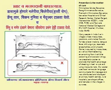
(e.g. in the Sundarbans). Ideally, avoid sleeping unprotected on the ground, but if you do choose, or are forced, to sleep on the ground, or are able to sleep on a raised bed, use an insecticide-impregnated mosquito net that is well tucked-in under the mattress or sleeping mat. This will protect against mosquitoes and other biting insects, centipedes, scorpions, and snakes (Chappuis et al., 2007). (Fig 001) No chemical has yet been discovered that is effectively repellent to snakes without being so toxic as to threaten the life of children and domestic animals.
In the farm yard, compound, or garden: Try not to provide hiding places for snakes. Clear away termite mounds, heaps of rubbish, building materials etc. from near the house. Do not have tree branches touching the house. Keep grass short or clear the ground around your house and clear underneath low bushes so that snakes cannot hide close to the
Figure 001: Protection from sleeping under a mosquito net (H Bawaskar)
Prevention is the mother of cure
Himmatroo Salube Bawaskar, Parag Himmatroo Bawaskar, Pramodini Himmatroo Bawaskar, bawasker Hospital and Clinical Research Center, Mahed Raigad, Maharashtra 402301, India (HSB, PrHB); and Topiwala National Medical College and BYL NAIR Hospital, Mumbai, India (PaHB)
Use a light when you walk outside the house, visit the latrine at night, or relieve yourself in the open.
In the countryside: firewood collection at night is a high risk activity. Watch where you walk. The value of wearing protective clothing is widely accepted, but there are many practical difficulties, including cost, habit, inconvenience and discomfort in a tropical climate as well as cultural and superstitious objections making this seem impracticable. However, in Myanmar, a specially developed light-weight boots proved acceptable to farmers for use during the high risk rice harvesting season, although these would be impracticable during rice planting when the fields are inundated (Tun Pe et al., 2002). Rather than walking bare-footed or wearing sandals, use proper shoes or boots and long trousers, especially when walking in the dark or in undergrowth. This advice may not be immediately acceptable in some communities because of cost, cultural attitudes, comfort and convenience. However, use of protective clothing (boots and gloves) should be promoted as the most obvious means of reducing occupational risk of snakebite. Step on to rocks or logs rather than straight over them – snakes may be sunning themselves on the sides. Do not put hands into holes or nests or any hidden places where snakes might be resting. Young boys often do this while hunting for rodents. Use a light (torch, flashlight or lamp) when walking at night especially after heavy rains and prod the ground ahead of you with a stick. Lamps placed at strategic locations such as at
the entrance to the house, in passages between houses, in front of the latrine outside the house or within the courtyard are valuable, provided that they are positioned so as to avoid casting shadows over corners and niches where snakes may be concealed. Be careful when handling dead or apparently dead snakes – even an accidental scratch from the fang of a snake’s severed head may inject venom. Snake restaurants pose a threat of bites to staff and customers. Many snakebites occur during ploughing, planting and harvesting and in the rainy season. Rain may wash snakes and debris into gutters at the edges of roads, and flush burrowing species out of their burrows, so be careful when walking on roads after heavy rain especially after dark.
On the road: Drivers or cyclists should never intentionally run over snakes on the road. The snake may not be instantly killed and may lie injured and pose a risk to pedestrians. The snake may also be injured and trapped under the vehicle, from where it will crawl out once the vehicle has stopped or has been parked in the house compound or garage.
In rivers, estuaries and the sea: To prevent sea snakebites, fishermen should avoid touching sea snakes caught in nets
are valuable, provided that they are
and on lines. The head and tail are not easily distinguishable. Sea snakes are air-breathing and are therefore drowned if caught in drift or trawl nets, but, unlike fish, may survive if laid on the beach. There is a risk of bites to bathers and those washing clothes in the muddy waters of estuaries, river mouths and some coastlines.
General: Avoid snakes as far as possible, including those displayed by snake charmers, who are frequently bitten. Displays by performers such as Austin Stevens and the late Steve Irwin on TV and social media have encouraged people to risk pursuing, attacking and handling wild snakes. This should be actively discouraged. Never handle, threaten or attack a snake and never intentionally trap or corner a snake in an enclosed space. In many parts of the Region there are dedicated snake catchers who will remove snakes found in houses and gardens. For example, in New Delhi, “if you find a snake hissing inside your house, just don’t panic or cause it any harm. All you need to do is dial a helpline (9871963535) and dedicated snake trappers will be at your doorstep to help you” Wildlife SOS (WSOS). Keep young children away from areas known to be snake-infested. In occupations that carry a high risk of snakebite, such as rice farming and fish farming, employers might be held responsible for providing protective clothing (boots). In Myanmar, farmers can take out special low-cost insurance to cover them specifically against snakebite, and India’s National Health Assurance Mission envisages free treatment for snakebite.
2.2 Implementing preventive
strategies for community
education
Community education has been proved to be an effective strategy for preventing snakebites. In the Terai region of Nepal, a group of villages was targeted for snakebite awareness sessions, involving political leaders, female health volunteers, community health workers, social workers, traditional healers and other villagers. Many leaflets, banners and posters were distributed. As a result, snakebite incidence decreased from 502 bites to 315 bites/100 000 population in targeted villages (relative risk reduction 0.373 (95% CI = 0.245–0.48) but it remained constant in the control untargeted villages (Sharma et al., 2013). A similar scheme in West Bengal, India involved the training of 800 health workers (Vishal Santra, Simultala Conservationists). Such programmes should be repeated every year, using well-designed posters with colour photographs of the medically significant snakes of the Region combined with clear recommendations for
a short time after the bite.
3 Consider snakebite in the differential diagnosis of unexplained altered sensorium, alteration to speech and swallowing, and abdominal pain, especially during the rainy season.
Patients can present with symptoms of envenomation, without any history of being bitten by a snake.
4 Bites by an unknown predator need to be taken seriously and observed for signs of envenomation.
14% of people, who were unable to identify what predator bit them were envenomed.
5 Don’t rely on the ability of patients or relatives to identify snakes.
Identification of snake species is poor, although it is better for cobras.
6 Antivenom should only be given to patients showing symptoms of envenomation
Envenomation of patients only occurred in 63.6% of the cases where cobras were brought. To give anti venom without symptoms of envenomation exposes people to the risk of adverse reactions and is costly, especially in a setting where demand exceeds supplies.
7 Consider the production of a bivalent ASV for regions of north India and Nepal.
Saw scaled and Russell's vipers are not present in this region and a bivalent anti venom is likely to cause less adverse reactions.
Recommendation for Community Education Rationale 1 A large part of the public health education
needs to be directed to children and young people to both genders.
The 10-19 year old age group is the peak age interval for bites. Numbers of males and females bitten are almost equal.
2 Encourage the use of footwear and long pants/trousers.
67% of bites occur on the feet and legs.
3 The use of a stick to sacare away snakes, prior to working in an area wth ones hands.
This would decrease the number of bites where hands are put into snake micro habitats without prior visualisation of the area.
4 Improved lighting using: • Torches when walking outside • Lighting in and around houses.
40% of bites occur between 1700-2200 hours. 59.2% of bites occur in and around the house. Lighting will enable better visualisation of snakes.
5 Provision of toilets and education regarding their use.
8% of bites occurred when people were going to the fields for the purpose of open defecation.
6 Encourage people to sleep on a bed and under a well tucked-in mosquito net.
10% of people were bitten while they were sleeping. Sleeping on the ground, inceases your risk of envenomation, sixfold. Sleeping under a mosquito net, decreases your risk of envenomation, six fold.
7 Provision of buffer zones between fields and housing areas.
59.2% of bites occur in and around the house. Snakes are attracted to the rodents who come for grain being grown. Keeping these distances separate may help in decreasing the encroachment of snakes in housing areas.
8 Make sleeping areas separate from food storage, preparation and sonsumption areas.
The presence of rodents in food related areas is prone to attract snakes. If people sleep in places away from areas of the house connected with food, theis may decrease the risk of people connecting with snakes. Recommendations for Community Education
The above recommendations for
preventing snakebite could be reformatted for national or local use as guidelines, training modules, leaflets, video clips and posters (Fig 001) that can be displayed on the walls of hospital and clinic waiting areas for the attention of patients and their families. At the village level, role-playing, drama and puppets have been used successfully to portray snakebite scenarios. Media such as radio and TV can be used for health promotion and advantage can be taken of FM radio phone-ins to publicise the problem. Increasingly, young people and advertisers use social networking (YouTube, Twitter) to communicate information and mobile phone messaging might also be employed.
Religious organizations and charities such as Rotary and Lions Clubs might be persuaded to promote snakebite awareness. It is especially valuable to win the support of high profile media figures such as sporting heroes, film stars, pop stars and politicians. Education departments should incorporate appropriate first-aid for snakebite in school textbooks.
2.3 Conclusion
Raising community awareness about prevention of snakebites is the most effective strategy for reducing snakebite morbidity and mortality. The current burden of snakebite morbidity can also be mitigated by providing region-specific guidelines and training protocols for effective case management. Support materials for health-care providers at all levels of the health service could help to disseminate appropriate scientific knowledge about snakebite management. Ensuring an efficient supply-chain for antivenom and other supplies can reduce preventable deaths from snakebite.
their venoms and the
Essentials:
The venom apparatus: venom glands of Elapidae and Viperidae are situated behind the eye, surrounded by compressor muscles. The venom duct opens at the base of the fang, conducting venom to its tip through a groove or canal. In Viperidae, but not Elapidae or Colubridae, fangs are mounted on a rotatable maxilla, making them erectile. In Colubridae, venom secreted by Duvernoy’s (supralabial) glands tracks down grooves in posterior maxillary fangs. Spitting cobras squeeze venom forward from tips of their fangs in a fine spray directed towards the eyes of an aggressor. Classification/taxonomy: there are three families of venomous snakes in South East Asia, Elapidae, Viperidae and Colubridae.
Elapidae are relatively long, thin, usually uniformly-coloured snakes with large smooth symmetrical scales on the top of the head. Cobras, raise the front part of their body off the ground and spread a hood. Venomous sea-snakes have flattened paddle-like tails. The
3
most important species medically are cobras (Naja), kraits (Bungarus), death adders, taipan, black and brown snakes (Acanthophis, Oxyuranus, Pseudechis, Pseudonaja) and sea-snakes.
Viperidae (vipers) are divided into typical vipers (Viperinae) and pit-vipers (Crotalinae) that have a heat-sensitive loreal pit organ situated between nostril and eye. Vipers are relatively short, thick-bodied, short-tailed snakes with many small rough scales on the top of the head and characteristic patterns of coloured markings on their backs. The most important species are: Russell’s vipers (Daboia), saw-scaled vipers (Echis), Malayan pit viper (Calloselasma), mamushis (Gloydius), hump-nosed pit- vipers
(Hypnale), Chinese habu (Protobothrops mucrosquamatus), and green pit vipers (Trimeresurus).
Among Colubridae, red-necked
keelback (Rhabdophis subminiatus) and yamakagashi (R. tigrinus) are dangerous. More than a dozen other species can cause mild local envenoming (e.g. cat snakes - Boiga).
Venomous snakes of the South-East
Asia Region, their venoms and the
Many bites are inflicted by non-venomous or minimally-venomous species of medical importance because they may be mistaken for venomous species, resulting in
unnecessary, expensive, risky and wasteful antivenom treatment. Large pythons (Boidae), occasionally attack and even swallow humans.
Identification: posters and picture cards, with vernacular names are useful. Expert identification of the snake responsible, or of a photo image, is recommended. Snake venoms 90% of dry weight comprises >100 different proteins: enzymes, non-enzymatic polypeptide toxins, and non-toxic proteins. Enzymes are digestive hydrolases, hyaluronidase (spreading factor), yellow L-amino acid oxidases, phospholipases A2, and peptidases. Snake venom metalloproteases (SVMPs) damage
basement membranes, causing endothelial cell damage and spontaneous systemic bleeding. Procoagulant enzymes are thrombin-like, splitting fibrinogen, or activators of factors V, X, prothrombin and other clotting factors, causing DIC, consumption coagulopathy and incoagulable blood. Phospholipases A2 damage mitochondria, red blood cells, leucocytes, platelets, peripheral nerve endings, skeletal muscle, vascular endothelium, and other membranes, producing presynaptic neurotoxic activity, cardiotoxicity, myotoxicity, necrosis, hypotension, haemolysis, anti-coagulation,
haemorrhage, plasma leakage (oedema-formation) and autopharmacological release of histamine and other autacoids. Polypeptide postsynaptic (α) neurotoxins bind to acetylcholine receptors at the motor endplate. Presynaptic (β) neurotoxins are phospholipases that damage nerve endings irreparably. Composition and antigenicity of snake venoms varies greatly between and within species, as snakes mature, seasonally, between sexes, and throughout the geographical range. Thus, envenoming by a particular species in one part of its geographical range may not be responsive to an antivenom raised against venom from this same species in another area. “Dry bites”: about 50% of venomous snakebites do not result in envenoming. Pathophysiology of human envenoming Swelling and bruising result from venom-induced increased vascular permeability and ischaemia caused by thrombosis, tight tourniquets applied as first-aid, or swollen muscle within tight fascial compartments. Hypotension and shock often results from hypovolaemia caused by leakage of plasma or blood into the bitten limb and elsewhere, vasodilatation and myocardial damage. Oligopeptides (ACE inhibitors and BPPs) and vasodilating autacoids cause early transient hypotension. Procoagulant enzymes cause defibrinogenation, DIC and consumption coagulopathy. Venom phospholipases are anticoagulant. Platelet activation/inhibition and
sequestration causes thrombocytopenia. Spontaneous systemic bleeding is attributable to Zn metalloproteases haemorrhagins. Complement activation affects platelets, blood coagulation and other humoral mediator. With elapid and some colubroid venoms the alternative pathway is triggered by
paralysis affects bulbar and respiratory muscle causing fatal upper airway obstruction, aspiration, or respiratory paralysis. Anticholinesterase drugs (e.g. neostigmine) prolong activity of ACh at neuromuscular junctions, improving paralytic symptoms when postsynaptic neurotoxins are involved. PLA2 myotoxins and metalloproteases in venoms of sea snakes, terrestrial Australasian elapids and some kraits and Russell’s vipers cause generalized rhabdomyolysis, myoglobinaemia, myoglobinuria and acute kidney injury. Acute kidney injury is associated with histopathological evidence of acute tubular necrosis, proliferative glomerulonephritis, bilateral renal cortical necrosis, acute interstitial nephritis, toxic mesangiolysis with platelet agglutination, fibrin deposition and ischaemic changes. Causes include prolonged hypotension, hypovolaemia, DIC, direct nephrotoxicity, haemoglobinuria, myoglobinuria, and hyperkalaemia. Antivenom can cause immune-complex-mediated kidney injury. DIC may result in deposition of fibrin on vascular endothelium that has been activated by metalloproteases, producing microangiopathic haemolysis and thrombotic microangiopathy, resembling haemolytic uraemic syndrome (HUS) and thrombotic thrombocytopenic purpura (TTP), but ADAMTS 13 levels are not depleted. Generalized increase in capillary permeability in Russell’s viper envenoming is attributable to metalloproteases that damage vascular endothelium.
3.1 The venom apparatus
(Fig 1) (Gans and Gans 1978; Junghanss and Bodio 1995; Weinstein et al. 2009)muscles (Fig 1). The venom duct opens within the sheath at the base of the fang and venom is conducted to its tip through a groove or canal, as through a hypodermic needle. In Elapidae, the (proteroglyph) fangs are mounted on a relatively fixed maxilla at the front of the mouth (Fig. 2a). In Viperidae, the (solenoglyph) fangs are mounted on a rotatable maxilla so that they can be folded flat against the roof of the mouth (Fig. 2b). In Colubridae (used here in the broad sense, including some subfamilies such as Natricinae, considered by some taxonomists to be separate families), venom secreted by Duvernoy’s (supralabial) glands tracks down grooves in the anterior
surfaces of (opisthoglyph) fangs at the posterior end of the maxilla (Fig 2c) (Weinstein et al., 2011) . Fangs allow the snake to introduced venom deep into the tissues of its natural prey. If a human is bitten, venom is usually injected subcutaneously or intramuscularly. Spitting cobras can squeeze the venom out of the tips of their fangs producing a fine spray directed towards the eyes of an aggressor.
3.2 Classification of
venomous snakes
3.2.1 Medically important species in South-East Asia Region countries
(Gopalakrishnakone and Chou 1990; Williams et al., 2009; WHO 2010)
There are three families of venomous snakes in South-East Asia, Elapidae, Viperidae and Colubridae.
Figure 2a: Short, permanently erect, front fangs of a typical elapid (Sri Lankan cobra - Naja naja) (Copyright DA Warrell)
Figure 2c: Rear fangs of a dangerously venomous Colubrid snake, the red-necked keelback (Rhabdophis subminiatus) ) (Copyright DA Warrell)
This family includes cobras, king cobra, kraits, coral snakes, Australasian snakes and sea snakes. Elapidae are relatively long, thin, uniformly-coloured snakes with large smooth symmetrical scales (plates) on the top (dorsum) of the head (Fig 3 and Fig 9). There is no loreal scale between the pre-ocular and nasal
Some important examples of the Elapidae inhabiting South-East Asia Region countries (References to reports or reviews of bites by these species are given in parenthesis):
• Cobras (genus Naja):
• Common spectacled (Indian) cobra
N. naja(Fig 4) (Theakston et al., 1990)
• North Indian or Oxus cobra N. oxiana (Fig 5)
• Monocellate cobra N. kaouthia (Fig 6a-c) (Reid 1964; Warrell 1986; Viravan et al., 1992)
• Andaman cobra Naja sagittifera (Fig 6d)
• Spitting cobras: N. siamensis (Fig 7) (Warrell 1986; Wüster et al., 1997),
• N. sumatrana (Fig 8), N. sputatrix, N. mandalayensis etc
• King cobra: Ophiophagus hannah (Fig 9) (Tin-Myint et al., 1991)
• Kraits (genus Bungarus):
• Common krait B. caeruleus(Fig 10) (Theakston et al., 1990; Ariaratnam et al., 2009)
• Malayan krait B. candidus(Fig 11)(Warrell et al., 1983; Kiem-Xuan-Trinh et al., 2010)
• Ceylon krait B. ceylonicus (Fig 12) (de Silva et al., 1993)
• Banded krait B. fasciatus (Fig 13)
• Red-headed krait B. flaviceps (Fig 14)
• Lesser black krait B. lividus (Kuch et al., 2011)
• Chinese krait B. multicinctus(Fig 15)
(Tun- Pe et al., 1997; Ha-Tran-Hung et al., 2010)
• Greater black krait B. niger(Fig 16) (Faiz et al., 2010)
• Sind krait B. sindanus (Pillai et al., 2012; in press) (Fig 17)
• Wall’s krait B. walli
• Spotted coral snake Calliophis maculiceps (Fig 18)
• MacClelland’s coral snake
Sinomicrurus maclellandi
(Kramer 1977)
• Australasian elapids inhabiting
Eastern Indonesia (Maluku and West Papua):
• Death adders (Genus Acanthophis):
A. rugosus (Fig 19a) (Lalloo et al., 1996)
• New Guinea small-eyed snake
Micropechis ikaheka (Fig 19b) (Warrell et al., 1996)
• Papuan Taipan Oxyuranus scutellatus canni(Fig 20) (Lalloo et al., 1995)
• Papuan black snake Pseudechis papuanus(Fig 21) (Lalloo et al., 1994)
• Brown snakes (Genus Pseudonaja) (Fig 22)
• Sea snakes (Reid 1975, 1979;
Reid and Lim 1957; Warrell 1994): important species include
Figures 4: Common spectacled cobra (Naja naja): (a) Sri Lanka; (b) Pune; (c) Kolkata; (d) Bardia, Nepal (a-c Copyright DA Warrell; d Copyright Mark O’Shea)
[c]
Figures 6: Monocellate cobra (Naja kaouthia): (a) Thailand nuchal pattern; (b) Thailand; (c) West Bengal (Fig a-c: Copyright DA Warrell); (d) Andamans cobra (Naja sagittifera) juvenile specimen (Fig d: Copyright Ashok Captain)
[d]
[b]
Figures 7: Indo-Chinese spitting cobra (Naja siamensis) Thailand: (a) Black and white specimen with ill-defined spectacle marking on hood; (b, c) Brown-coloured specimen showing spectacle marking on hood (Copyright DA Warrell)
[c]
Figures 9: King cobra or hamadryad (Ophiophagus hannah) (Copyright DA Warrell)
(a) The famous king cobra dance in Yangon, Myanmar; (b) Specimen from Trang, southern Thailand more than 3.5 metres in total length; (c, d) Dorsal and lateral views of head of Thai specimens showing the two large occipital scales which distinguish this species from cobras (Naja); (e) Pune, India (Copyright DA Warrell)
[a]
[b]
[d]
Figures 11: Malayan krait (Bungarus candidus) Thai specimen: (a) Showing dorsal black saddle-shaped markings; (b) Showing pure white ventrals (Copyright DA Warrell).
[a]
[b]
Figures 12: Ceylon krait (Bungarus ceylonicus): (a) showing incomplete white bands (spots); (b) head; (c) showing white bands and dark ventral scales. Perideniya, Sri Lanka (Copyright DA Warrell)
[c]
[a]
Figure 15: Chinese krait (Bungarus multicinctus) (Copyright DA Warrell)
Figure 16: Greater black krait (Bungarus niger) Nepal (Copyright F. Tillack)
Figures 17: Sind krait (Bungarus sindanus): (a) specimen from Bikaner (Copyright Rom Whitaker); (b) head of specimen from Maharashtra, India (Copyright DA Warrell)
[a]
Figures 19: Death adders (Acanthophis rugosus): (a) Specimen from West Irian, Indonesia; (b) Specimen from Seram, Indonesia
[b]
Figure 19c: New Guinea small-eyed snake (Micropechis ikaheka). Specimen from Arso, West Irian, Indonesia 1.69m in total length responsible for a case of envenoming (see Warrell et al., 1996).; (Copyright DA Warrell)
Figure 22: Eastern brown snake (Pseudechis textilis) Australia (Copyright DA Warrell)
Figure 23: Beaked sea snake (Enhydrina schistose or Hydrophis schistosus) Bunapas Mission, Ramu River, Papua New Guinea (Copyright DA Warrell)
Figure 24a:
Figure 24b: Banded sea snake (Hydrophis fasciatus atriceps) Thailand (Copyright DA Warrell)
Figure 24c: Flattened paddle-like tail of sea snakes: Hydrophis cyanocinctus (above); Lapemis curtus (below) Thailand (Copyright DA Warrell)
Figures 27: Sea krait (Laticauda colubrina) Madang, Papua New Guinea: (a) Showing blue and banded pattern and amphibious behaviour; (b) Showing fangs (Copyright DA Warrell)
Viperidae have relatively long fangs (solenoglyph) which are normally folded flat against the upper jaw but, when the snake strikes, are erected (Fig 2b). There are two subfamilies, typical vipers (Viperinae) and pit-vipers (Crotalinae). The Crotalinae possess a special infra-red heat-sensing organ, the loreal pit organ,
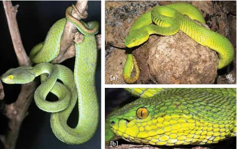
Figure 28: Head of a typical pit viper - dark green pit viper (Trimeresurus T. macrops) showing the pit organ situated between the nostril and the eye (red arrow) (Copyright DA Warrell)
to detect their warm-blooded prey. This is situated between the nostril and the eye (Fig. 28). Viperidae are relatively short, thick-bodied snakes with many small rough scales on the top (dorsum) of the head and characteristic patterns of coloured markings on the dorsal surface of the body (Fig 29).
Pit Viper (Trimeresurus T. Macrops)
Figures 29: Western Russell’s viper (Daboia russelii): (a) Specimen from Sri Lanka; (b, c) Specimens from Tamil Nadu, southern India; (d, e) specimens from Pune, India (Copyright DA Warrell)
[d]
[e]
Some important examples of the Viperidae inhabiting South-East Asia Region countries (References to reports of reviews of bites by these species are given in parenthesis): Typical vipers (subfamily Viperinae): Russell’s vipers, Western, Daboia russelii
(Fig 29 a-e) (Phillips et al., 1988; Warrell 1989; Gawarammana et al., 2009); and Eastern, D. siamensis(Fig 30) (Myint-Lwin et al., 1985; Tun-Pe et al., 1987; Than-Than et al., 1987; Than-Than-Than-Than et al., 1988; Warrell 1989; Than- Than et al., 1989; Thein-Than et al., 1991; Tin-Nu-Swe et al., 1993; Belt et al., 1997)
Saw-scaled or carpet vipers Echis c. carinatus (Fig 31) (Bhat 1974; Warrell and Arnett 1976) and E. c. sochureki (Fig 31c) (Kochar et al., 2007).
Levantine or blunt-nosed viper
Macrovipera lebetina (Fig 32) (Sharma et al., 2008)
Pit vipers (subfamily Crotalinae):
Malayan pit viper Calloselasma
rhodostoma (Fig 33) (Reid et al., 1963a; Reid et al., 1963b;
Reid 1968; Warrell et al., 1986)
Mount Kinabalu pit viper Garthia chaseni (Fig 34) (Haile 1963; Warrell 1995) Mamushis (Genus Gloydius): G. brevicaudus (Fig 35) (Warrell 1995) Hump-nosed pit viper Hypnale hypnale (Fig 36) (de Silva et al., 1994; Joseph et al., 2007;
Ariaratnam et al., 2008; Premawardhena et al, 1998) and other Hypnale species (Maduwage et al., 2011).
Chinese habu Protobothrops
mucrosquamatus (Fig 37) (Warrell 1995) Green pit vipers, bamboo vipers, palm vipers and habus (formerly all genus Trimeresurus)
Indian bamboo viperTrimeresurus
(Craspedocephalus) gramineus (Fig 38) Malabar rock pit viper Trimeresurus (Crapedocephalus) malabaricus (Fig 39) (Gowda et al., 2006)
Palm viper Trimeresurus
(Craspedocephalus) puniceus (Fig 40) Sri Lankan viper Trimeresurus
(Craspedocephalus) trigonocephalus (Fig 41)(Warrell 1995)
Hagen’s pit viper Trimeresurus (Parias) hageni (Fig 42)
Pope’s pit viper Trimeresurus (Popeia) popeiorum(Fig 43)
Chinese bamboo viperTrimeresurus (Viridovipera) stejnegeri(Fig 44) (Warrell 1995)
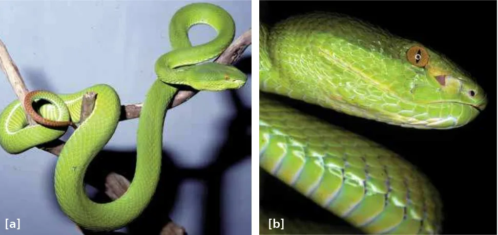
White-lipped green pit viper Trimeresurus (Trimeresurus) albolabris(Fig 45) (Hutton et al., 1990; Rojnuckarin et al., 1998)
Dark green pit viper Trimeresurus (Trimeresurus) macrops (Fig 28, 46) (Hutton et al., 1990; Warrell 1990b)
Spot-tailed green pit viper Trimeresurus (Trimeresurus) erythrurus (Fig 47) (Warrell 1995)
White-lipped island viper Trimeresurus (Trimeresurus) insularis (Fig 48) Kanchanburi pit viper Trimeresurus (Trimeresurus) kanburiensis (Warrell et al., 1992)
Mangrove pit viper Trimeresurus
(Trimeresurus) purpureomaculatus (Fig 49) (Warrell 1995)
Northern white-lipped pit viper
Trimeresurus (Trimeresurus) septentrionalis
(Whitaker)
Beautiful pit viper Trimeresurus (Trimeresurus) venustus (Fig 50)
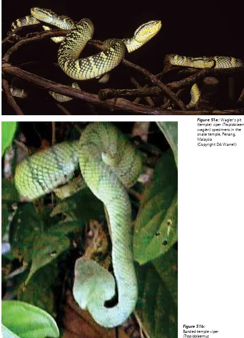
Wagler’s (temple) pit viper Tropidolaemus wagleri (Fig 51a) (Reid 1968)
Figures 30: Eastern Russell’s viper (Daboia siamensis): (a) Specimen from Myanmar; (b) Specimen from Thailand; (c) Specimen from East Java, Indonesia; (d) Specimen from Flores, Indonesia (Copyright DA Warrell)
Figures 31: Saw-scaled viper (Echis carinatus): (a) Echis carinatus carinatus Specimen from southern India; (b) Echis carinatus carinatus Specimen from Sri Lanka; (c) Echis carinatus sochureki Specimen from Chhattagharh Rajisthan (Copyright DA Warrell)
[c]
[d]
[a]
[c]
Figure 32: Levantine viper (Macrovipera lebetina) Specimen from Cyprus (Copyright DA Warrell)
Figures 33: Malayan pit viper (Calloselasma rhodostoma) Thai specimens: (a) Showing characteristic posture and triangular dorsal markings; (b) Showing supralabial markings (Copyright DA Warrell)
Figure 34: Mount Kinabalu pit viper (Garthia chaseni) (Copyright Prof RS Thorpe)
Figure 35: Mamushi or Fu-she (Gloydius brevicaudus) from China (Copyright DA Warrell)
[b]
Figures 36 a-d: Hump-nosed viper (Hynpale hypnale) (Copyright DA Warrell)
a) Specimen from Sri Lanka; (b) Specimen from Sri Lanka showing long fangs; (c) Specimen from south western India; (d) Specimen from south western India showing upturned snout
Figure 37:
Chinese habu (Protobothrops mucrosquamatus) Specimen from China (Copyright DA Warrell)
Figure 38: Indian bamboo viper (Trimeresurus Craspedocephalus gramineus)
(Copyright DA Warrell)
Figure 44: Chinese bamboo viper (Viridovipera stejnegeri) Specimen from China (Copyright DA Warrell)
Figures 45 a,b: White-lipped green pit viper (Cryptelytrops albolabris) Thai specimen: (a) Showing colouring and distinctive brown-topped tail; (b) Showing details of the head: note smooth temporal scales (Copyright DA Warrell)
Figure 48: White-lipped island viper (Trimeresurus T. insularis) specimen from Tutuala Sub-district Lautem District Timor-Leste (Copyright Mark O'Shea)
[b]
Figures 47 a, b: Spot-tailed green pit viper (Cryptelytrops erythrurus) Specimen from near Yangon, Myanmar: (a) Showing colouring and brown spotted tail; (b) Showing details of head; note keeled temporal scales (Copyright DA Warrell)
Figures 49 a,b:
Mangrove pit viper (Trimeresurus T. purpureomaculatus): (a) Specimen from Kanchanburi, Thailand; (b) Specimen from upper Myanmar (Copyright DA Warrell)
Figure 50: Beautiful pit viper (Cryptelytrops venustus) specimen from Thung Song, Thailand (Copyright DA Warrell)
Figure 51b:
3.2.2 Other medically important venomous snakes
Several species of medically important Colubridae (sensu lato) have been identified in the South-East Asia Region. The red-necked keelback
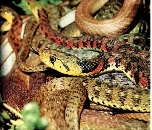
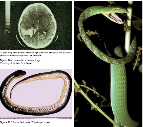
Rhabdophis subminiatus(Fig 2c and 52a) and yamakagashi R. tigrinus (Fig 52b) (Warrell 1995) can cause severe life-threatening anti-haemostatic disturbances and acute kidney injury (Fig 52c, 52d). A case of systemic envenoming by the Sri Lankan keelback
(Balanophis ceylonicus) with similar
features has also been reported Figure 52a: subminiatus) Thai specimen (Copyright DA Red-necked keelback (Rhabdophis
Warrell)
faced water snake (Cerberus rhynchops); and water snakes/paddy field snakes (Enhydris species) (Fig 52f and 53c). Undoubtedly more will be described as the concept of “non-venomous” is further
reviewed (Weinstein et al., 2011) Figure 52c: Extensive bruising from envenoming by R. tigrinus in Japan
Figure 52d: Intracerebral haemorrhage (Courtesy of the late Dr Y Sawai)
Figure 52e: Long-nosed whip snake (Ahaetulla nasuta) Sri Lanka (Copyright DA Warrell)
Figure 52f: Paddy field snake (Enhydris plumbea)
3.2.3 Non-venomous snakes
Many species of non-venomous or only trivially-venomous species are responsible for bites, especially those that are aggressive, irritable or prone to strike at humans who approach to closely or that commonly inhabit urban and rural gardens and compounds.
Apart from the taxa mentioned above, these include paradise or flying snakes
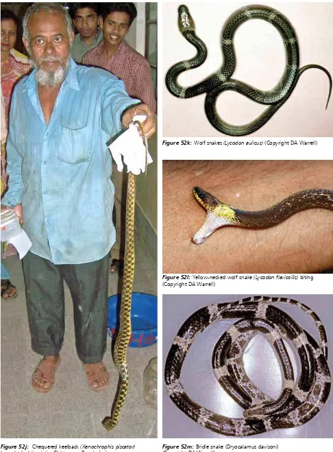
(Chrysopelea species) (Fig 52g), striped keelbacks (Amphiesma species) (Fig 52h), kukri snakes (Oligodon species) (Fig 52i), checkered keelbacks or Asian water snake (Xenochrophis species) (Fig 52j), wolf snakes (Lycodon or Dinodon species) (Fig 52k), bridle snakes (Dryocalamus) (Fig 52m) and rat snakes (Ptyas, Elaphe, Coelognathus, Goniosoma etc.)(Fig 52n). Their medical importance is that they may be mistaken for venomous species (notably the krait mimics Lycodon and
Dryocalamus), resulting in unnecessary and wasteful antivenom treatment (Viravan et al., 1992; Ariaratnam et al., 2009).
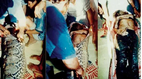
Large pythons (Boidae), notably the reticulated python Python reticularis in Indonesia, have been reported to attack and even ingest humans, usually inebriated farmers (Fig 52o).
Figure 52g: Paradise or flying snakes (Chrysopelea ornata) (Copyright Mark O’Shea)
Figure 52j: Chequered keelback (Xenochrophis piscator) brought by bite victim Chittagong Bangladesh
Figure 52l: Yellow-necked wolf snake (Lycodon flavicollis) biting (Copyright DA Warrell)
Figure 52o: Reticulated python (Python reticularis) containing the body of a farmer it had swallowed at Palu, Sulawesi, Indonesia (Copyright Excel Sawuwu)
region with short one line or two line notes of identifying features below the pictures in the local language will be very useful. In a dead snake, a needle may be drawn along the upper jaw from the angle of the jaw to the snout to snag and reveal the fangs. Some harmless snakes have evolved to look almost identical to venomous ones. Examples are various species of Lycodon, Dryocalamus and
Cercaspis that mimic the appearance of the kraits B. candidus, B. caeruleus and
B. ceylonicus; Cylindropis ruffus, whose tail raising display and colouring may mimic coral snakes (Calliophis species), and Boiga multomaculata that mimics Daboia siamensis. However, some of the most notorious venomous snakes can be recognized by their size, shape, colour, pattern of markings, behaviour and the sound they make when they feel threatened. For example, the defensive behaviour of the cobras is well known (Fig 4-9): they rear up, spread a hood, hiss and make repeated strikes towards the aggressor. Colouring can vary enormously. However, some patterns, like the
longitudinal rows of large, dark-rimmed, pale-centred spots of the Russell’s vipers (Figs 29, 30), or the alternating black and yellow circumferential bands of the banded krait (Fig 13), are distinctive. The blowing hiss of the Russell’s viper and the grating rasp of the saw-scaled viper are warning and identifying sounds, even in the dark.
3.4 Snake venoms
(Bucherl et al., 1968,1971; Gans and Gans 1978; Lee 1979; Harvey 1991; Ménez 2003; Mackessy 2009; Warrell 2010)
than 90% of snake venom (dry weight) is protein. Each venom contains more than a hundred different proteins: enzymes (constituting 80-90 % of viperid and 25-70 % of elapid venoms), non-enzymatic polypeptide toxins, and non-toxic proteins such as nerve growth factor. Nonprotein ingredients include carbohydrates and metals (often part of glycoprotein
metalloprotein enzymes), lipids, free amino acids, nucleosides, and biogenic amines such as serotonin and acetylcholine.
Venom enzymes
These include digestive hydrolases (proteinases, exopeptidase,
endopeptidases, phosphodiesterases, metalloproteinases, and phospholipases), hyaluronidase (spreading fator), and activators or inactivators of physiological processes, such as kininogenases. Most venoms contain l-amino acid oxidase (containing a riboflavin 5’-phosphate prosthetic group that confers the yellow colour of many venoms), phospho mono- and di- esterases, 5'-nucleotidase, DNAase, NAD-nucleosidase, phospholipase A2, and peptidases.
Zinc metalloproteinases/
metalloproteases (metalloproteinase-like, disintegrin-(metalloproteinase-like, cysteine-rich) haemorrhagins (snake venom metalloproteinases, SVMPs): degrade basement membrane components, leading to endothelial cell damage and contributing to spontaneous systemic bleeding.
Colubridae contain serine proteases and other procoagulant enzymes that are thrombin-like or activate factors V, X, prothrombin and other clotting factors. These enzymes stimulate blood clotting with formation of fibrin in the blood stream. Paradoxically, this process results in incoagulable blood because most of the fibrin clot is broken down immediately by the body’s own plasmin fibrinolytic system. Sometimes within 30 minutes of the bite, the levels of clotting factors have been so depleted that the blood will not clot (“consumption coagulopathy”). Some venoms contain multiple anti-haemostatic factors. For example, Russell’s viper venom contains toxins that activate factors II (prothrombin), V, X, IX and XIII, fibrinolysis and protein C, and cause platelet aggregation, anticoagulation and haemorrhage.
Phospholipases A2 (lecithinase): are most widespread and extensively studied of all venom enzymes. They damages mitochondria, red blood cells, leucocytes, platelets, peripheral nerve endings, skeletal muscle, vascular endothelium, and other membranes, producing presynaptic neurotoxic activity, cardiotoxicity, myotoxicity, necrosis, hypotension, haemolysis, haemorrhage, plasma leakage (oedema-induction), opiate-like sedative effects and autopharmacological release of histamine and other autacoids. They are anti-coagulant, either by hydrolysing plasma or platelet membrane phospholipids, or by interacting with different coagulation factors.
Acetylcholinesterases: although found in most elapid venoms, may cause fasciculation.
Hyaluronidase: promotes the spread of venom through tissues by increasing permeability but can also contribute to tissue damage.
Proteolytic enzymes
(metalloproteinases, endopeptidases or hydrolases) and polypetide
cytotoxins (“cardiotoxins”): increase vascular permeability causing oedema, blistering, bruising and necrosis at the site of the bite.
Venom polypeptide toxins (“neurotoxins”)
Postsynaptic (α) neurotoxins such as
α-bungarotoxin and cobrotoxin, consist of 60-62 or 66- 74 amino acids. They bind to acetylcholine receptors at the motor endplate. Presynaptic (β) neurotoxins such as β-bungarotoxin, and taipoxin, contain 120-140 amino-acids and a phospholipase A subunit. These release acetylcholine at the nerve endings at neuromuscular junctions and then damage the endings, preventing further release of transmitter.
Variation in venom composition within species
The composition of snake venoms, and hence their antigenicities in inducing specific neutralizing antibodies during antivenom manufacture, varies greatly between different species, but also within an individual species, as the snake matures (ontogenic variation), seasonally, between sexes, and throughout the geographical range (Warrell, 1997; Mackessy et al., 2003). The two important implications of venom variation are 1- envenoming by juvenile and adult snakes may cause qualitatively different clinical effects and 2- envenoming by a snake in one part of its geographical range may not be neutralized