PHYTOSTEROL CONTENT IN BENGKOANG (Pachyrhizus erosus)
KANDUNGAN PHYTOSTEROL DALAM BENGKOANG (Pachyrhizus erosus)
Endang Lukitaningsih
Pharmacy Faculty, Gadjah Mada University, Jogjakarta [email protected]
ABSTRACT
Bengkoang has long been used by ancestors as a raw material for cosmetics. In Indonesia, it is usually eaten raw, sometimes with salt, lemon juice and powdered chili. However, scientific evidences that support the use of bengkoang have not been widely published. Phytosterols from the bengkoang root have been isolated and identified based on their NMR spectrum data and mass spectrum. The result of the structure elucidation informed that ß-sitosterol and stigmasterol are major components of phytosterol in bengkoang root. Phytosterol content was about 0.02% of dry Indonesia, bengkoang biasa dimakan secara langsung bersama dengan garam, jus jeruk atau cabe. Namun hingga saat ini bukti ilmiah yang mendukung pemanfaatan bengkoang tersebut belum banyak dipublikasikan. Phytosterol dari umbi bengkoang telah berhasil diisolasi dan diidentifikasi berdasarkan data spektra NMR dan spektra massa. Hasil elusidasi struktur menginformasikan bahwa ß-sitosterol dan stigmasterol merupakan komponen utama phytosterol dalam umbi bengkoang. Kandungan phytosterol berkisar 0,02% per berat kering bengkoang atau 2,76% dalam ekstrak petroleum eter, dengan perbandingan ß-sitosterol dan stigmasterol (65:35). Dengan demikian bengkoang dapat dimanfaatkan lebih lanjut sebagai bahan baku suplemen phytosterol.
INTRODUCTION
Phytosterol is a class of sterol
compounds contained in plants. It is an integral component of the membrane lipid bilayer (Demel and DeKruyff, 1976; Schuleretal., 1991). Phytosterols are different from the cholesterol on their side chain substitution and the position of double bonds intheir cyclic chain. Because they have a double bond in their cyclic group, then either the phytosterol or cholesterol are susceptible to oxidation (Zhang, et al., 2005). Each plant species has its own characteristic distribution of phytosterols. There are three phytosterol constituents are most
commonly found, namely beta-sitosterol,
campesterol and stigmasterol (Benveniste, 1986; Benveniste, 2004).
Now, phytosterol is not only used as ingredients such as anti-inflammatory drugs (Parra-Delgado et al. 2004; Dicksonetal. 2007), hormones and vitamins (Dewick, 2002), but also as a nutritional supplement (anti-cholesterol additives) and cosmetics (as a cream and lipsticks) (Berezin et al., 2001).
Gomes et al. (2007) reported that
beta-sitosterol and stigmasterol isolated from Plucheaindica Less plant was able to against the snake venom.
In addition, phytosterols are also able to
reduce cholesterol absorption, so the
cancer can be reduced by dietary phytosterol.
However, until now the mechanism of inhibition of cell growth and stimulate apoptosis of the sitosterol is not well known (Moon, etal., 2007).
METHODOLOGY Plant materials
The bengkoang belongs to the taxonomic class of Magnoliopsida; order Fabales; family
Fabaceae; subfamily Faboideae; genus
Pachyrhizus; species Pachyrhizus erosus. The
bengkoang Pachyrhizus erosus (L) Urb roots
were collected from Purworejo, Central Java, Indonesia in dry season.
Chemicals and solvents
The chemicals used in the detection and isolation methods were anisaldehyde (4-methoxybenzaldehyde), glacial acetic acid, aluminium chloride, hydrochloric acid and concentrated sulphuric acid (all purchased from Merck, Darmstadt, Germany), Sephadex LH20 (Aldrich, Steinheim, Germany), Silica gel 60
(particle sizes 0.063-0.200mm, Merck,
48
PHARMACON, Vol. 13, No. 2, Desember 2012, Lukitaningsih,E. (47-54) silica gel 60 F254 (layer thickness 0.2 mm,Merck, Darmstadt, Germany).
Solvents for separation techniques were petroleum ether, ethyl acetate (Fisher Scientific,
Leichestershire, UK), methanol (Merck,
Darmstadt, Germany), chloroform,
dichloromethane, from the Fluka, Seelze, Germany.
Instruments
Melting point SMP3 Stuart® apparatus
(Staffordshire, UK), Cary 50 Bio UV-Visible spectrophotometer (Varian, California, USA), JASCO FT/IR-6100 Spectrophotometer (Gross-Umstadt, Germany), ALPHA II-12 Freeze dryer (Osterode, Germany), Bruker Avance 400 NMR
spectrometer (Rheinstetten, Germany),
Shimadzu GC/MS-QP 20105 gas
chromatography (Kyoto, Japan).
Extraction and isolation of phytosterol from bengkoang
The bengkoang roots (45 kg) were peeled and washed with water, subsequently dried at 60°C and milled into fine powder. The fine powder (4.75 kg) was extracted by soxhlet using petroleum ether.
The petroleum ether extract (26.3 gram)
was further subjected to silica gel
chromatography and eluted using gradient mixture of PE-EtOAc (from 100% of PE to 100% of EtOAc) and followed by gradient mixture of EtOAc-MeOH (from 100% of EtOAc to 100% of MeOH). 29 fractions of 100 ml were collected according to the TLC results. TLC was conducted to each fraction respectively using silica gel as a stationary phase, chloroform-ethyl acetate (6:4) as a mobile phase. Detection was performed with UV light at 254 and 366 nm and followed by spraying the TLC plates with
anisaldehyde-H2SO4 reagent and subsequent
heating at 110°C. Phytosterol will react positive with anisaldehide and give blue-purple colour.
Fractions 8, 9, 10, 11 have the same spot on TLC with an Rf value of 0.48. Therefore, they were combined and then subjected to another silica gel column chromatography using dichloromethane-ethyl acetate (60:40) as a mobile phase. Ten millilitre fractions were collected and evaluated by TLC using
dichloromethane-ethyl acetate (60:40) as
Tabel 1-1
H NMR and 13
C NMR spectroscopic data of the isolated compound
C Compound 109a (Stigmasterol) δH δC δC* Compound 109b (ß-sitosterol) δH δC δC*
1 1.84 (1H, m);
1.30 (1H, m) 37.4 37.2
1.84 (1H, m);
1.30 (1H, m) 37.4 37.3
2 1.50 (2H, m) 28.4 31.3 1.50 (2H, m) 28.4 31.6
3 3.50(3H, m) 71.9 70.3 3.50 (3H, m) 71.9 71.7
4 2.27 (2H, m) 42.5 42.0 2.27 (2H, m) 42.5 42.3
5 - 140.9 141.6 - 140.9 140.8
6 5.34 (1H, dd) 121.9 120.2 5.34 (1H, dd) 121.9 121.6
7 1.98 (2H, m) 32.0 31.6 1.98 (2H, m) 32.0 31.9
8 1.70 (1H, m) 32.1 31.8 1.70 (1H, m) 32.1 31.9
9 0.92 (1H, m) 50.3 50.1 0.92 (1H, m) 50.3 50.2
10 - 36.7 36.3 - 36.7 36.5
11 1.50 (2H, m) 20.0 20.8 1.50 (2H, m) 20.0 21.1
12 2.01 (2H, m) 39.9 39.4 2.01 (2H, m) 39.9 39.8
13 - 46.0 42.4 - 46.0 42.3
14 1.10 (1H, m) 56.9 56.5 1.10 (1H, m) 56.9 56.8
15 1.09 (1H, m),
1.70 (1H,m) 23.2 23.9
1.09 (1H, m),
1.70 (1H,m) 23.2 24.3
16 1.70 (2H, m) 26.3 28.1 1.70 (2H, m) 26.3 28.3
17 1.20 (1H, m) 56.2 55.9 1.20 (1H, m) 56.2 56.1
18 0.68 (3H, s) 12.0 11.8 0.68 (3H, s) 12.0 11.9
19 1.01 (3H, s) 19.2 19.1 1.01 (3H, s) 19.2 19.4
20 1.28 (1H, m) 40.6 40.3 1.28 (1H, m) 40.6 36.2
21 0.92 (3H, s) 19.6 20.6 0.92 (3H, s) 19.6 18.8
22 5.01 (1H, dd) 138.5 137.7 1.20 (2H, m) 33.90 33.9
23 5.16 (1H, dd) 129.4 129.3 1.7 (2H, m) 28.30 26.1
24 1.50 (2H, m) 51.4 50.6 1.50 (2H, m) 51.4 45.9
25 1.20 (2H, m) 24.5 24.7 1.2 (2H, m) 24.5 23.1
26 0.83 (3H, d) 12.3 11.9 0.83 (3H, d) 12.3 12.3
27 1.90 (1H, m) 29.3 31.50 1.90 (1H, m) 29.3 29.2
28 0.83 (3H, d) 18.9 18.90 0.83 (3H, d) 18.9 19.1
29 0.83 (3H, d) 21.2 21.04 0.83 (3H, d) 21.2 19.8
Note : δC* is data base of carbon chemical shift from SDBS AIST Japan
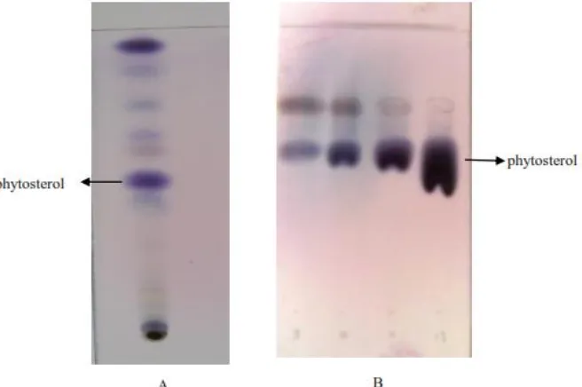
The TLC analysis of petroleum ether extract of bengkoang has been carried out to find out how many compounds are in the extract and their distribution. This step was
necessary before doing isolation and
determination. Figure 1 displayed the TLC chromatogram of bengkoang extract and fraction containing phytosterol.
Fig.1-The TLC chromatogram of petroleum extract (A) and fraction 8, 9,10,11 containing phytosterols (B). The TLC system consists of silica as stationary phase and a mixture of chloroform-methanol (60:40) as mobile phase and detected by anisaldehyde
The isolated compound from fractions 8, 9, 10, 11 of the petroleum ether extract as white needle crystals having a melting point of
50
PHARMACON, Vol. 13, No. 2, Desember 2012, Lukitaningsih,E. (47-54)cm-1 (-CH group) and 1051 cm-1 (-C-O-C
group).
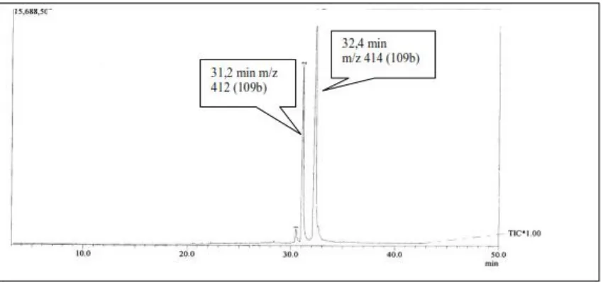
An analysis using gas chromatogram has been conducted and the chromatogram can be seen in Figure 2.The chromatogram showed two peaks with retention times of 31.2 and 32.4
min, corresponding to the molecular ion peaks at m/z 412 and 414, respectively. They are consistent with the molecular formula C29H48O (e.g. stigmasterol) and C29H50O (e.g. ß-sitosterol).
Figure 2. Gas chromatogram Mass Spectroscopy of the sample, the chromatogram showed two peaks with retention times of 31.2 and 32.4 min, corresponding to the molecular ion peaks at m/z 412 and 414, respectively.
The hypothesis that the isolated sitosterol in literature reported (Kovganko et al. 2000) and also with the database in SDBS AIST Japan.
The downfield signal in the 1H-NMR
spectrum at δ 5.34 (1H, dd) was due to an
olefinic proton at C6 and a methine proton at C3 was represented by multiplet signal at 3.50 (1H,
m). The presence of a pair of doublets at δ 5.01 the structures of P-1 and P-2 were stigmasterol and ß-sitosterol.
The mass fragmentation pattern above was in accordance with previously paper. Chaves et al. (2004) found in GC/MS the fragments with m/z value of 412 (M+.), 271 and 273. Based on the
above fragmentation, this substance P-1 was
identified as stigmasterol.
The Figure 6 displayed a mass
fragmentation pattern of P-2 having a molecular ion m/z 414. The fragment ions at m/z 396 and 381 were due to the loss of water and followed by a methyl group from the molecular ion. The characteristic observed in the mass spectrum of P-2 was the presence of a fragment ion peak at m/z 273. This signal was in a close agreement with the loss of side chain caused by the fission of C17-C20 bond. This was further fragmented producing water. From this process, the signal at m/z 255 fragmentation of ß-sitosterol and stigmasetrol using HPLC-MS equipped with APCI. The result showed that ß-sitosterol and stigmasterol were protonated by a reactive species in the plasma of
ion source. ß-Sitosterol had an exact mass of 414.39, which became 397.38 after protonation and loss of water. While stigmasetrol had an exact mass of 412.41 which became 395.4 after protonation and loss of water.
All these spectroscopy data proved the structure of the substance P-1 as stigmasterol and
P-2 as ß-sitosterol. Based on the gas
chromatogram at Figure 2, the concentration ratio of stigmasterol and ß-sitosterol from this fraction was 35 : 65.
This research proved that the total phytosterol content in petroleum ether extract of bengkoang was relatively high (2,76%) or about 0.02 % of dry weight bengkoang. Therefore, bengkoang can be used as a good source of phytosterol and potentially used as nutritional supplement of cholesterol replacement.
CONCLUSSION
Bengkoang contained a relatively high of phytosterols consisted of two major compounds, namely ß-sitosterol and stigmasterol. Therefore, bengkoang can be further exploited not only as cosmetics material, but also as nutritional supplement especially for cholesterol replacement.
ACKNOWLEDGMENT
We are grateful to the Germany Academic Exchange Service (DAAD) for financial support.
REFERENCES
Awad, A.B., Chen, Y.C., Fink, C.S., Hennessey, T., 1996, β-Sitosterol inhibits HT-29 human colon cancer
cell growth and alters membrane lipids, Anticancer Res, 16, 2797–2804.
Benveniste P, 1986, Sterol biosynthesis, Annu Rev Plant Physiol, 37, 275–308
Benveniste P, 2004, Biosynthesis and accumulation of sterols, Annu Rev Plant Bio, 55, 429–457
Benveniste, P., Schaller, H., Bouvier-Nave, P., 2005, Cellular sterol ester synthesis in plants is performed by an enzyme (phospholipid: sterol acyltransferase) different from the yeast and mammalian acyl-CoA:
sterol acyltransferases. J Biol Chem280: 34626–34634
Berezin, M.Y., Dzenitis, J.M., Hughes, B.M., Ho, S.V., 2001, Separation of sterols using zeolites, Phys. Chem., 3, 2184-2189
Demel, R.A. and De Kruyff, B. 1976, The function of sterols in membranes, Biochim. Biophys. Acta, 457,
109–132
Dewick, P.M., 2002, Medicinal Natural Products: a biosynthethic approach, 2nd Ed., John Wiley and Sons
54
PHARMACON, Vol. 13, No. 2, Desember 2012, Lukitaningsih,E. (47-54)Dickson, R.A., Houghton, P.J., Hylands, P.J., 2007, Antibacterial and antioxidant cassane diterpenoids
from Caesalpinia benthamiana, Phytochem., 68, , 1436-1441
Downie, A., Fink, S.C., Awad, A.B., 1999, Effect of phytosterols on MDAMB-231 human breast cancer cell
growth, FASEB J., 113, A333 [abs.].
Gomes, A., Saha, A., Chatterjee, I., Chakravarty, A.K., 2007, Viper and cobra venom neutralization by
ß-sitosterol and stigmasterol isolated from the root extract of Pluchea indica Less. (Asteraceae),
Phytomedicine, 14, , 637-643
Moon, D-O., Lee, K-J., Choi, Y.H., Kim, G-Y., 2007, ß-sitosterol-induced-apoptosis is mediated by the
activation of ERK and the downregulation of Akt in MCA-102 murine fibrosarcoma cells, Internatl.
Immunopharmacol., 7, 1044-1053
Parra-Delgado, H., Ruiz, G.G., Camacho, A.N., Martinez-Vazquez, M.M., 2004, Anti-inflamatory activity of
some extracts and isolates from Leonotis nepetaefolia on TPA-induced edema model, Rev. Soc. Quim.
Mex., 48, , 293-295
Schuler I., Milon, A., Nakatani, Y., Ourisson, G., Albrecht, A., Benveniste, P., Hartman, M., 1991, Differential effects of plant sterols on water permeability and on acyl chain ordering of soybean
phospatidylcholine bilayers, Proc. Natl. Acad. Sci. USA, 88, 6926–6930
von Holtz, R.L., Fink, C.S., Awad, .A.B., 1998, β-Sitosterol activates the sphingomyelin cycle and induces
apoptosis in LNCaP human prostate cancer cells, Nutr. Cancer, 32, 8–12.
Zhang, X., Julien-David, D., Miesch, M., Geoffroy, P., Raul, F., Roussi, S., Aoud´e-Werner, D., Marchioni,. E., 2005, Identification and quantitative analysis of sitosterol oxides in vegetable oils by capillary gas