MICROBIAL COMMUNITY ASSOCIATED WITH AMBROSIA BEETLE,
Euplatypus parallelus ON SONOKEMBANG, Pterocarpus indicus IN MALANG
Hagus Tarno 1*), Erfan Dani Septia 2) and Luqman Qurata Aini 1)
1) Plant Pest and Disease Department Faculty of Agriculture University of Brawijaya Jl. Veteran Malang 65145 East Java Indonesia
2) Faculty of Agriculture, University of Muhammadiyah Malang Jl. Raya Tlogomas 246 Malang 65145 East Java Indonesia
*) Corresponding author E-mail: [email protected]
Received: August 8, 2015 /Accepted: August 1, 2016
ABSTRACT
Recently, most of sonokembang, Pterocarpus indicus trees are dying in Malang. In 2012, the death rate of trees reached ca. 11%. In addition, death of trees spread to other regencies in East Java. Euplatypus parallelus is a specific species of ambrosia beetles that were the causal agents to the dying and wilting of sonokembang trees in Malang. Wilting is caused mainly by the pathogenic fungi carried by ambrosia beetles. To confirm the microbial communities related to E. parallelus that attack sonokembang, E. parallelus and some attacked trees were collected in Malang city. Isolation and identification of these species were conducted at the Laboratory of Mycology, Faculty of Agriculture, University of Brawijaya and Laboratory of Molecular Biology, Islamic State University, Malang. Results showed that there were nine microbes including five genera of fungi, two genera of yeasts and one genus of bacterium were identified. The microbial communities that were found namely Aspergillus spp., Penicillium spp., Trichoderma spp., Fusarium spp., Acremonium spp., Gliocladium spp. (fungi), Streptomyces spp. (bacteria), Saccharomyces spp., and Candida spp. (yeast).
Keywords: ambrosia beetle; Euplatypus parallelus; microbial community; Pterocarpus indicus
INTRODUCTION
Recently, most of sonokembang trees are dying in Malang. Tarno, Suprapto, & Himawan, (2014) reported that a death rate of trees reached ca. 11%. In addition, such occurrence had spread to the other regencies in East Java. Wilting is commonly caused by pathogenic fungi carried by
Euplatypus parallelus, an ambrosia beetle that was responsible to wilting and dying of yeast or bacteria (Endoh, Suzuki, Benno, & Futai, 2008). However, the ambrosia beetle (Platypus quercivorus) could carry both pathogenic and non-pathogenic microbes.
Some microbes are advantageous to ambrosia beetles (Harris et al., 2009) as they can serve as food for the ambrosia beetles and aid in their growth and development (Moon, Park, Oh, & Kim, 2008). Several ambrosia beetles feed on yeasts; however, some of these microbes are classified as pathogens that can reduce plant resistance and eventually leads to wilting and dying of trees (Henriques, Inácio, & Sousa, 2009).
Infected trees by ambrosia fungi showed general symptoms such as fallen leaves, wilting and dying, including discoloration of sapwood (Kubono & Ito, 2002). In this study, the microbial communities related to E. parallelus that attack sonokembang were investigated.
MATERIALS AND METHODS
Research was conducted from early December 2013 to the end of June 2014 in Malang City. Ambrosia beetle, wood and frass samples were collected randomly from five points of attacked coordinate sites. Stratified Random Sampling as sampling method was used in this research (Singarimbun & Effendi, 2005). Each collected sample was identified and then compared between three groups of samples such as insect body, gallery in the wood and frass. Identification Cite this as: Tarno, H., Septia, E. D. & Aini, L. Q. (2016). Microbial community associated with ambrosia beetle, Euplatypus parallelus on sonokembang, Pterocarpus indicus in Malang. AGRIVITA Journal of Agricultural Science, 38(3), 312-320. http://doi.org/10.17503/agrivita.v38i3.628
Accredited: SK No. 81/DIKTI/Kep/2011
was conducted at Laboratory of Mycology, Faculty of Agriculture, University of Brawijaya and Laboratory of Molecular Biology, Islamic State University, Malang.
Microbial Isolation, Collected from Insect Body, Gallery and Frass
Thorax of female beetles was incised to get microbes from the mycangia. For the gallery, wood samples were cut in small parts (ca. 1 cm). In the case of frass, two types of frass such as fibrous and powdery frass were collected from the mouth of tree tunnels. All of the samples were isolated according to the method of Larran, Rollán, Ángeles, Alippi, & Urrutia (2002).
Purification
All microbial colonies were purified on Potato Dextrose Agar (PDA) medium. Nutrient Agar (NA) medium was used for bacteria and Yeast Mannitol Agar (YMA) medium was used for yeast. Each microbial colony characterizing a different color and form based on macroscopic morphology was purified. Specific microbes were separated from each other, taken by oose, and then cultured on Petridish on their respective medium.
Microbial Preparates
Microbial preparates were made on object glass. Fungal spores were taken by oose and placed on small PDA media on object glass, then covered by cover glass. Preparates were placed into tray with sterile paper and then incubated for 23 days.
Identification
Macroscopic and microscopic observations were used during the identification process. Color, form (concentric or non-concentric), texture and growth (cm day-1) of colonies were used to dis-tinguish each colony. Macroscopic observation was conducted every day after period of inoculation until microbe covered all of space on Petri dish ( ca. 9 cm). Microscopic observation was conducted within 57 days by microscope. Types of hyphae, growth of hyphae, color of hyphae, color of conidia, form of conidia, form of mycelia, size of conidia, conidiophores and form of spores were used to identify each microbe as microscopic variables. In addition, form, color, growth and development of colonies were used to identify as macroscopic variables.
Data Analysis
Based on the data of each variable, some ecological indices, such as Simpson Index (D), Shannon-Wiener Indices (H`), Species Evenness Indices, and Simpson's dominance index were calculated:
1. Simpson Index (D) and species diversity indices, Shannon-Wiener Indices (H`) are described as (Davari, Jouri, & Ariapour,
index, index of species diversity, proportion of individuals of species i in the community, number of species in the sample, and logarithm respectively (Table 1).
Table 1. Value and description of Shannon-Wiener Indices (H')
Index
values Description
< 1 low level of diversity, low individual distribution of each species
1-3 middle level of diversity, middle level of individual distribution for each species
>3 high level of diversity, high level of individual distribution for each species
2. Species Evenness Indices (E) (Table 2) is formulated as (Pawhestri, Hidayat, & Putro, 2015):
𝐄 =
ln (𝑆)𝐻′ ……….…(3)Remarks: H’, ln and S are index of species diversity, exponential logarithm, and proportion of individuals of species.
Table 2. Value and description of index for species evenness indices (E)
Index values Description
0.00<E<0.50 Evenness is low, Community is underpressure
0.50<E<0.75 Evenness is medium level, Community is unstable
3. Simpson's dominance index (C) (Table 3) is used to measure the dominance of microbes in the community. Formula of Simpson's dominance index is described as (Davari, Jouri, & Ariapour, 2011):
𝐂 = ∑
𝑛[
𝑛𝑖𝑁]
2𝑖=1
.………(4)
Remarks: C, ni and N are Simpson's
dominance index, population of i-species, and population total of all species, respectively.
Table 3. Value and description of dominance index
Index values Description
0.00<C<0.50 Low dominance of species 0.50<C<0.75 Middle dominance of species 0.75<C<1.00 High dominance of species
RESULTS AND DISCUSSION
Symptom and Mortality of Sonokembang Trees in Malang City Caused by Ambrosia Beetle, E. parallelus
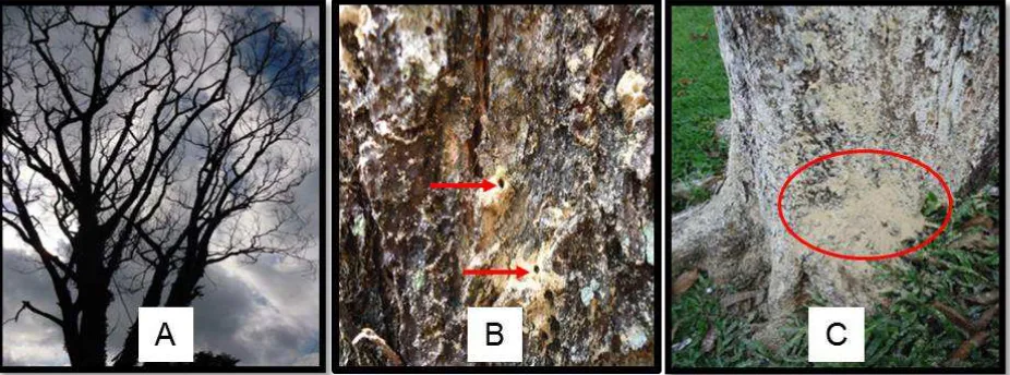
Sonokembang that attacked by E. parallelus showed distinguished characteristics. High amounts of fallen leaves indicated wilting and dying of trees. In addition, a high number of frass as a special sign was also produced during Ambrosia
beetle attacks. Tarno et al. (2010) reported that there are two types of frass produced by ambrosia beetles, namely: fibrous and powdery frass. Fibrous and powdery frass are caused by adults and larva, respectively (Tarno et al., 2010). Powdery frass was much higher produced by Ambrosia beetle than fibrous frass. Both of frass are expelled by male adult to keep the gallery clean (Tarno, Qi, Yamasaki, Kobayashi, & Futai, 2016).
Ambrosia beetle made hole on the stem and then construct longer and complicated tunnels inside the wood as known as gallery. Tarno, Suprapto, & Himawan (2014) explained that the diameter of entrance holes is ca. 1.90 ± 0.21 mm. During constructing tunnels, ambrosia beetle ejects frass from their tunnels (Tarno et al., 2010; Tarno, Qi, Yamasaki, Kobayashi, & Futai, 2016). Figure 1 describes the signs and symptoms of sonokembang that were attacked by the Ambrosia beetle, E. parallelus.
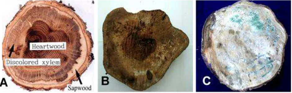
In case of an attacked tree, a cross-section of the wood will show a vivid discoloration of the xylem as shown in Figure 2A (Kuroda & Yamada, 1996), which indicate that the xylem tissue already died. Pattern of gallery is easily observed if the wood were incubated. Likewise, the growing fungi can also be seen during incubation. In this study, the conditions of wood before and after incubation are shown in Figure 2B and 2C, respectively.
Figure 2. Morphological characteristics in the stem of tree before and after incubation. (A) Discoloration on xylem (Kuroda & Yamada, 1996) (B) condition of wood before incubation and (C) condition of wood after incubation.
Diversity of Microbial Community on Insect Body, Gallery and Frass
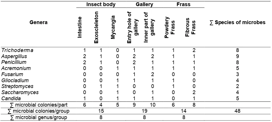
Based on the identified isolates, there were 48 isolates of microbes which were associated with insect body of ambrosia beetle, gallery of tunnels and frass. Genus of isolates are described in Table 4.
Nine genera of microbes were identified from the insect body, gallery of tunnel and frass such as Trichoderma, Aspergillus, Acremonium, Fusarium, Gliocladium, Streptomyces, Saccharomyces and Candida.
There were 15 species of microbes that were identified from the insect body of ambrosia beetle, E. parallelus. Five species were isolated from mycangia such as Acremonium spp., Gliocladium spp. (fungi), Streptomyces spp. (bacteria), Saccharomyces spp. and Candida spp. (yeasts). However, Fusarium was not found in the insect body of E. parallelus.
In the tunnel gallery, there were 19 identified species of microbes. Nine and ten species were identified from the entry holes and the inner part of galleries. One species of Trichoderma, two species of Aspergilus, two species of Penicilium, one species of Acremonium, one species of Gliocladium, and one species of Candida were found in the entry holes of galleries. One species
of Trichoderma, two species of Aspergilus, one species of Penicilium, one species of Acremonium, two species of Fusarium, one species of Gliocladium, one species of Saccharomyces and one species of Candida were found in the inner part of galleries.
In the case of frass, 14 species of microbes were identified. Six and eight species were identified in powdery and fibrous frass, respectively. Only Fusarium was not found on both powdery and fibrous frass. Two species of yeasts were not found on the powdery frass and three species of yeasts were found in the fibrous frass.
Based on the composition of microbes in the three different samples (insect body, gallery and frass), species of microbes in gallery were the highest. Frass was lowest in terms of number of microbes. Fusarium species were found only in the gallery. In addition, Simpson diversity index (D), Shannon (H'), Evenness (E) and Dominance index (C) of microbial communities were described in Table 5.
Table 4. Genera of microbial community isolated from insect body, gallery and frass
∑ microbial genus/group 8 8 8
Table 5. Simpson's diversity index (D), Diversity index of Shannon (H'), Evenness (E) and dominance index (C) of microbial communities between insect body, gallery and frass
Sources of isolates D H’Index valuesE C ∑ genus ∑ species ∑ colony
Insect body 0.085 1.991 0.957 0.062 8 15 23
Gallery 0.090 2.013 0.969 0.065 8 19 27
Frass 0.076 2.007 0.968 0.063 8 14 22
Total 0.251 6.011 2.895 0.190 24 48 72
Average 0.084 2.004 0.965 0.063 8 16 24
Macroscopic and Microscopic Characteristic of Microbial Isolates on Insect Body, Gallery and Frass
Based on the macroscopic and microscopic characteristic of microbial isolates, description of each genus is explained in Table 6, Table 7 and Figure 3. Most of the ambrosia fungi belong to four mitosporic genera: Ambrosiella, Raffaelea, Monacrosporium and Phialophoropsis. However, more genera have been reported to be involved with ambrosia beetles including Fusarium, Acremonium, Candida and Graphium (Batra, 1963; 1967; Baker & Norris, 1968).
Table 6. Description of each genus of microbial isolates that associated with ambrosia beetle, E. parallelus in Malang city based on macroscopic characteristics
Genera
Upper surface
Lower surface
Textures Densities colonies Color of Patterns of growth
Aspergillus Smooth,
powdery
high Green or black Concentric Similar to upper face, orange medium
Penicillium Smooth, thick,
powdery
high Green to brown
Symmetry Similar to upper face, Yellowish medium
Trichoderma Rough, thick,
powdery
high Early white to dark green
Concentric Similar to upper face
Fusarium Smooth and
thick like cotton
high White Concentric Similar to upper face
Gliocladium Feathered high Yellowish with
white
Concentric & wavy
Yellowish medium
Streptomyces Slick, powdery high grey Radial Yellowish medium
Saccharomyces Slick shiny,
thick.
high White to grey Radial Yellowish medium
Candida Slick not shiny high White to grey Radial Yellowish medium
Table 7. Description of each genus of microbial isolates associated with the ambrosia beetle, E. parallelus in Malang city based on microscopic characteristics
Genera
Microscopic characteristics of microbes
Observations
Mycelia Spores / Conidia
Shape Color Shape Color (μm)
Aspergillus Elongated,
unbranched
Hyaline Spherical serrated of conidia
Hyaline Elliptical clustered conidia
Hyaline 3.7 – 4.2 Tip of conidia clustered phialide
Fusarium Sectional,
branched
Hyaline Elongated oval conidia
Hyaline 3.7 - 4.3 Conidia taper at both ends
Acremonium Branched has
septa
Hyaline Elliptical cylindrical conidia
Hyaline 3.3 – 7.0 Conidia groups such as ball
Gliocladium Branched has
septa
Hyaline Ovoid conidia Blackish brown
4.6 – 5.2 Conidiophores branches such as brush
Streptomyces Branched Hyaline Chain spherical spore Blackish
brown
0.5 – 2.0 Spore chain consists of three or more
Saccharomyces Pseudohyphae Hyaline Conidiogenous cells
poliblastic, cylindric & thick
Hyaline 4.5 – 5.0 Did not form hyphae
Candida Pseudohyphae Hyaline Conidiogenous cells
poliblastic, oval
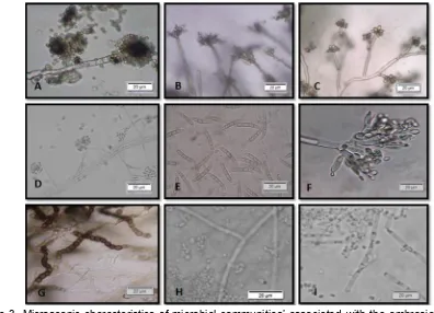
Figure 3. Microscopic characteristics of microbial communities’ associated with the ambrosia beetle, E. parallelus on sonokembang in Malang. (A) Aspergilus (B) Penicillium (C) Trichoderma (D) Acremonium (E) Fusarium (F) Gliocladium (G) Streptomyces (H) Saccharomyces and (I) Candida.
In Singapore, Sanderson, Fong, Yik, Ong, & Anuar (1997) considered that P. indicus attract E. parallelus when they became stressed by lightning strike. If the beetles were carrying spores of F. oxysporum, infection by the fungus is likely to follow, and result in the death of the trees (Sanderson, Fong, Yik, Ong, & Anuar, 1997).
Fusarium and Acremonium are plant pathogenic fungi. Kiffer & Morelet (1997) stated that Acremonium is plant pathogen for woody plants. In case of Fusarium, it commonly attacks on xylem and phloem of plants (Kiffer & Morelet, 1997). Streptomyces is classified as actionbacteria that have gained high commercial interest for the production of a variety of metabolites acting as potential insecticides (Ruiu, 2015). In addition, Penicilium and Gliocladium are potential anta-gonistic fungi (Gouli, V., Gouli, S., Marcelino, Skinner, & Parker, 2013; Vázquez-Martínez, Cirerol-Cruz, Torres-Estrada, & López, 2014).
CONCLUSION
From nine identified microbes, there were five genera of fungi, two genera of yeasts and one genus of bacterium. The microbial communities
that were associated with E. parallelus namely Aspergillus sp., Penicillium sp., Trichoderma sp., Fusarium sp., Acremonium sp., Gliocladium sp. (fungi), Streptomyces sp. (bacteria), Saccharomyces sp., and Candida sp. (yeast).
ACKNOWLEDGEMENT
The Authors would like to gratitude PHB Batch II, 2013, University of Brawijaya for financial support. In addition, the Authors also thank to Dr. Hideaki Tanaka, The University of Miyazaki for correction and suggestion.
REFERENCES
Baker, J. M., & Norris, D. M. (1968). A complex of fungi mutualistically involved in the nutrition of the ambrosia beetle Xyleborus ferrugineus. Journal of Invertebrate Pathology, 11(2), 246–250. http://doi.org/10.1016/0022-2011(68)901 57-2
2307/3626562
Batra, L. R. (1967). Ambrosia fungi: A taxonomic revision, and nutritional studies of some species. Mycologia, 59(6), 976–1017. http://doi.org/10.2307/3757271
Bumrungsri, S., Beaver, R., Phongpaichit, S., & Sittichaya, W. (2008). The infestation by an exotic ambrosia beetle, Euplatypus parallelus (F.) (Coleoptera: Curculionidae: Platypodinae) of Angsana trees (Pterocarpus indicus Willd.) in southern Thailand. Songklanakarin Journal of Sci-ence and Technology, 30(5), 579–582. Retrieved from http://rdo.psu.ac.th/sjstweb/ journal/30-5/0125-3395-30-5-579-582.pdf Davari, N., Jouri, M. H., & Ariapour, A. (2011).
Comparison of measurement indices of diversity, richness, dominance, and even-ness in rangeland ecosystem (case study: Jvaherdeh-Ramesar). Journal of Rangeland Science, 2(1), 389–398. Re-trieved from http://www.sid.ir/en/vewssid/ j_pdf/1004020110105.pdf
Endoh, R., Suzuki, M., Benno, Y., & Futai, K. (2008). Candida kashinagacola sp. nov., C. pseudovanderkliftii sp. nov. and C. vanderkliftii sp. nov., three new yeasts from ambrosia beetle-associated sources. Antonie van Leeuwenhoek, International Journal of General and Molecular Micro-biology, 94(3), 389–402. http://doi.org/ 10.1007/s10482-008-9256-9
Gouli, V., Gouli, S., Marcelino, J. A. P., Skinner, M. & Parker, B. L. (2013). Entomo-pathogenic fungi associated with exotic invasive insect pests in Northeastern forests of the USA. Insects, 4(4), 631-645. http://doi.org/10.3390/insects4040631 Harris, R. N., Brucker, R. M., Walke, J. B., Becker,
M. H., Schwantes, C. R., Flaherty, D. C., … Minbiole, K. P. C. (2009). Skin micro-bes on frogs prevent morbidity and mortality caused by a lethal skin fungus. The ISME Journal, 3(7), 818–24. http:// doi.org/10.1038/ismej.2009.27
Henriques, J., Inácio, M. de L., & Sousa, E. (2009). Fungi associated to Platypus cylindrus Fab. (Coleoptera: Platypodi-dae) in cork oak. Revista de Ciências Agrárias, 32(2), 56–66. Retrieved from http://www.scielo.mec.pt/pdf/rca/v32n2/v 32n2a07.pdf
Kiffer, E. & Morelet, M. (1997). The
deutero-mycetes mitosporic fungi: Classification and generic keys. Enfield, NH: Science Publishers.
Krebs, C. J. (2014). Ecological Methodology (3rd ed.). London: Addison Wesley Longman. Kubono, T., & Ito, S. (2002). Raffaelea quercivora
sp. nov. associated with mass mortality of Japanese oak, and the ambrosia beetle (Platypus quercivorus). Mycoscience, 43(3), 255–260. http://doi.org/10.1007/ s102670200037
Kuroda, K. & Yamada, T. (1996). Discoloration of sapwood and blockage of xylem sap ascent in the trunks of wilting Quercus spp. following attack by Platypus quercivorus. Journal of the Japanese Forestry Society, 78(1), 84-88. Retrieved from https://www. jstage.jst.go.jp/article/jjfs1953/78/1/78_1 _84/_pdf
Larran, S., Rollán, C., Ángeles, H. B., Alippi, H. E., & Urrutia, M. I. (2002). Endophytic fungi in healthy soybean leaves. Producción Y Protección Vegetales, 17(1), 173–178. Retrieved from http:// www.inia.es/GCONTREC/PUB/fungi2_1 161160547796.pdf
Moon, M. J., Park, J. G., Oh, E., & Kim, K. H. (2008). External microstructure of the ambrosia beetle Platypus koryoensis (Coleoptera: Curculionidae: Platypodi-nae). Entomological Research, 38(3), 202–210. http://doi.org/10.1111/j.1748-5 967.2008.00166.x
Pawhestri, S. W., Hidayat, J. W., & Putro, S. P. (2015). Assessment of water quality using macrobenthos as bioindicator and its application on Abundance-Biomass Comparison (ABC) curves. International Journal of Science and Engineering, 8(2), 84–87. Retrieved from http://www. ejournal.undip.ac.id/index.php/ijse/article /view/7972
Philip, E. (1999). Wilt disease of angsana (Pterocarpus indicus) in Peninsular Malaysia and its possible control. Journal of Tropical Forest Science, 11(3), 519– 527. Retrieved from http://www.jstor.org/ stable/pdf/43582560.pdf
Ruiu, L. (2015). Insect pathogenic bacteria in integrated pest management. Insects, 6(2), 352-367. http://doi.org/10.3390/ insects6020352
& Anuar, S. (1997). A fusarium wilt (Fusarium oxysporum) of angsana (Pterocarpus indicus) in Singapore. I. Epidemiology and identification of the causal organism. Arboricultural Journal: The International Journal of Urban Forestry, 21(3), 187-204. http://doi.org/ 10.1080/03071375.1997.9747165
Singarimbun, M. & Effendi, S. (1995). Metode Penelitian Survai [Survey research methods]. Jakarta: LP3S.
Tarno, H., Qi, H., Endoh, R., Kobayashi, M., Goto, H. & Futai, K. (2010). Types of frass produced by the ambrosia beetle Platypus quercivorus during gallery construction, and host suitability of five tree species for the beetle. Journal of Forest Research, 16(1), 68-75. http://doi.org/10.1007/s10 310-010-0211-z
Tarno, H., Qi, H., Yamasaki, M., Kobayashi, M., & Futai, K. (2016). The behavioral role of males of Platypus quercivorus
Murayama in their subsocial colonies. AGRIVITA Journal of Agricultural Science, 38(1), 47–54. http://doi.org/10.17503/ agrivita.v38i1.778
Tarno, H., Suprapto, H., & Himawan, T. (2014). First record of ambrosia beetle (Euplatypus paralellus Fabricius) infestation on sonokembang (Pterocarpus indicus Willd.) from Malang Indonesia. AGRIVITA Journal of Agricultural Science, 36(2), 189–200. http://doi.org/10.17503/Agrivita-2014-36-2-p189-200