H I G H L I G H T S
of the 2015 American Heart Association
Introduction. . . .1
Ethical Issues . . . .3
Systems of Care and Continuous Quality Improvement . . . .3
Adult Basic Life Support and CPR Quality: Lay Rescuer CPR . . . .5
Adult Basic Life Support and CPR Quality: HCP BLS . . . .8
Alternative Techniques and Ancillary Devices for CPR . . . 11
Adult Advanced Cardiovascular Life Support . . . .13
Post–Cardiac Arrest Care . . . .15
Acute Coronary Syndromes . . . .16
Special Circumstances of Resuscitation . . . .18
Pediatric Basic Life Support and CPR Quality . . . .20
Pediatric Advanced Life Support. . . .23
Neonatal Resuscitation. . . .25
Education . . . .27
First Aid. . . .29
References . . . .32
Contents
Acknowledgments
The American Heart Association thanks the following people for their contributions to the development of this publication: Mary Fran Hazinski, RN, MSN; Michael Shuster, MD; Michael W. Donnino, MD; Andrew H. Travers, MD, MSc; Ricardo A. Samson, MD; Steven M. Schexnayder, MD; Elizabeth H. Sinz, MD; Jeff A. Woodin, NREMT-P; Dianne L. Atkins, MD; Farhan Bhanji, MD; Steven C. Brooks, MHSc, MD; Clifton W. Callaway, MD, PhD; Allan R. de Caen, MD; Monica E. Kleinman, MD; Steven L. Kronick, MD, MS; Eric J. Lavonas, MD; Mark S. Link, MD; Mary E. Mancini, RN, PhD; Laurie J. Morrison, MD, MSc; Robert W. Neumar, MD, PhD; Robert E. O’Connor, MD, MPH; Eunice M. Singletary, MD; Myra H. Wyckoff, MD; and the AHA Guidelines Highlights Project Team.
Highlights of the 2015 AHA Guidelines Update for CPR and ECC 1
Introduction
This “Guidelines Highlights” publication summarizes the
key issues and changes in the 2015 American Heart
Association (AHA) Guidelines Update for Cardiopulmonary Resuscitation (CPR) and Emergency Cardiovascular Care
(ECC). It has been developed for resuscitation providers and for AHA instructors to focus on the resuscitation science and guidelines recommendations that are most significant or controversial or those that will result in changes in resuscitation practice or resuscitation training. In addition, it provides the rationale for the recommendations.
Because this publication is designed as a summary, it does not reference the supporting published studies and does not list Classes of Recommendation or Levels of Evidence.
For more detailed information and references, readers are
encouraged to read the 2015 AHA Guidelines Update for
CPR and ECC, including the Executive Summary,1 published
in Circulation in October 2015, and to consult the detailed
summary of resuscitation science in the 2015 International
Consensus on CPR and ECC Science With Treatment Recommendations, published simultaneously in Circulation2
and Resuscitation.3
The 2015 AHA Guidelines Update for CPR and ECC is based on an international evidence evaluation process that involved 250 evidence reviewers from 39 countries. The process for the 2015 International Liaison Committee on Resuscitation (ILCOR) systematic review was quite different when compared with the process used in 2010. For the 2015 systematic review process, the ILCOR task forces prioritized topics for review, selecting those where there was
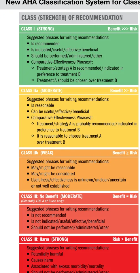
Figure 1
CLASS I (STRONG) Benefit >>> Risk
Suggested phrases for writing recommendations:
■ Is recommended
■ Is indicated/useful/effective/beneficial ■ Should be performed/administered/other ■ Comparative-Effectiveness Phrases†:
º Treatment/strategy A is recommended/indicated in preference to treatment B
º Treatment A should be chosen over treatment B
CLASS IIa (MODERATE) Benefit >> Risk
Suggested phrases for writing recommendations:
■ Is reasonable
■ Can be useful/effective/beneficial ■ Comparative-Effectiveness Phrases†:
º Treatment/strategy A is probably recommended/indicated in preference to treatment B
º It is reasonable to choose treatment A over treatment B
CLASS IIb (WEAK) Benefit ≥ Risk
Suggested phrases for writing recommendations:
■ May/might be reasonable ■ May/might be considered
■ Usefulness/effectiveness is unknown/unclear/uncertain
or not well established
CLASS III: No Benefit (MODERATE) Benefit = Risk
(Generally, LOE A or B use only)
Suggested phrases for writing recommendations:
■ Is not recommended
■ Is not indicated/useful/effective/beneficial ■ Should not be performed/administered/other
CLASS III: Harm (STRONG) Risk > Benefit
Suggested phrases for writing recommendations:
■ Potentially harmful ■ Causes harm
■ Associated with excess morbidity/mortality ■ Should not be performed/administered/other
LEVEL A
■ High-quality evidence‡ from more than 1 RCTs ■ Meta-analyses of high-quality RCTs
■ One or more RCTs corroborated by high-quality registry studies
LEVEL B-R (Randomized)
■ Moderate-quality evidence‡ from 1 or more RCTs ■ Meta-analyses of moderate-quality RCTs
LEVEL B-NR (Nonrandomized)
■ Moderate-quality evidence‡ from 1 or more well-designed,
well-executed nonrandomized studies, observational studies, or registry studies
■ Meta-analyses of such studies
LEVEL C-LD (Limited Data)
■ Randomized or nonrandomized observational or registry
studies with limitations of design or execution
■ Meta-analyses of such studies
■ Physiological or mechanistic studies in human subjects
LEVEL C-EO (Expert Opinion)
Consensus of expert opinion based on clinical experience
COR and LOE are determined independently (any COR may be paired with any LOE). A recommendation with LOE C does not imply that the recommendation is weak. Many important clinical questions addressed in guidelines do not lend themselves to clinical trials. Although RCTs are unavailable, there may be a very clear clinical consensus that a particular test or therapy is useful or effective.
* The outcome or result of the intervention should be specified (an improved clinical outcome or increased diagnostic accuracy or incremental prognostic information). † For comparative-effectiveness recommendations (COR I and IIa; LOE A and B only),
studies that support the use of comparator verbs should involve direct comparisons of the treatments or strategies being evaluated.
‡ The method of assessing quality is evolving, including the application of standardized, widely used, and preferably validated evidence grading tools; and for systematic reviews, the incorporation of an Evidence Review Committee.
COR indicates Class of Recommendation; EO, expert opinion; LD, limited data; LOE, Level of Evidence; NR, nonrandomized; R, randomized; and RCT, randomized controlled trial.
CLASS (STRENGTH) OF RECOMMENDATION
LEVEL (QUALITY) OF EVIDENCE‡
sufficient new science or controversy to prompt a systematic review. As a result of this prioritization, there were fewer reviews completed in 2015 (166) than in 2010 (274).
Once the topics were selected, there were 2 important additions to the 2015 process of review itself. First, reviewers used Grading of Recommendations Assessment, Development, and Evaluation (GRADE;
www.gradeworkinggroup.org), a highly structured and reproducible evidence review system, to improve the consistency and quality of the 2015 systematic reviews. Second, reviewers from around the world were able to work together virtually to complete the systematic reviews through the use of a purpose-built AHA Web-based platform, the Systematic Evidence Evaluation and Review System (SEERS), designed to support the many steps of the evaluation process. This SEERS site was used to provide public disclosure of
drafts of the ILCOR 2015 International Consensus on CPR
and ECC Science With Treatment Recommendations and to receive public comment. To learn more about SEERS and to see a comprehensive list of all systematic reviews conducted by ILCOR, visit www.ilcor.org/seers.
The 2015 AHA Guidelines Update for CPR and ECC
is very different from previous editions of the AHA Guidelines for CPR and ECC. The ECC Committee
determined that this 2015 version would be an update,
addressing only those topics addressed by the 2015 ILCOR
evidence review or those requested by the training network. This decision ensures that we have only one standard for evidence evaluation, and that is the process created by
ILCOR. As a result, the 2015 AHA Guidelines Update for CPR
and ECC is not a comprehensive revision of the 2010 AHA Guidelines for CPR and ECC. Such an integrated version is
available online at ECCguidelines.heart.org.
The publication of the 2015 International Consensus on CPR
and ECC Science With Treatment Recommendations begins a process of ongoing review of resuscitation science. The topics reviewed in 2015 will be updated as needed and new topics will be added. Readers will want to monitor the SEERS site to keep up-to-date on the newest resuscitation science and the ILCOR evaluation of that science. When sufficient evidence emerges that indicates the need to change the AHA Guidelines for CPR and ECC, such changes will be made and communicated to clinicians and to the training network.
The 2015 Guidelines Update used the most recent version of the AHA definitions for the Classes of Recommendation and Levels of Evidence (Figure 1). Readers will note that this version contains a modified Class III recommendation, Class III: No Benefit, to be used infrequently when evidence suggests a strategy is demonstrated by a high- or moderate-quality study (Level of Evidence [LOE] A or B, respectively) to be no better than the control. The Levels of Evidence have also been modified. LOE B is now divided into LOE B-R (randomized studies) and LOE B-NR (nonrandomized studies). LOE C is now divided into LOE C-LD (limited data) and C-EO (expert opinion).
As outlined in the recently published Institute of Medicine report4
and the AHA ECC consensus response to this report,5 more
needs to be done to advance the science and practice of
resuscitation. There must be a concerted effort to fund cardiac arrest resuscitation research similar to what has driven cancer and stroke research over the past 2 decades. The gaps in the science are clear when the recommendations contained within the 2015 Guidelines Update are scrutinized (Figure 2). Collectively, the Levels of Evidence and the Classes of Recommendation in resuscitation are low, with only 1% of the total recommendations in 2015 (3 of 315) based on the highest Level of Evidence (LOE A) and only 25% of the recommendations (78 of 315) designated as Class I (strong recommendation). Most (69%) of the 2015 Guidelines Update recommendations are supported by the lowest Levels of Evidence (LOE C-LD or C-EO), and nearly half (144 of 315; 45%) are categorized as Class IIb (weak recommendation).
Throughout the ILCOR evidence evaluation process and the 2015 Guidelines Update development, participants adhered strictly to the AHA conflict of interest disclosure requirements. The AHA staff processed more than 1000 conflict of interest disclosures, and all Guidelines writing group chairs and at least 50% of Guidelines writing group members were required to be free of relevant conflicts of interest.
Figure 2
Distribution of Classes of
Recommendation and Levels of
Evidence as Percent of 315 Total
Highlights of the 2015 AHA Guidelines Update for CPR and ECC 3
Ethical Issues
As resuscitation practice evolves, ethical considerations must also evolve. Managing the multiple decisions associated with resuscitation is challenging from many perspectives, no more so than when healthcare providers (HCPs) are dealing with the ethics surrounding decisions to provide or withhold emergency cardiovascular interventions.
Ethical issues surrounding whether to start or when to terminate CPR are complex and may vary across settings (in- or out-of-hospital), providers (basic or advanced), and patient population (neonatal, pediatrics, adult). Although ethical principles have not changed since the 2010
Guidelines were published, the data that inform many ethical discussions have been updated through the evidence review process. The 2015 ILCOR evidence review process and resultant AHA Guidelines Update include several science updates that have implications for ethical decision making for periarrest, arrest, and postarrest patients.
Significant New and Updated
Recommendations That May Inform
Ethical Decisions
•The use of extracorporeal CPR (ECPR) for cardiac arrest
•Intra-arrest prognostic factors
•Review of evidence about prognostic scores for preterm infants •Prognostication for children and adults after cardiac arrest
•Function of transplanted organs recovered after cardiac arrest
New resuscitation strategies such as ECPR have made decisions to discontinue resuscitation measures more complicated (see the Adult Advanced Cardiovascular Life Support section in this publication). Understanding the appropriate use, implications, and likely benefits related to such new treatments
will have an impact on decision making. There is new information about prognostication for neonates, children, and adults in cardiac arrest and after cardiac arrest (see Neonatal Resuscitation, Pediatric Advanced Life
Support, and Post– Cardiac Arrest Care). The increased use of targeted temperature management (TTM) has led to new challenges for predicting neurologic outcomes in comatose post–cardiac arrest patients, and the latest data about the
usefulness of particular tests and studies should inform decisions about goals of care and limiting interventions.
There is greater awareness that although children and adolescents cannot make legally binding decisions, information should be shared with them to the extent possible, using appropriate language and information for each patient’s level of development. In addition, the
phrase limitations of care has been changed to limitations
of interventions, and there is increasing availability of the Physician Orders for Life-Sustaining Treatment (POLST) form, a new method of legally identifying people with specific limits on interventions at the end of life, both in and out of healthcare facilities. Even with new information that the success of kidney and liver transplants from adult donors is unrelated to whether the donor receives CPR, the donation of organs after resuscitation remains controversial. Viewpoints on several important ethical concerns that are the topics of ongoing debate around organ donation in an emergency setting are summarized in “Part 3: Ethical Issues” of the 2015 Guidelines Update.
Systems of Care and Continuous
Quality Improvement
The 2015 Guidelines Update provides stakeholders with a new perspective on systems of care, differentiating in-hospital cardiac arrests (IHCAs) from out-of-in-hospital cardiac arrests (OHCAs). Major highlights include
• A universal taxonomy of systems of care
• Separation of the AHA adult Chain of Survival into 2 chains: one for in-hospital and one for out-of-hospital systems of care
• Review of best evidence on how these cardiac arrest systems of care are reviewed, with a focus on cardiac arrest, ST-segment elevation myocardial infarction (STEMI), and stroke
Figure 3
Taxonomy of Systems of Care: SPSO
Structure Process System Patient
Outcome
Continuous Quality Improvement
Integration, Collaboration, Measurement, Benchmarking, Feedback
People Education Equipment
Protocols Policies Procedures
Programs Organization Culture
Satisfaction
Quality Safety
Components of a System of Care
2015 (New): Universal elements of a system of care have been identified to provide stakeholders with a common framework with which to assemble an integrated resuscitation system (Figure 3).
Why: Healthcare delivery requires structure (eg, people,
equipment, education) and process (eg, policies, protocols, procedures) that, when integrated, produce a system (eg, programs, organizations, cultures) that leads to optimal outcomes (eg, patient survival and safety, quality, satisfaction). An effective system of care comprises all of these elements— structure, process, system, and patient outcomes—in a framework of continuous quality improvement.
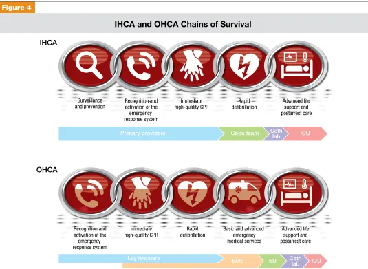
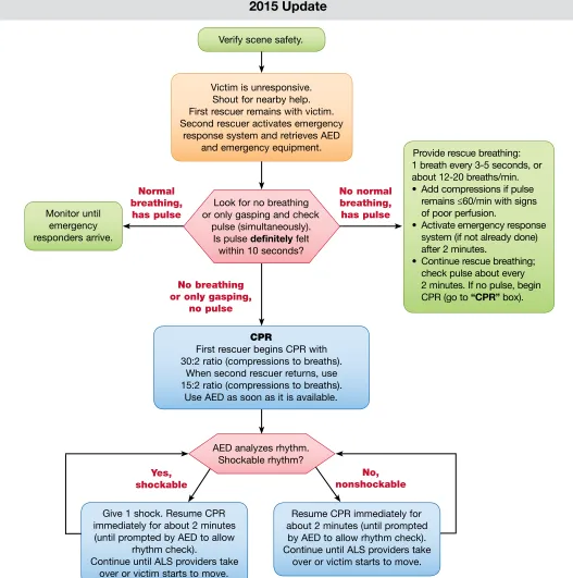
Chains of Survival
2015 (New): Separate Chains of Survival (Figure 4) have been recommended that identify the different pathways of care for patients who experience cardiac arrest in the hospital as distinct from out-of-hospital settings.
Why: The care for all post–cardiac arrest patients, regardless
of where their arrests occur, converges in the hospital, generally in an intensive care unit where post–cardiac arrest care is provided. The elements of structure and process
that are required before that convergence are very different for the 2 settings. Patients who have an OHCA depend on their community for support. Lay rescuers must recognize the arrest, call for help, and initiate CPR and provide defibrillation (ie, public-access defibrillation [PAD]) until a team of professionally trained emergency medical service (EMS) providers assumes responsibility and then transports the patient to an emergency department and/or cardiac catheterization lab. The patient is ultimately transferred to a critical care unit for continued care. In contrast, patients who have an IHCA depend on a system of appropriate surveillance (eg, rapid response or early warning system) to prevent cardiac arrest. If cardiac arrest occurs, patients depend on the smooth interaction of the institution’s various departments and services and on a multidisciplinary team of professional providers, including physicians, nurses, respiratory therapists, and others.
Use of Social Media to Summon Rescuers
2015 (New): It may be reasonable for communities to incorporate social media technologies that summon rescuers who are in close proximity to a victim of suspected OHCA and are willing and able to perform CPR.
Why: There is limited evidence to support the use of social
media by dispatchers to notify potential rescuers of a possible
Primary providers Code team Cathlab ICU
IHCA
OHCA
Lay rescuers EMS Cath ICU
lab ED
Surveillance and prevention
Recognition and activation of the emergency response system
Immediate high-quality CPR
Rapid defibrillation
Advanced life support and postarrest care
Recognition and activation of the emergency response system
Immediate high-quality CPR
Rapid defibrillation
Basic and advanced emergency medical services
Advanced life support and postarrest care Figure 4
Highlights of the 2015 AHA Guidelines Update for CPR and ECC 5
cardiac arrest nearby, and activation of social media has not been shown to improve survival from OHCA. However, in a recent study in Sweden, there was a significant increase in the rate of bystander-initiated CPR when a mobile-phone
dispatch system was used.6 Given the low harm and the
potential benefit, as well as the ubiquitous presence of digital devices, municipalities could consider incorporating these technologies into their OHCA systems of care.
Team Resuscitation: Early Warning Sign
Systems, Rapid Response Teams, and Medical
Emergency Team Systems
2015 (Updated): For adult patients, rapid response team (RRT) or medical emergency team (MET) systems can be effective in reducing the incidence of cardiac arrest, particularly in the general care wards. Pediatric MET/RRT systems may be considered in facilities where children with high-risk illnesses are cared for in general in-patient units. The use of early warning sign systems may be considered for adults and children.
2010 (Old): Although conflicting evidence exists, expert consensus recommended the systematic identification of patients at risk of cardiac arrest, an organized response to such patients, and an evaluation of outcomes to foster continuous quality improvement.
Why: RRTs or METs were established to provide early
intervention for patients with clinical deterioration, with the goal of preventing IHCA. Teams can be composed of varying combinations of physicians, nurses, and respiratory therapists. These teams are usually summoned to a patient bedside when acute deterioration is identified by hospital staff. The team typically brings emergency monitoring and resuscitation equipment and drugs. Although the evidence is still evolving, there is face validity in the concept of having teams trained in the complex choreography of resuscitation.
Continuous Quality Improvement for
Resuscitation Programs
2015 (Reaffirmation of 2010): Resuscitation systems should establish ongoing assessment and improvement of systems of care.
Why: There is evidence of considerable regional variation
in the reported incidence and outcome of cardiac arrest in the United States. This variation underscores the need for communities and systems to accurately identify each occurrence of treated cardiac arrest and to record outcomes. There are likely to be opportunities to improve survival rates in many communities.
Community- and hospital-based resuscitation programs should systematically monitor cardiac arrests, the level of resuscitation care provided, and outcome. Continuous quality improvement includes systematic evaluation and feedback, measurement or benchmarking, and analysis. Continuous efforts are needed to optimize resuscitation care so that the gaps between ideal and actual resuscitation performance can be narrowed.
Regionalization of Care
2015 (Reaffirmation of 2010): A regionalized approach to OHCA resuscitation that includes the use of cardiac resuscitation centers may be considered.
Why: A cardiac resuscitation center is a hospital that
provides evidence-based care in resuscitation and post– cardiac arrest care, including 24-hour, 7-day percutaneous coronary intervention (PCI) capability, TTM with an adequate annual volume of cases, and commitment to ongoing performance improvement that includes measurement, benchmarking, and both feedback and process change. It is hoped that resuscitation systems of care will achieve the improved survival rates that followed establishment of other systems of care, such as trauma.
Adult Basic Life Support and CPR
Quality: Lay Rescuer CPR
Summary of Key Issues and Major Changes
Key issues and major changes in the 2015 Guidelines Update recommendations for adult CPR by lay rescuers include the following:
• The crucial links in the out-of-hospital adult Chain of Survival are unchanged from 2010, with continued emphasis on the simplified universal Adult Basic Life Support (BLS) Algorithm.
• The Adult BLS Algorithm has been modified to reflect the fact that rescuers can activate an emergency response (ie, through use of a mobile telephone) without leaving the victim’s side.
• It is recommended that communities with people at risk for cardiac arrest implement PAD programs.
• Recommendations have been strengthened to encourage immediate recognition of unresponsiveness, activation of the emergency response system, and initiation of CPR if the lay rescuer finds an unresponsive victim is not breathing or not breathing normally (eg, gasping).
• Emphasis has been increased about the rapid identification of potential cardiac arrest by dispatchers, with immediate provision of CPR instructions to the caller (ie, dispatch-guided CPR).
• The recommended sequence for a single rescuer has been confirmed: the single rescuer is to initiate chest compressions before giving rescue breaths (C-A-B rather than A-B-C) to reduce delay to first compression. The single rescuer should begin CPR with 30 chest compressions followed by 2 breaths.
• There is continued emphasis on the characteristics of high-quality CPR: compressing the chest at an adequate rate and depth, allowing complete chest recoil after each compression, minimizing interruptions in compressions, and avoiding excessive ventilation. • The recommended chest compression rate is 100 to 120/min
(updated from at least 100/min).
• The clarified recommendation for chest compression depth for adults is at least 2 inches (5 cm) but not greater than 2.4 inches (6 cm).
These changes are designed to simplify lay rescuer training and to emphasize the need for early chest compressions for victims of sudden cardiac arrest. More information about these changes appears below.
In the following topics, changes or points of emphasis that are similar for lay rescuers and HCPs are noted with an asterisk (*).
Community Lay Rescuer AED Programs
2015 (Updated): It is recommended that PAD programs for patients with OHCA be implemented in public locations where there is a relatively high likelihood of witnessed cardiac arrest (eg, airports, casinos, sports facilities).
2010 (Old): CPR and the use of automated external defibrillators (AEDs) by public safety first responders were recommended to increase survival rates for out-of-hospital sudden cardiac arrest. The 2010 Guidelines recommended the establishment of AED programs in public locations where there is a relatively high likelihood of witnessed cardiac arrest (eg, airports, casinos, sports facilities).
Why: There is clear and consistent evidence of improved
survival from cardiac arrest when a bystander performs CPR and rapidly uses an AED. Thus, immediate access to a defibrillator is a primary component of the system of care. The implementation of a PAD program requires 4 essential components: (1) a planned and practiced response, which ideally includes identification of locations and neighborhoods where there is high risk of cardiac arrest, placement of AEDs in those areas and ensuring that bystanders are aware of the location of the AEDs, and, typically, oversight by an HCP; (2) training of anticipated rescuers in CPR and use of the AED; (3) an integrated link with the local EMS system; and (4) a program of ongoing quality improvement.
A system-of-care approach for OHCA might include public policy that encourages reporting of public AED locations
to public service access points (PSAPs; the term public
service access point has replaced the less-precise EMS dispatch center). Such a policy would enable PSAPs to direct bystanders to retrieve nearby AEDs and assist in their use when OHCA occurs. Many municipalities as well as the US federal government have enacted legislation to place AEDs in municipal buildings, large public venues, airports, casinos, and schools. For the 20% of OHCAs that occur in public areas, these community programs represent an important link in the Chain of Survival between recognition and
activation of the PSAPs. This information is expanded in “Part 4: Systems of Care and Continuous Quality Improvement” in the 2015 Guidelines Update.
There is insufficient evidence to recommend for or against the deployment of AEDs in homes. Victims of OHCAs that occur in private residences are much less likely to receive chest compressions than are patients who experience cardiac arrest in public settings. Real-time instructions provided by emergency dispatchers may help potential in-home rescuers to initiate action. Robust community CPR training programs for cardiac arrest, along with effective, prearrival dispatch protocols, can improve outcomes.
Dispatcher Identification of Agonal Gasps
Cardiac arrest victims sometimes present with seizure-like activity or agonal gasps that can confuse potential rescuers. Dispatchers should be specifically trained to identify these presentations of cardiac arrest to enable prompt recognition and immediate dispatcher-guided CPR.
2015 (Updated): To help bystanders recognize cardiac arrest, dispatchers should inquire about a victim’s absence of responsiveness and quality of breathing (normal versus not normal). If the victim is unresponsive with absent or abnormal breathing, the rescuer and the dispatcher should assume that the victim is in cardiac arrest. Dispatchers should be educated to identify unresponsiveness with abnormal and agonal gasps across a range of clinical presentations and descriptions.
2010 (Old): To help bystanders recognize cardiac arrest, dispatchers should ask about an adult victim’s responsiveness, if the victim is breathing, and if the breathing is normal, in an attempt to distinguish victims with agonal gasps (ie, in those who need CPR) from victims who are breathing normally and do not need CPR.
Why: This change from the 2010 Guidelines emphasizes the
role that emergency dispatchers can play in helping the lay rescuer recognize absent or abnormal breathing.
Dispatchers should be specifically educated to help bystanders recognize that agonal gasps are a sign of cardiac arrest. Dispatchers should also be aware that brief generalized seizures may be the first manifestation of cardiac arrest. In summary, in addition to activating professional emergency responders, the dispatcher should ask straightforward questions about whether the patient is unresponsive and if breathing is normal or abnormal in order to identify patients with possible cardiac arrest and enable dispatcher-guided CPR.
Emphasis on Chest Compressions*
2015 (Updated): Untrained lay rescuers should provide compression-only (Hands-Only) CPR, with or without dispatcher guidance, for adult victims of cardiac arrest. The rescuer should continue compression-only CPR until the arrival of an AED or rescuers with additional training. All lay rescuers should, at a minimum, provide chest compressions for victims of cardiac arrest. In addition, if the trained lay rescuer is able to perform rescue breaths, he or she should add rescue breaths in a ratio of 30 compressions to 2 breaths. The rescuer should continue CPR until an AED arrives and is ready for use, EMS providers take over care of the victim, or the victim starts to move.
Highlights of the 2015 AHA Guidelines Update for CPR and ECC 7
provide chest compressions for victims of cardiac arrest. In addition, if the trained lay rescuer is able to perform rescue breaths, compressions and breaths should be provided in a ratio of 30 compressions to 2 breaths. The rescuer should continue CPR until an AED arrives and is ready for use or EMS providers take over care of the victim.
Why: Compression-only CPR is easy for an untrained rescuer
to perform and can be more effectively guided by dispatchers over the telephone. Moreover, survival rates from adult cardiac arrests of cardiac etiology are similar with either compression-only CPR or CPR with both compressions and rescue breaths when provided before EMS arrival. However, for the trained lay rescuer who is able, the recommendation remains for the rescuer to perform both compressions and breaths.
Chest Compression Rate*
2015 (Updated): In adult victims of cardiac arrest, it is reasonable for rescuers to perform chest compressions at a rate of 100 to 120/min.
2010 (Old): It is reasonable for lay rescuers and HCPs to perform chest compressions at a rate of at least 100/min.
Why: The number of chest compressions delivered per
minute during CPR is an important determinant of return of spontaneous circulation (ROSC) and survival with good neurologic function. The actual number of chest compressions delivered per minute is determined by the rate of chest
compressions and the number and duration of interruptions in
compressions (eg, to open the airway, deliver rescue breaths, allow AED analysis). In most studies, more compressions are associated with higher survival rates, and fewer compressions are associated with lower survival rates. Provision of adequate chest compressions requires an emphasis not only on an adequate compression rate but also on minimizing interruptions to this critical component of CPR. An inadequate compression rate or frequent interruptions (or both) will reduce the total number of compressions delivered per minute. New to the 2015 Guidelines Update are upper limits of recommended compression rate and compression depth, based on preliminary data suggesting that excessive compression rate and depth adversely affect outcomes. The addition of an upper limit of compression rate is based on 1 large registry study analysis associating extremely rapid compression rates (greater than 140/min) with inadequate compression depth. Box 1 uses the analogy of automobile travel to explain the effect of compression rate and interruptions on total number of compressions delivered during resuscitation.
Chest Compression Depth*
2015 (Updated): During manual CPR, rescuers should perform chest compressions to a depth of at least 2 inches (5 cm) for an average adult, while avoiding excessive chest compression depths (greater than 2.4 inches [6 cm]).
2010 (Old): The adult sternum should be depressed at least 2 inches (5 cm).
Why: Compressions create blood flow primarily by increasing
intrathoracic pressure and directly compressing the heart, which in turn results in critical blood flow and oxygen delivery to the heart and brain. Rescuers often do not compress the chest deeply enough despite the recommendation to “push hard.” While a compression depth of at least 2 inches (5 cm) is recommended, the 2015 Guidelines Update incorporates new evidence about the potential for an upper threshold of compression depth (greater than 2.4 inches [6 cm]), beyond which complications may occur. Compression depth may be difficult to judge without use of feedback devices, and identification of upper limits of compression depth may be challenging. It is important for rescuers to know that the recommendation about the upper limit of compression depth is based on 1 very small study that reported an association between excessive compression depth and injuries that were not life-threatening. Most monitoring via CPR feedback devices suggests that compressions are more often too shallow than they are too deep.
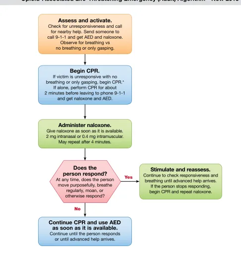
Bystander Naloxone in Opioid-Associated
Life-Threatening Emergencies*
2015 (New): For patients with known or suspected opioid addiction who are unresponsive with no normal breathing but a pulse, it is reasonable for appropriately trained lay rescuers and BLS providers, in addition to providing standard BLS care, to administer intramuscular (IM) or intranasal (IN) naloxone. Opioid overdose response education with or without naloxone distribution to persons at risk for opioid overdose in any setting may be considered. This topic is also addressed in the Special Circumstances of Resuscitation section.
Box 1
Number of Compressions Delivered
Affected by Compression
Rate and by Interruptions
The total number of compressions delivered during resuscitation is an important determinant of survival from cardiac arrest.
• The number of compressions delivered is affected by the compression
rate (the frequency of chest compressions per minute) and by the compression fraction (the portion of total CPR time during which compressions are performed). Increases in compression rate and fraction increase the total number of compressions delivered. Compression fraction is improved by reducing the number and duration of any interruptions in compressions.
• An analogy can be found in automobile travel. When traveling in an automobile, the number of miles traveled in a day is affected not only by the speed (rate of travel) but also by the number and duration of any stops (interruptions in travel). Traveling 60 mph without interruptions translates to an actual travel distance of 60 miles in an hour. Traveling 60 mph except for a 10-minute stop translates to an actual travel of 50 miles in that hour. The more frequent and the more prolonged the stops, the lower the actual miles traveled.
• During CPR, rescuers should deliver effective compressions at an appropriate rate (100 to 120/min) and depth while minimizing the number and duration of interruptions in chest compressions. Additional
Why: There is substantial epidemiologic data demonstrating the large burden of disease from lethal opioid overdoses, as well as some documented success in targeted national strategies for bystander-administered naloxone for people at risk. In 2014, the naloxone autoinjector was approved by the US Food and Drug Administration for use by lay
rescuers and HCPs.7 The resuscitation training network has
requested information about the best way to incorporate such a device into the adult BLS guidelines and training. This recommendation incorporates the newly approved treatment.
Adult Basic Life Support and
CPR Quality: HCP BLS
Summary of Key Issues and Major Changes
Key issues and major changes in the 2015 Guidelines Update recommendations for HCPs include the following:
• These recommendations allow flexibility for activation of the
emergency response system to better match the HCP’s clinical setting.
• Trained rescuers are encouraged to simultaneously perform some steps (ie, checking for breathing and pulse at the same time), in an effort to reduce the time to first chest compression.
• Integrated teams of highly trained rescuers may use a choreographed approach that accomplishes multiple steps and assessments simultaneously rather than the sequential manner used by individual rescuers (eg, one rescuer activates the emergency response system while another begins chest compressions, a third either provides ventilation or retrieves the bag-mask device for rescue breaths, and a fourth retrieves and sets up a defibrillator).
• Increased emphasis has been placed on high-quality CPR using performance targets (compressions of adequate rate and depth, allowing complete chest recoil between compressions, minimizing interruptions in compressions, and avoiding excessive ventilation). See Table 1.
• Compression rate is modified to a range of 100 to 120/min.
• Compression depth for adults is modified to at least 2 inches (5 cm) but should not exceed 2.4 inches (6 cm).
• To allow full chest wall recoil after each compression, rescuers must avoid leaning on the chest between compressions.
• Criteria for minimizing interruptions is clarified with a goal of
chest compression fraction as high as possible, with a target of at least 60%.
• Where EMS systems have adopted bundles of care involving continuous chest compressions, the use of passive ventilation techniques may be considered as part of that bundle for victims of OHCA.
• For patients with ongoing CPR and an advanced airway in place, a simplified ventilation rate of 1 breath every 6 seconds (10 breaths per minute) is recommended.
These changes are designed to simplify training for HCPs and to continue to emphasize the need to provide early and high-quality CPR for victims of cardiac arrest. More information about these changes follows.
In the following topics for HCPs, an asterisk (*) marks those that are similar for HCPs and lay rescuers.
Immediate Recognition and Activation of
Emergency Response System
2015 (Updated): HCPs must call for nearby help upon finding the victim unresponsive, but it would be practical for an HCP to continue to assess the breathing and pulse simultaneously before fully activating the emergency response system (or calling for backup).
2010 (Old): The HCP should check for response while looking at the patient to determine if breathing is absent or not normal.
Why: The intent of the recommendation change is to
minimize delay and to encourage fast, efficient simultaneous assessment and response, rather than a slow, methodical, step-by-step approach.
Emphasis on Chest Compressions*
2015 (Updated): It is reasonable for HCPs to provide chest compressions and ventilation for all adult patients in cardiac arrest, whether from a cardiac or noncardiac cause. Moreover, it is realistic for HCPs to tailor the sequence of rescue actions to the most likely cause of arrest.
2010 (Old): It is reasonable for both EMS and in-hospital professional rescuers to provide chest compressions and rescue breaths for cardiac arrest victims.
BLS Dos and Don’ts of Adult High-Quality CPR Table 1
Rescuers Should Rescuers Should Not
Perform chest compressions at a rate of 100-120/min Compress at a rate slower than 100/min or faster than 120/min
Compress to a depth of at least 2 inches (5 cm) Compress to a depth of less than 2 inches (5 cm) or greater than 2.4 inches (6 cm)
Allow full recoil after each compression Lean on the chest between compressions
Minimize pauses in compressions Interrupt compressions for greater than 10 seconds
Ventilate adequately (2 breaths after 30 compressions, each breath delivered over 1 second, each causing chest rise)
Provide excessive ventilation
Highlights of the 2015 AHA Guidelines Update for CPR and ECC 9
Why: Compression-only CPR is recommended for untrained
rescuers because it is relatively easy for dispatchers to guide with telephone instructions. It is expected that HCPs are trained in CPR and can effectively perform both compressions and ventilation. However, the priority for the provider, especially if acting alone, should still be to activate the emergency response system and to provide chest compressions. There may be circumstances that warrant a change of sequence, such as the availability of an AED that the provider can quickly retrieve and use.
Shock First vs CPR First
2015 (Updated): For witnessed adult cardiac arrest when an AED is immediately available, it is reasonable that the defibrillator be used as soon as possible. For adults with unmonitored cardiac arrest or for whom an AED is not immediately available, it is reasonable that CPR be initiated while the defibrillator equipment is being retrieved and applied and that defibrillation, if indicated, be attempted as soon as the device is ready for use.
2010 (Old): When any rescuer witnesses an out-of-hospital arrest and an AED is immediately available on-site, the rescuer should start CPR with chest compressions and use the AED as soon as possible. HCPs who treat cardiac arrest in hospitals and other facilities with on-site AEDs or defibrillators should provide immediate CPR and should use the AED/defibrillator as soon as it is available. These recommendations are designed to support early CPR and early defibrillation, particularly when an AED or defibrillator is available within moments of the onset of sudden cardiac arrest. When an OHCA is not witnessed by EMS personnel, EMS may initiate CPR while checking the rhythm with the AED or on the electrocardiogram (ECG) and preparing for defibrillation. In such instances, 1½ to 3 minutes of CPR may be considered before attempted defibrillation. Whenever 2 or more rescuers are present, CPR should be provided while the defibrillator is retrieved.
With in-hospital sudden cardiac arrest, there is insufficient evidence to support or refute CPR before defibrillation. However, in monitored patients, the time from ventricular fibrillation (VF) to shock delivery should be under 3 minutes, and CPR should be performed while the defibrillator is readied.
Why: While numerous studies have addressed the question
of whether a benefit is conferred by providing a specified period (typically 1½ to 3 minutes) of chest compressions before shock delivery, as compared with delivering a shock as soon as the AED can be readied, no difference in outcome has been shown. CPR should be provided while the AED pads are applied and until the AED is ready to analyze the rhythm.
Chest Compression Rate: 100 to 120/min*
2015 (Updated): In adult victims of cardiac arrest, it is reasonable for rescuers to perform chest compressions at a rate of 100 to 120/min.
2010 (Old): It is reasonable for lay rescuers and HCPs to perform chest compressions at a rate of at least 100/min.
Why: The minimum recommended compression rate
remains 100/min. The upper limit rate of 120/min has been added because 1 large registry series suggested that as the compression rate increases to more than 120/min, compression depth decreases in a dose-dependent manner. For example, the proportion of compressions of inadequate depth was about 35% for a compression rate of 100 to 119/min but increased to inadequate depth in 50% of compressions when the compression rate was 120 to 139/min and to inadequate depth in 70% of compressions when compression rate was more than 140/min.
Chest Compression Depth*
2015 (Updated): During manual CPR, rescuers should perform chest compressions to a depth of at least 2 inches (5 cm) for an average adult while avoiding excessive chest compression depths (greater than 2.4 inches [6 cm]).
2010 (Old): The adult sternum should be depressed at least 2 inches (5 cm).
Why: A compression depth of approximately 5 cm is
associated with greater likelihood of favorable outcomes compared with shallower compressions. While there is less evidence about whether there is an upper threshold beyond which compressions may be too deep, a recent very small study suggests potential injuries (none life-threatening) from excessive chest compression depth (greater than 2.4 inches [6 cm]). Compression depth may be difficult to judge without use of feedback devices, and identification of upper limits of compression depth may be challenging. It is important for rescuers to know that chest compression depth is more often too shallow than too deep.
Chest Recoil*
2015 (Updated): It is reasonable for rescuers to avoid leaning on the chest between compressions, to allow full chest wall recoil for adults in cardiac arrest.
2010 (Old): Rescuers should allow complete recoil of the chest after each compression, to allow the heart to fill completely before the next compression.
Why: Full chest wall recoil occurs when the sternum returns
to its natural or neutral position during the decompression phase of CPR. Chest wall recoil creates a relative negative intrathoracic pressure that promotes venous return and cardiopulmonary blood flow. Leaning on the chest wall between compressions precludes full chest wall recoil. Incomplete recoil raises intrathoracic pressure and reduces venous return, coronary perfusion pressure, and myocardial blood flow and can influence resuscitation outcomes.
Minimizing Interruptions in Chest
Compressions*
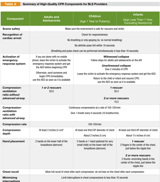
Component Adults and Adolescents
Children
(Age 1 Year to Puberty)
Infants
(Age Less Than 1 Year, Excluding Newborns)
Scene safety Make sure the environment is safe for rescuers and victim
Recognition of cardiac arrest
Check for responsiveness
No breathing or only gasping (ie, no normal breathing)
No definite pulse felt within 10 seconds
(Breathing and pulse check can be performed simultaneously in less than 10 seconds)
Activation of emergency response system
If you are alone with no mobile phone, leave the victim to activate the
emergency response system and get the AED before beginning CPR
Otherwise, send someone and begin CPR immediately; use the AED as soon as it is available
Witnessed collapse
Follow steps for adults and adolescents on the left
Unwitnessed collapse
Give 2 minutes of CPR
Leave the victim to activate the emergency response system and get the AED
Return to the child or infant and resume CPR; use the AED as soon as it is available
Compression-ventilation ratio without advanced airway
1 or 2 rescuers
30:2
1 rescuer
30:2
2 or more rescuers
15:2
Compression-ventilation ratio with advanced airway
Continuous compressions at a rate of 100-120/min
Give 1 breath every 6 seconds (10 breaths/min)
Compression rate 100-120/min
Compression depth
At least 2 inches (5 cm)* At least one third AP diameter of chest
About 2 inches (5 cm)
At least one third AP diameter of chest
About 1½ inches (4 cm)
Hand placement 2 hands on the lower half of the breastbone (sternum)
2 hands or 1 hand (optional for very small child) on the lower half of the
breastbone (sternum)
1 rescuer
2 fingers in the center of the chest, just below the nipple line
2 or more rescuers
2 thumb–encircling hands in the center of the chest, just below the
nipple line
Chest recoil Allow full recoil of chest after each compression; do not lean on the chest after each compression
Minimizing interruptions
Limit interruptions in chest compressions to less than 10 seconds
Summary of High-Quality CPR Components for BLS Providers Table 2
*Compression depth should be no more than 2.4 inches (6 cm).
Highlights of the 2015 AHA Guidelines Update for CPR and ECC 11
2015 (New): For adults in cardiac arrest who receive CPR without an advanced airway, it may be reasonable to perform CPR with the goal of a chest compression fraction as high as possible, with a target of at least 60%.
Why: Interruptions in chest compressions can be intended
as part of required care (ie, rhythm analysis and ventilation) or unintended (ie, rescuer distraction). Chest compression fraction is a measurement of the proportion of total resuscitation time that compressions are performed. An increase in chest compression fraction can be achieved by minimizing pauses in chest compressions. The optimal goal for chest compression fraction has not been defined. The addition of a target compression fraction is intended to limit interruptions in compressions and to maximize coronary perfusion and blood flow during CPR.
Comparison of Key Elements of Adult,
Child, and Infant BLS
Table 2 lists the 2015 key elements of adult, child, and infant BLS (excluding CPR for newly born infants).
Chest Compression Feedback
2015 (Updated): It may be reasonable to use audiovisual feedback devices during CPR for real-time optimization of CPR performance.
2010 (Old): New CPR prompt and feedback devices may be useful for training rescuers and as part of an overall strategy to improve the quality of CPR in actual resuscitations. Training for the complex combination of skills required to perform adequate chest compressions should focus on demonstrating mastery.
Why: Technology allows for real-time monitoring, recording,
and feedback about CPR quality, including both physiologic patient parameters and rescuer performance metrics. These important data can be used in real time during resuscitation, for debriefing after resuscitation, and for system-wide quality improvement programs. Maintaining focus during CPR on the characteristics of compression rate and depth and chest recoil while minimizing interruptions is a complex challenge even for highly trained professionals. There is some evidence that the use of CPR feedback may be effective in modifying chest compression rates that are too fast, and there is separate evidence that CPR feedback decreases the leaning force during chest compressions. However, studies to date have not demonstrated a significant improvement in favorable neurologic outcome or survival to hospital discharge with the use of CPR feedback devices during actual cardiac arrest events.
Delayed Ventilation
2015 (New): For witnessed OHCA with a shockable rhythm, it may be reasonable for EMS systems with
priority-based, multitiered response to delay positive-pressure ventilation (PPV) by using a strategy of up to 3 cycles of 200 continuous compressions with passive oxygen insufflation and airway adjuncts.
Why: Several EMS systems have tested a strategy of
providing initial continuous chest compressions with delayed PPV for adult victims of OHCA. In all of these EMS systems, the providers received additional training with emphasis on provision of high-quality chest compressions. Three studies in systems that use priority-based, multitiered response in both urban and rural communities, and provide a bundled package of care that includes up to 3 cycles of passive oxygen insufflation, airway adjunct insertion, and 200 continuous chest compressions with interposed shocks, showed improved survival with favorable neurologic status for victims with witnessed arrest or shockable rhythm.
Ventilation During CPR With an
Advanced Airway
2015 (Updated): It may be reasonable for the provider to deliver 1 breath every 6 seconds (10 breaths per minute) while continuous chest compressions are being performed (ie, during CPR with an advanced airway).
2010 (Old): When an advanced airway (ie, endotracheal tube, Combitube, or laryngeal mask airway) is in place during 2-person CPR, give 1 breath every 6 to 8 seconds without attempting to synchronize breaths between compressions (this will result in delivery of 8 to 10 breaths per minute).
Why: This simple single rate for adults, children, and
infants—rather than a range of breaths per minute—should be easier to learn, remember, and perform.
Team Resuscitation: Basic Principles
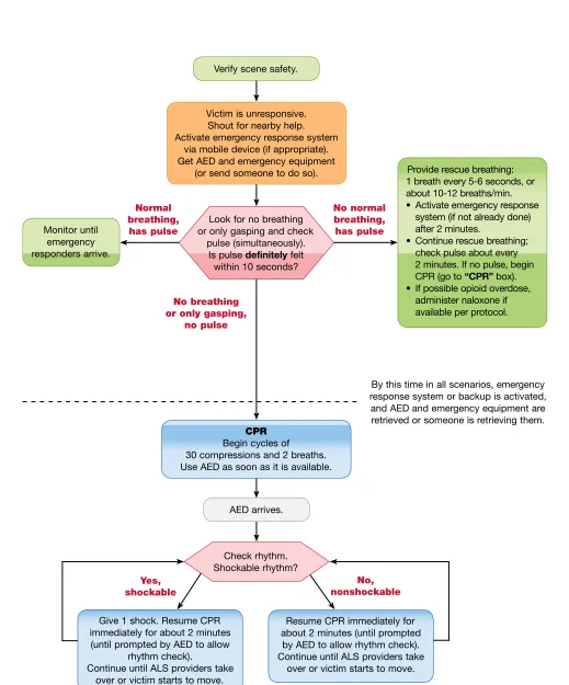
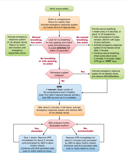
2015 (New): For HCPs, the 2015 Guidelines Update allows flexibility for activation of the emergency response and subsequent management in order to better match the provider’s clinical setting (Figure 5).
Why: The steps in the BLS algorithms have traditionally
been presented as a sequence in order to help a single rescuer prioritize actions. However, there are several factors in any resuscitation (eg, type of arrest, location, whether trained providers are nearby, whether the rescuer must leave a victim to activate the emergency response system) that may require modifications in the BLS sequence. The updated BLS HCP algorithms aim to communicate when and where flexibility in sequence is appropriate.
Alternative Techniques and
Ancillary Devices for CPR
Summary of Key Issues and Major Changes
Figure 5
No, nonshockable Yes,
shockable
No normal breathing, has pulse
AED arrives.
Check rhythm. Shockable rhythm?
Give 1 shock. Resume CPR immediately for about 2 minutes (until prompted by AED to allow
rhythm check).
Continue until ALS providers take over or victim starts to move.
Provide rescue breathing: 1 breath every 5-6 seconds, or about 10-12 breaths/min. • Activate emergency response
system (if not already done) after 2 minutes.
• Continue rescue breathing; check pulse about every 2 minutes. If no pulse, begin CPR (go to “CPR” box). • If possible opioid overdose,
administer naloxone if available per protocol.
Resume CPR immediately for about 2 minutes (until prompted
by AED to allow rhythm check). Continue until ALS providers take
over or victim starts to move. CPR
Begin cycles of 30 compressions and 2 breaths. Use AED as soon as it is available. Monitor until
emergency responders arrive.
Verify scene safety.
Victim is unresponsive. Shout for nearby help. Activate emergency response system
via mobile device (if appropriate). Get AED and emergency equipment
(or send someone to do so).
Look for no breathing or only gasping and check
pulse (simultaneously). Is pulse definitely felt
within 10 seconds?
Normal breathing, has pulse
No breathing or only gasping,
no pulse
Highlights of the 2015 AHA Guidelines Update for CPR and ECC 13
Compared with conventional CPR, many of these techniques and devices require specialized equipment and training. When rescuers or healthcare systems are considering implementation, it must be noted that some techniques and devices have been tested only in highly selected subgroups of cardiac arrest patients.
• The routine use of the impedance threshold device (ITD) as an adjunct to conventional CPR is not recommended.
• A recent randomized controlled trial suggests that the use of the ITD plus active compression-decompression CPR is associated with improved neurologically intact survival for patients with OHCA.
• The routine use of mechanical chest compression devices is not recommended, but special settings where this technology may be useful are identified.
• The use of ECPR may be considered for selected patients in settings where a reversible cause of cardiac arrest is suspected.
Impedance Threshold Devices
2015 (Updated): The routine use of the ITD as an adjunct during conventional CPR is not recommended. The combination of ITD with active compression-decompression CPR may be a reasonable alternative to conventional CPR in settings with available equipment and properly trained personnel.
2010 (Old): The use of the ITD may be considered by trained personnel as a CPR adjunct in adult cardiac arrest.
Why: Two large randomized controlled trials have provided
new information about the use of the ITD in OHCA. One large multicenter randomized clinical trial failed to demonstrate any improvement associated with the use of an ITD (compared with a sham device) as an adjunct to conventional CPR. Another clinical trial demonstrated a benefit with the use of active compression-decompression CPR plus an ITD when compared with conventional CPR and no ITD. However, confidence intervals around the primary outcome point estimate were very broad, and there is a high risk of bias on the basis of co-intervention (the group receiving active compression-decompression CPR plus the ITD also had CPR delivered using CPR quality feedback devices, while the control arm did not have the use of such feedback devices).
Mechanical Chest Compression Devices
2015 (Updated): The evidence does not demonstrate a benefit with the use of mechanical piston devices for chest compressions versus manual chest compressions in patients with cardiac arrest. Manual chest compressions remain the standard of care for the treatment of cardiac arrest. However, such a device may be a reasonable alternative to conventional CPR in specific settings where the delivery of high-quality manual compressions may be challenging or dangerous for the provider (eg, limited rescuers available, prolonged CPR, CPR during hypothermic cardiac arrest, CPR in a moving ambulance, CPR in the angiography suite, CPR during preparation for ECPR).
2010 (Old): Mechanical piston devices may be considered for use by properly trained personnel in specific settings for the treatment of adult cardiac arrest in circumstances (eg,
during diagnostic and interventional procedures) that make manual resuscitation difficult. The load-distributing band may be considered for use by properly trained personnel in specific settings for the treatment of cardiac arrest.
Why: Three large randomized controlled trials comparing
mechanical chest compression devices have not
demonstrated improved outcomes for patients with OHCA when compared with manual chest compressions. For this reason, manual chest compressions remain the standard of care.
Extracorporeal Techniques and Invasive
Perfusion Devices
2015 (Updated): ECPR may be considered an alternative to conventional CPR for select patients who have a cardiac arrest and for whom the suspected etiology of the cardiac arrest is potentially reversible.
2010 (Old): There was insufficient evidence to recommend the routine use of ECPR for patients in cardiac arrest. However, in settings where ECPR is readily available, it may be considered when the time without blood flow is brief and the condition leading to the cardiac arrest is reversible (eg, accidental hypothermia, drug intoxication) or amenable to heart transplantation (eg, myocarditis) or revascularization (eg, acute myocardial infarction).
Why: The term extracorporeal CPR is used to describe the
initiation of extracorporeal circulation and oxygenation during the resuscitation of a patient in cardiac arrest. ECPR involves the emergency cannulation of a large vein and artery (eg, femoral vessels). The goal of ECPR is to support patients in cardiac arrest while potentially reversible conditions are treated. ECPR is a complex process that requires a highly trained team, specialized equipment, and multidisciplinary support within the local healthcare system. There are no clinical trials on ECPR, and available published series have used rigorous inclusion and exclusion criteria to select patients for ECPR. Although these inclusion criteria are highly variable, most included only patients aged 18 to 75 years with limited comorbidities, with arrest of cardiac origin, after conventional CPR for more than 10 minutes without ROSC. These inclusion criteria should be considered in a provider’s selection of potential candidates for ECPR.
Adult Advanced Cardiovascular
Life Support
Summary of Key Issues and Major Changes
Key issues and major changes in the 2015 Guidelines Update recommendations for advanced cardiac life support include the following:
• Low end-tidal carbon dioxide (ETCO2) in intubated patients after 20 minutes of CPR is associated with a very low likelihood of resuscitation. While this parameter should not be used in isolation for decision making, providers may consider low ETCO2 after 20 minutes of CPR in combination with other factors to help determine when to terminate resuscitation.
• Steroids may provide some benefit when bundled with vasopressin and epinephrine in treating IHCA. While routine use is not
recommended pending follow-up studies, it would be reasonable for a provider to administer the bundle for IHCA.
• When rapidly implemented, ECPR can prolong viability, as it may provide time to treat potentially reversible conditions or arrange for cardiac transplantation for patients who are not resuscitated by conventional CPR.
• I n cardiac arrest patients with nonshockable rhythm and who are otherwise receiving epinephrine, the early provision of epinephrine is suggested.
• Studies about the use of lidocaine after ROSC are conflicting, and routine lidocaine use is not recommended. However, the initiation or continuation of lidocaine may be considered immediately after ROSC from VF/pulseless ventricular tachycardia (pVT) cardiac arrest.
• One observational study suggests that ß-blocker use after cardiac arrest may be associated with better outcomes than when ß-blockers are not used. Although this observational study is not strong-enough evidence to recommend routine use, the initiation or continuation of an oral or intravenous (IV) ß-blocker may be considered early after hospitalization from cardiac arrest due to VF/pVT.
Vasopressors for Resuscitation: Vasopressin
2015 (Updated): Vasopressin in combination with epinephrine offers no advantage as a substitute for standard-dose epinephrine in cardiac arrest.
2010 (Old): One dose of vasopressin 40 units IV/
intraosseously may replace either the first or second dose of epinephrine in the treatment of cardiac arrest.
Why: Both epinephrine and vasopressin administration
during cardiac arrest have been shown to improve ROSC. Review of the available evidence shows that efficacy of the 2 drugs is similar and that there is no demonstrable benefit from administering both epinephrine and vasopressin as compared with epinephrine alone. In the interest of simplicity, vasopressin has been removed from the Adult Cardiac Arrest Algorithm.
Vasopressors for Resuscitation: Epinephrine
2015 (New): It may be reasonable to administer epinephrine as soon as feasible after the onset of cardiac arrest due to an initial nonshockable rhythm.
Why: A very large observational study of cardiac arrest with
nonshockable rhythm compared epinephrine given at 1 to 3 minutes with epinephrine given at 3 later time intervals (4 to 6, 7 to 9, and greater than 9 minutes). The study found an association between early administration of epinephrine and increased ROSC, survival to hospital discharge, and neurologically intact survival.
ETCO
2for Prediction of Failed Resuscitation
2015 (New): In intubated patients, failure to achieve an
ETCO2 of greater than 10 mm Hg by waveform capnography
after 20 minutes of CPR may be considered as one
component of a multimodal approach to decide when to end resuscitative efforts but should not be used in isolation.
Why: Failure to achieve an ETCO2 of 10 mm Hg by
waveform capnography after 20 minutes of resuscitation has been associated with an extremely poor chance of ROSC and survival. However, the studies to date are limited in that they have potential confounders and have included relatively small numbers of patients, so it is inadvisable to rely solely on
ETCO2 in determining when to terminate resuscitation.
Extracorporeal CPR
2015 (New): ECPR may be considered among select cardiac arrest patients who have not responded to initial conventional CPR, in settings where it can be rapidly implemented.
Why: Although no high-quality studies have compared
ECPR to conventional CPR, a number of lower-quality studies suggest improved survival with good neurologic outcome for select patient populations. Because ECPR is resource intensive and costly, it should be considered only when the patient has a reasonably high likelihood of benefit— in cases where the patient has a potentially reversible illness or to support a patient while waiting for a cardiac transplant.
Post–Cardiac Arrest Drug Therapy: Lidocaine
2015 (New): There is inadequate evidence to support the routine use of lidocaine after cardiac arrest. However, the initiation or continuation of lidocaine may be considered immediately after ROSC from cardiac arrest due to VF/pVT.
Why: While earlier studies showed an association between
giving lidocaine after myocardial infarction and increased mortality, a recent study of lidocaine in cardiac arrest survivors showed a decrease in the incidence of recurrent VF/pVT but did not show either long-term benefit or harm.
Post–Cardiac Arrest Drug Therapy: ß-Blockers
2015 (New): There is inadequate evidence to support the
routine use of a ß-blocker after cardiac arrest. However, the
initiation or continuation of an oral or IV ß-blocker may be
considered early after hospitalization from cardiac arrest due to VF/pVT.
Why: In an observational study of patients who had ROSC
after VF/pVT cardiac arrest, ß-blocker administration
was associated with higher survival rates. However, this finding is only an associative relationship, and the routine use of ß-blockers after cardiac arrest is potentially
hazardous because ß-blockers can cause or worsen
Highlights of the 2015 AHA Guidelines Update for CPR and ECC 15
Post–Cardiac Arrest Care
Summary of Key Issues and Major Changes
Key issues and major changes in the 2015 Guidelines Update recommendations for post–cardiac arrest care include the following:
• Emergency coronary angiography is recommended for all patients with ST elevation and for hemodynamically or electrically unstable patients without ST elevation for whom a cardiovascular lesion is suspected.
• TTM recommendations have been updated with new evidence suggesting that a range of temperatures may be acceptable to target in the post–cardiac arrest period.
• After TTM is complete, fever may develop. While there are conflicting observational data about the harm of fever after TTM, the prevention of fever is considered benign and therefore is reasonable to pursue.
• Identification and correction of hypotension is recommended in the immediate post–cardiac arrest period.
• Prognostication is now recommended no sooner than 72 hours after the completion of TTM; for those who do not have TTM, prognostication is not recommended any sooner than 72 hours after ROSC.
• All patients who progress to brain death or circulatory death after initial cardiac arrest should be considered potential organ donors.
Coronary Angiography
2015 (Updated): Coronary angiography should be performed emergently (rather than later in the hospital stay or not at all) for OHCA patients with suspected cardiac etiology of arrest and ST elevation on ECG. Emergency coronary angiography is reasonable for select (eg, electrically or hemodynamically unstable) adult patients who are comatose after OHCA of suspected cardiac origin but without ST elevation on ECG. Coronary angiography is reasonable in post–cardiac arrest patients for whom coronary angiography is indicated, regardless of whether the patient is comatose or awake.
2010 (Old): Primary PCI (PPCI) after ROSC in subjects with arrest of presumed ischemic cardiac etiology may be reasonable, even in the absence of a clearly defined STEMI. Appropriate treatment of acute coronary syndromes (ACS) or STEMI, including PCI or fibrinolysis, should be initiated regardless of coma.
Why: Multiple observational studies found positive
associations between emergency coronary revascularization and both survival and favorable functional outcome. In the absence of cardiac arrest, guidelines already recommend emergency treatment of STEMI and emergency treatment of non–ST-segment elevation ACS with electrical or hemodynamic instability. Because the outcome of coma may be improved by correction of cardiac instability, and the prognosis of coma cannot be reliably determined in the first few hours after cardiac arrest, emergency treatment of post– cardiac arrest patients should follow identical guidelines.
Targeted Temperature Management
2015 (Updated): All comatose (ie, lacking meaningful response to verbal commands) adult patients with ROSC after cardiac arrest should have TTM, with a target
temperature between 32°C and 36°C selected and achieved, then maintained constantly for at least 24 hours.
2010 (Old): Comatose (ie, lacking of meaningful response to verbal commands) adult patients with ROSC after out-of-hospital VF cardiac arrest should be cooled to 32°C to 34°C for 12 to 24 hours. Induced hypothermia also may be considered for comatose adult patients with ROSC after IHCA of any initial rhythm or after OHCA with an initial rhythm of pulseless electrical activity or asystole.
Why: Initial studies of TTM examined cooling to
temperatures between 32°C and 34°C compared with no well-defined TTM and found improvement in neurologic outcome for those in whom hypothermia was induced. A recent high-quality study compared temperature management at 36°C and at 33°C and found outcomes to be similar for both. Taken together, the initial studies suggest that TTM is beneficial, so the recommendation remains to select a single target temperature and perform TTM. Given that 33°C is no better than 36°C, clinicians can select from a wider range of target temperatures. The selected temperature may be determined by clinician preference or clinical factors.
Continuing Temperature Management Beyond
24 Hours
2015 (New): Actively preventing fever in comatose patients after TTM is reasonable.
Why: In some observational studies, fever after rewarming
from TTM is associated with worsened neurologic injury, although studies are conflicting. Because preventing fever after TTM is relatively benign and fever may be associated with harm, preventing fever is suggested.
Out-of-Hospital Cooling
2015 (New): The routine prehospital cooling of patients with rapid infusion of cold IV fluids after ROSC is not recommended.
Why: Before 2010, cooling patients in the prehospital setting
had not been extensively evaluated. It had been assumed that earlier initiation of cooling might provide added benefits and also that prehospital initiation might facilitate and encourage continued in-hospital cooling. Recently published high-quality studies demonstrated no benefit to prehospital cooling and also identified potential complications when using cold IV fluids for prehospital cooling.