Hindawi Publishing Corporation Case Reports in Emergency Medicine Volume 2013, Article ID 125043,3pages http://dx.doi.org/10.1155/2013/125043
Case Report
Critical Pertussis in a Young Infant Requiring
Mechanical Ventilation
Heda Melinda Nataprawira, Dadang Hudaya Somasetia, Sri Sudarwati,
Minerva Kadir, and Nanan Sekarwana
Department of Child Health, Faculty of Medicine, Universitas Padjadjaran, Hasan Sadikin General Hospital, Pasteur 38, Bandung, West Java 40161, Indonesia
Correspondence should be addressed to Heda Melinda Nataprawira; heda [email protected]
Received 13 March 2013; Accepted 9 April 2013
Academic Editors: L. Boji´c, A. K. Exadaktylos, P. Iannone, K. Imanaka, and H. P. Wu
Copyright © 2013 Heda Melinda Nataprawira et al. his is an open access article distributed under the Creative Commons Attribution License, which permits unrestricted use, distribution, and reproduction in any medium, provided the original work is properly cited.
Pertussis may likely be misdiagnosed in its initial or catarrhal phase as a common respiratory infection. he earlier diagnosis of pertussis really depends on the capability of the medical professional especially in the irst line public health services. he lack of awareness in diagnosis of severe pertussis as one of the causes of severe respiratory problems may likely misdiagnose pertussis as respiratory failure or even septic shock. In fact, pertussis may manifest as a critical pertussis which can be fatal due to the respiratory failure that require pediatric intensive care unit using mechanical ventilation. We reported a conirmed pertussis case of a 7-weeks-old female infant referred to our tertiary hospital with gasping leading to respiratory failure and septic shock requiring mechanical ventilation, aggressive luid therapy, and antibiotics. Pertussis was diagnosed late during the course of illness when the patient was hospitalized. Improvement was noted ater administering macrolide which gave a good response.Bordetella pertussisisolation from Bordet-Gengou media culture yielded positive result.
1. Introduction
Pertussis can afect infant, children, and adolescence, but mostly children younger than 10 years [1]. In 2004, 35 percent cases occurred in the 10–14 year age group and only 18 percent cases in infants which reported an increase in infants group in 2005 [2].
Manifestation in infants is usually catastrophic. Severe pertussis leads to critical pertussis may manifest fatal for infants under three months of life because the symptoms may present themselves as other causes of illness such as sepsis, very severe pneumonia, and encephalopathy, which may result in respiratory and cardiovascular disturbances [3]. In reality, infection due to Bordetella pertussis (B. per-tussis)can mimic other respiratory pathogens infection such asrespiratory syncytial virus(RSV), adenovirus, rhinovirus, parainluenza virus,Mycoplasma pneumonia,andChlamydia pneumonia, so it is nearly impossible to distinguish them without microbiological conirmation [4]. his happens due to the nonspeciic symptoms in early catarrhal phase and/or doctor’s unawareness of pertussis diagnosis. It was reported
2 Case Reports in Emergency Medicine
decreased sensitivity and increased false negative ater the irst two weeks [10]. We illustrated a case of a 7-week-old female infant without the history of diphtheria-pertussis-tetanus (DPT) vaccination who presented with respiratory failure and circulation disturbance and inally treated suc-cessfully.
2. Case Report
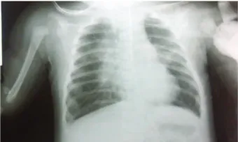
A 7-week-old female infant was admitted to our hospital with gasping and mottling along with perioral cyanosis at presentation. hese symptoms which have been associated with coughing and fever 8 days before admission led parents to bring her to public health medical service who gave antibi-otics and symptomatic medication. here was no history of postcough vomiting or breath cessation before. Since there was no clinical improvement and the patient’s condition was worsening, she was referred to our tertiary hospital. She had no history of diphtheria-pertussis-tetanus (DPT) vaccination. She came from a low social economic family and nonsupporting environment with poor ventilation. On admission, she looked severely ill with fever (40∘C), gasping, and bounding tachycardia with O2 saturation decreased to 84%. Physical examination revealed perioral cyanosis, nasal laring, chest retraction, and crackles in both lung on auscultation. Abdominal indings were normal. Mechanical ventilation was set, and she got aggressive luid therapy, cephalosporin antibiotic, and dopamine as well. Blood test result showed anemia (9.7 g/dL), leukocytosis (68,000/mm3) with absolute lymphocytosis (54%), and thrombocytosis (690,000/mm3). Blood smear result showed toxic granules and hypersegmentation. Blood gas analysis result showed metabolic acidosis and hypoxemia. Suggested bilateral pneu-monia was noted on chest radiograph (Figure 1). Severe bronchopneumonia with respiratory and cardiovascular fail-ure (septic shock) and anemia due to underlying disease was diagnosed. She was on ventilator for 5 days. On the 7th day of hospitalization, whooping cough was noted, and macrolide was administered. Nasopharyngeal swab for B. pertussisisolation in Bordet-Gengou media was taken, and it yielded positive result. Severe probable pertussis was then diagnosed. We conducted echocardiography to detect the probable pulmonary hypertension resulting in no pulmonary hypertension and good ventricular function. She was placed in isolation ward at the 9th day of hospitalization, showing clinical improvement and being discharged with better con-dition.
3. Discussions
From further medical history, we found that this patient had been treated in peripheral health care facilities for twice and treated acute respiratory infection and then further referred to our tertiary hospital as a severe pneumonia. In the emergency department, she presented with respiratory failure along with vascular disturbance and she was given emergence luid therapy, mechanical ventilation and antibiotics as well. Initially she was not diagnosed with pertussis as a cause of her condition. According to CDC, classic pertussis can
Figure 1: Anteroposterior thorax imaging showed bilateral pneu-monia and no cardiac enlargement.
be deined into three phases such as catarrhal phase with rhinorrhea with unspeciic cough as symptoms, paroxysmal phase showing increasing severity of cough and coughing spells, frequently followed by whooping and post-cough vomiting, and convalescent phase [10]. In this inal phase, the frequency and severity of cough will decrease. In this patient, the symptoms led to misdiagnosis of pertussis infection, and in fact, she further presented in advanced severe acute respiratory infection disease, which was severe pertussis. he deinition of severe pertussis itself is still unclear although it can be classiied as more severe and less severe [9]. Paroxysmal phase oten ends in respiratory failure, apnea, seizure, and cardiogenic circulation disturbance. Our critical ill patient admitted to our hospital in paroxysmal phase needed intensive care unit and it is called is critical pertussis [3]. In Germany, it was reported that 12 of 234 babies died because of sudden infant death syndrome (SIDS) caused by B. pertussis[11].
Case Reports in Emergency Medicine 3
leukocytosis might likely as a cause of respiratory failure. his critical pertussis caused hypotension and shock that mimicked severe sepsis. In critical pertussis, intensive care unit is required and several cases require assistance of ventila-tion, especially in infants with apnea, severe pneumonia and respiratory failure [3]. Treatment of pertussis should include the management of symptomatic signs and direct to the causative organism with macrolide antibiotic. Symptomatic treatment aims at decreasing coughing and support nutrition. Erythromycin is the traditional treatment, however, multi-centered randomized controlled trials in children reported that the newer macrolides azithromycin and clarithromycin have similar efectiveness, better compliance with less side efects compared with erythromycin [15, 16]. Dosage uses in treatment erithromycin can be 40–50 mg/kg of body weight/day (maximum two g per day) in four divided doses for 14 days. Clarithromycin dosage is 15 mg/kg of body weight/day (maximum one g per day) in two divided doses each day for 7 days. Azithromycin was given 10 mg/kg of body weight/day for ive days [16,17].
Immunization can reduce the severity of breakthrough disease in children who receive three doses of vaccine compared with that in unvaccinated children. In our case, the patient has not received DPT vaccination as her age was under 2 months of age when sufering from the disease. So, it is likely that she got the infection from adults surrounding her. he predominant factors for this patient were no history of immunization, low social economy, and nonsupporting household environment [18].
4. Conclusions
Pertussis can be presented in fatal respiratory failure if it is diagnosed late and not treated appropriately. he clinical presentation is oten diicult to observe by parents and peripheral medical services, and it causes delay in diagnosis and treatment. It is noteworthy that we concern about prob-able pertussis in young infants with long-lasting cough par-ticularly associated with postcough vomiting and/or perioral cyanosis, having no DPT immunization, apnea, or whooping cough, and for this disease, macrolides are drug of choice.
References
[1] D. S. Gregory, “Pertussis: a disease afecting all ages,”American Family Physician, vol. 74, no. 3, pp. 420–426, 2006.
[2] Manitoba Health Communicable Disease Control Unit, Pertus-sis/Parapertussis, Communicable Disease Management Proto-col, Manitoba, Canada, 2001.
[3] J. S. Burr, T. L. Jenkins, R. Harrison et al., “he collaborative pediatric critical care research network critical pertussis study: collaborative research in pediatric critical care medicine,” Pedi-atric Critical Care Medicine, vol. 12, no. 4, pp. 387–392, 2011. [4] U. Heininger, “Update on pertussis in children,”Expert Review
of Anti-Infective herapy, vol. 8, no. 2, pp. 163–173, 2010.
[5] H. M. Nataprawira, F. Cahayasari, and A. Kashmir, “Pertussis-like syndrome or pertussis: a delay diagnosis,”Paediatr Indones, vol. 52, pp. 28–31, 2012.
[6] E. L. Murray, D. Nieves, J. S. Bradley et al., “Characteristics of severeBordetella pertussisinfection among infants 90 days of age admitted to pediatric intensive care unit-Southern Cali-fornia, September 2009-June 2011,”Pediatric Infectious Disease Journal, vol. 2, no. 1, pp. 1–6, 2013.
[7] J. D. Cherry, R. Harrison, J. S. Bradley et al., “Pertussis in young infants. Guidance for Clinicians,” 2010.
[8] Centers for Disease Control and Prevention, “Pertussis (whoop-ing cough): Surveillance & Report(whoop-ing,” 2010,http://www.cdc
.gov/pertussis/surv-reporting.html.
[9] World Health Organization and Department of Vaccines and Biologicals, “Pertussis surveillance. A global meeting,” Tech. Rep. WHO/V&B/01.19, World Health Organization, Geneva, Switzerland, 2001.
[10] C. D. Paddock, G. N. Sanden, J. D. Cherry et al., “Pathology and pathogenesis of fatalBordetella pertussisinfection in infants,”
Clinical Infectious Diseases, vol. 47, no. 3, pp. 328–338, 2008. [11] U. Heininger, W. J. Kleemann, and J. D. Cherry, “A controlled
study of the relationship betweenBordetella pertussisinfections and sudden unexpected deaths among German infants,” Pedi-atrics, vol. 114, no. 1, pp. e9–15, 2004.
[12] C. Pierce, N. Klein, and M. Peters, “Is leukocytosis a predictor of mortality in severe pertussis infection?” Intensive Care Medicine, vol. 26, no. 10, pp. 1512–1514, 2000.
[13] A. Donoso, J. Le´on, M. Ram´ırez, G. Rojas, and B. Oberpaur, “Pertussis and fatal pulmonary hypertension: a discouraged entity,”Scandinavian Journal of Infectious Diseases, vol. 37, no. 2, pp. 145–148, 2005.
[14] Centers for Disease Control and Prevention,National Immu-nization Program: Epidemiology and Prevention of Vaccine-Preventable Diseases, “he Pink Book”, Pertussis, Centers for Disease Control and Prevention, Atlanta, Ga, USA, 8th edition, 2003.
[15] J. M. Langley, S. A. Halperin, F. D. Boucher, and B. Smith, “Azithromycin is as efective as and better tolerated than erythromycin estolate for the treatment of pertussis,”Pediatrics, vol. 114, no. 1, pp. e96–101, 2004.
[16] J. M. Zuckerman, “Macrolides and ketolides: azithromycin, clarithromycin, telithromycin,” Infectious Disease Clinics of North America, vol. 18, no. 3, pp. 621–649, 2004.
[17] Centers for Disease Control and Prevention, “Recommended antimicrobial agents for the treatment and postexposure prophylaxis of pertussis,” 2005, http://www.cdc.gov/mmwr/
preview/mmwrhtml.
[18] M.-P. Preziosi and M. E. Halloran, “Efects of pertussis vacci-nation on disease: vaccine eicacy in reducing clinical severity,”
Submit your manuscripts at
http://www.hindawi.com
Stem Cells
International
Hindawi Publishing Corporationhttp://www.hindawi.com Volume 2014
Hindawi Publishing Corporation
http://www.hindawi.com Volume 2014
MEDIATORS
INFLAMMATIONof
Hindawi Publishing Corporation
http://www.hindawi.com Volume 2014
Behavioural
Neurology
Endocrinology
International Journal of Hindawi Publishing Corporationhttp://www.hindawi.com Volume 2014
Hindawi Publishing Corporation
http://www.hindawi.com Volume 2014
Disease Markers
Hindawi Publishing Corporation
http://www.hindawi.com Volume 2014
BioMed
Research International
Oncology
Journal ofHindawi Publishing Corporation
http://www.hindawi.com Volume 2014
Hindawi Publishing Corporation
http://www.hindawi.com Volume 2014 Oxidative Medicine and Cellular Longevity Hindawi Publishing Corporation
http://www.hindawi.com Volume 2014
PPAR Research
The Scientiic
World Journal
Hindawi Publishing Corporation
http://www.hindawi.com Volume 2014
Immunology Research
Hindawi Publishing Corporation
http://www.hindawi.com Volume 2014
Journal of
Obesity
Journal ofHindawi Publishing Corporation
http://www.hindawi.com Volume 2014
Hindawi Publishing Corporation
http://www.hindawi.com Volume 2014
Computational and Mathematical Methods in Medicine
Ophthalmology
Journal ofHindawi Publishing Corporation
http://www.hindawi.com Volume 2014
Diabetes Research
Journal ofHindawi Publishing Corporation
http://www.hindawi.com Volume 2014
Hindawi Publishing Corporation
http://www.hindawi.com Volume 2014
Research and Treatment
AIDS
Hindawi Publishing Corporation
http://www.hindawi.com Volume 2014
Gastroenterology Research and Practice
Hindawi Publishing Corporation
http://www.hindawi.com Volume 2014
Parkinson’s
Disease
Evidence-Based Complementary and Alternative Medicine
Volume 2014 Hindawi Publishing Corporation