Affect Melatonin or Bilirubin Levels in Humans
Niki Lindblom, Taina Ha¨to¨nen, Maija-Liisa Laakso, Aino Alila-Johansson,
Marja-Leena Laipio, and Ursula Turpeinen
Background:Light treatment through the eyes is effective in alleviating the symptoms of some psychiatric disorders. A recent report suggested that skin light exposure can affect human circadian rhythms. Bilirubin can serve as a hypothetical blood-borne mediator of skin illumination into the brain. We studied whether bright light directed to a large body area could suppress the pineal melatonin secretion or decrease serum total bilirubin in conditions that could be used for therapeutic purposes.
Methods: Seven healthy volunteers participated in two consecutive overnight sessions that were identical except for a light exposure on the chest and abdomen in the second night from 12:00 AM to 6:00 AM (10,000-lux, 32 W/m2cool white for six subjects and 3000-lux, 15 W/m2 blue light for one subject). Hourly blood samples were collected from 7:00PMto 7:00AMfor melatonin radioim-munoassays. Bilirubin was measured by a modified diazo method in blood samples taken at 12:00AMand 6:00AM and in urine samples collected from 7:00PMto 11:00PM and from 11:00PM to 7:00AM.
Results: The skin light exposure did not cause any significant changes in serum melatonin or bilirubin levels. The excretion of bilirubin in urine was also the same in both sessions.
Conclusions: Significant melatonin suppression by ex-traocular light does not occur in humans. Robust concen-tration changes of serum total bilirubin do not have a role in mediating light information from the skin to the central nervous system. Biol Psychiatry 2000;48:1098 –1104 ©2000 Society of Biological Psychiatry
Key Words: Extraocular phototransduction, skin light exposure, light treatment, pineal gland, melatonin suppres-sion, bilirubin
Introduction
T
he mechanisms by which light affects the regulation of sleep, circadian rhythms, and mood are poorly understood. Nevertheless, since the 1980s numerous stud-ies have shown that light therapy has beneficial effects when applied in certain types of sleep and mood disorders (e.g., Eastman et al 1998; Lewy et al 1982; Rosenthal et al 1984; Terman et al 1995, 1998). Today, research is in progress for defining the optimum quality and quantity of light, and the proper timing of exposure in various disorders (e.g., Chesson et al 1999; Lewy et al 1998).To date, it was thought that in adult mammals light can affect brain functions only through the eyes (Nelson and Zucker 1981; Underwood and Groos 1982); however, this concept has to be re-evaluated, since it was reported that the human body temperature rhythm and melatonin onset time were shifted by bright light directed to the skin behind the knee (Campbell and Murphy 1998). The finding is theoretically interesting, and in addition, it might have practical applications.
The level of effective illuminance in light treatment directed to the eyes is highly dependent on the subject’s behavior. For example, the threshold illuminance needed to suppress melatonin varies greatly according to the experimental conditions. Illuminances as low as 6 –17 photopic lux have been reported to suppress melatonin in strictly controlled conditions (monochromatic light of 509 nm, light beam directed uniformly on the retina, dilated pupils, and subject’s head kept motionless; Brainard et al 1988). In another study, when the subjects were sitting in front of a light source and gazed at the source for 10 sec every 2 min, a significant suppression was produced by 500-lux but not by 200-lux illuminance (Hashimoto et al 1996). This variability does not cause problems if the patient participating in the light therapy is well motivated and able to follow the instructions; however, sleep disor-ders are extremely common and harmful in, for example, mentally retarded people and in patients with Alzheimer’s disease, and some of them can benefit from light therapy (Guilleminault et al 1993; Van Someren et al 1997). In their case, it would be useful if the light treatment could be
From Pediatric Neurology, Hospital for Children and Adolescents (NL) and Institute of Biomedicine, Department of Physiology (TH, M-LLaa, AA-J), University of Helsinki, Helsinki; Neural Networks Research Centre, Helsinki University of Technology, Espoo (M-LLaa); the Department of Clinical Chemistry (M-LLai) and Laboratory (UT), Helsinki University Central Hospi-tal, Helsinki, Finland.
Address reprint requests to Niki Lindblom, M.D., Rinnekoti Foundation, Kumputie 1, FIN-02980 Espoo, Finland.
Received January 3, 2000; revised April 14, 2000; accepted April 20, 2000.
© 2000 Society of Biological Psychiatry 0006-3223/00/$20.00
delivered without worrying about the gaze direction or openness of the eyes.
In attempts to determine the effectiveness of light, the melatonin suppression test is often used. Although it is quite possible that light affects the brain functions without changing the secretion of the pineal hormone, the suppres-sion of nocturnal melatonin synthesis can be a sign of the light stimulus reaching the hypothalamus, the site of the main body clock. This assumption is valid at least if the light stimulus is directed to the eyes and mediated through the retinohypothalamic pathways. Two independent stud-ies have shown that bright light directed to the small skin area behind the knee for 3 hours does not suppress melatonin secretion in humans (He´bert et al 1999; Lockley et al 1998). It is possible, however, that the efficacy of skin light exposure depends on the size of the exposed area and the duration of the exposure. Therefore, we decided to study whether extraocular light exposure delivered to a larger body area and for a longer period would produce melatonin suppression.
The mechanisms involved in the possible humoral phototransduction are completely unknown; however, an attractive hypothesis has been presented. According to the original model, some light-sensitive molecules, such as bilirubin or hemoglobin, circulating in retinal blood ves-sels could mediate the effects of light to the circadian regulatory machinery (Oren 1996). Later this hypothesis was further developed to include the concept of extraoc-ular phototransduction through the skin (Oren and Terman 1998).
Bilirubin is formed mainly in the cells of the reticuloen-dothelial system as a product of heme catabolism. Despite seemingly only a waste product, it has been shown to posses strong antioxidant activity (Stocker et al 1987). This and its well-known sensitivity to light may be in favor of the assumption that it might function as a blood-borne mediator of extraocular phototransduction. In hyperbilirubinemic newborns the renal excretion of bili-rubin can be substantially increased by skin phototherapy (McDonagh and Lightener 1985). The effect of photother-apy is based on the light-induced formation of hydrophilic molecules from the lipophilic bilirubin.
There is minimal information available about the effects of light on normobilirubinemic adults. According to a case study, molecular changes similar to those in newborns occurred in the structure of bilirubin in an adult subject wearing only shorts and exposed to natural sunlight for 90 min (McDonagh 1986); however, no changes in the serum level of total bilirubin could be found. This may be due to the short exposure time and the fact that the bilirubin concentration changes caused by the excretion of photo-degradation products may be detectable only after a longer period. Therefore, we studied whether a 6-hour skin light
exposure might cause changes in total serum bilirubin levels or in the amounts of bilirubin excreted in urine of healthy adult volunteers.
Methods and Materials
Seven healthy unmedicated subjects (two female and five male, all white, age range 19 – 43 years) gave informed consent after the nature of the study had been explained. The study protocol was accepted by the ethical committee of the institute. During the 5 days preceding the study the subjects were told to avoid bright lights at night, strenuous physical excercise, and beverages containing alcohol.
For the sham light laboratory session the subjects arrived between 5:00PM and 6:30 PM and were asked to empty their bladders, after which a venous cannula for hourly blood samples was inserted in the antecubital vein. Thereafter the subjects were in a dimly lit room (,10 lux, Chroma Meter CL-100, Minolta, Osaka, Japan). During the period from 7:00PMto 11:00PMthe subjects were allowed to play games and listen to music but not to lie down. A standard snack was served from 10:00PMto 11:00
PM. Urine was collected at 11:00 PM (not at 12:00 AM) for practical reasons.
At 11:00PMthe subjects lay down under the light sources and their eyes were covered with black cloth (tied four times around the head and extending from the upper forehead to cheeks) impermeable to the illuminance of 10,000 lux (tested by the authors). The subjects were also specifically asked to report immediately if any light entered their eyes during the light exposure. No such report was given by any one of the subjects. In addition, the light sources were covered with multiple dark blankets to prevent light from escaping to the surroundings. During the sham light session, the lights of the panels were off, but fans were on from 12:00AMto 6:00AMto make the sound conditions similar to those during the light exposure session. At 7:00 AM the last blood sample was drawn, the eyes were uncovered, and the urine was collected.
On the same day between 5:00PMand 6:30PM the subjects returned to the laboratory for the skin light session, which was carried out in a manner similar to that of the sham light session except that from 12:00AMto 6:00AMthe lights were on and the fans were directed under the panels to cool the air. The temperature under the panels was continuously monitored by the researchers during both sessions. Most of the time it was 24 –26°C, but rose transiently to 29°C during the light exposure session. The subjects were allowed to sleep from 11:00PM to 7:00 AM while lying, but their sleep was disturbed when the researchers monitored the posture, eye covers, and temperature under the light panels and collected the hourly blood samples. The sleep was similarily disturbed during both nights.
spectroradiometri-cally—Bentham Instruments, Reading, UK) to the subjects’ naked skin between the neck and hip levels (the size of the illuminated body area was estimated to be approximately 50 cm340 cm, distance from the lamps ca. 20 cm). For one subject a commercial fluorescent blue light device (Figure 1B) was used as the light source (Phototherapy lamp Medela Ag, Baar, Switzerland, 90 W, 3000 lux, 15 W/m2, distance 40 cm from the
subject). The other conditions and protocol for this subject were similar to those of the other subjects. All calculations and statistical evaluations were performed both by including and by excluding the subject exposed to blue light. Because the results and interpretations were the same for both sets of subjects, only the results from all seven subjects are presented.
The blood samples were centrifuged and the serum stored at
224°C. Melatonin was extracted from 1.0 mL of serum with chloroform and measured in duplicate by radioimmunoassay (Vakkuri et al 1984). The nonspecific binding of the tracer was 5– 6%. The least detectable concentration, defined as apparent concentration at 2 SDs from the counts at maximum binding (n56 in each assay), was smaller than the lowest standard (1.95 ng/L). Intra-assay variability was,10%. The interassay variabil-ity during 24 months in 16 assays including the assays of this study was 15–18%, depending on the concentration. All samples of each pattern were measured in the same assay.
The assay used for the analysis of total bilirubin was a modified diazo method (Doumas et al 1982; Jendrassik and Gro´f 1938). Total bilirubin was measured in both sessions from serum samples taken at 12:00 AM and 6:00 AM and from urinary specimens taken as described above. The intra-assay variability was ,7%. All bilirubin samples were placed in dark tubes, protected from light during all procedures, and measured in the same assay.
Two-way analysis of variance (ANOVA) for repeated mea-sures was used in statistical evaluations of the serum melatonin and bilirubin levels, the excretion rate of urine, and the excretion of bilirubin in urine. In addition, the secretion of melatonin during the 2 nights was compared by applying the area under the curve (AUC) analysis. The “prelight” and “during light” AUCs and the peak and postlight levels of melatonin were expressed as percentages of the corresponding values in the sham light session and evaluated by two-tailed one-sample ttest (deviation from 100%). The minimum detectable deviation from 100% was calculated by a method described previously (Rosner 1986). The same method was used for the calculations of the minimum detectable difference in serum total bilirubin levels between the samples taken at 12:00AMand 6:00AM.
Results
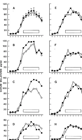
All seven subjects had a clear melatonin rhythm with peak values during the night (Figure 2, B–H). The average serum level profiles did not differ significantly between the sham light and skin light sessions (two-way ANOVA; Figure 2A). In most subjects the profiles were very similar during both nights. However, two of the seven subjects (Figure 2, B and C) had somewhat lower serum melatonin concentrations during the light exposure than during the sham light, one from the beginning of the treatment for 3– 4 hours and the other during the latter part of the light exposure from 3:00AM to the end of the experiment.
In AUC analysis it was found that subject B had similarily decreased melatonin levels before and during the light treatment as compared with the corresponding intervals in the sham light night (85% vs. 86%; Table 1). The decrease of serum melatonin in subject C during light exposure seemed more pronounced (AUC during light 78% of sham light vs. AUC prelight 102% of sham light; Table 1); however, statistical evaluation of the mean AUCs of all seven subjects did not disclose any differ-ences between the sessions. Furthermore, the melatonin peak level, found at any time from 12:00AMto 6:00 AM, did not differ between the sessions, nor did the postlight melatonin concentration (Table 1).
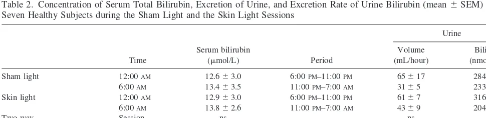
The mean serum bilirubin levels were equal in both sessions at 12:00AMand of the same magnitude at 6:00AM (Table 2). The standard deviation of the individual differ-ences between the values at 12:00 AM and 6:00 AM was 63.4 mmol/L. Thus, the minimum detectable difference was 3.7 mmol/L (n5 7,p5 .05, power 80%). Bilirubin Figure 1. Spectral power distributions of the light sources used
excretion in urine was equally low during both sampling intervals and similar in both sessions. Thus, no effect of skin light exposure was found on serum total bilirubin concentration or excretion rate in urine. There were no significant correlations between the individual serum bil-irubin levels and the excretion rates in urine, probably due to the large variation of urine bilirubin excretion rates.
Discussion
The 6-hour bright light exposure on a large skin area had no significant influence on the average melatonin
secre-tion profile in our subjects. The result is in line with the findings that illumination of the popliteal skin with bright white light (Lockley et al 1998) or blue light (400 –550 nm; He´bert et al 1999) for 3 hours did not suppress melatonin in healthy adults. The illuminated skin areas in the previous studies were 10 and 50 –100 cm2, respec-tively. In our study, a rough estimation for the skin area illuminated effectively was about 50 cm3 40 cm. Thus, prolonging the exposure period twofold and extending the illuminated area at least 20-fold did not make the treat-ment more effective.
Furthermore, the conclusion that melatonin suppression Figure 2. Serum melatonin concentrations in 7 healthy subjects during the sham light session (■) and the skin light session (E). (A) Means with SEMs (two-way analysis of variance: session, ns; time,p,.001; interaction, ns).(B–H)Individual curves. Light was directed to the chest and abdo-men of the subjects with the eyes covered for 6 hours, with 10,000-lux, 32 W/m2cool white light
for six subjects (B–D and F–H) and 3000-lux, 15 W/m2blue light for one subject(E). The period of
in humans does not occur through skin illumination is supported by the finding that in hyperbilirubinemic new-borns exposed to phototherapy with the eyes covered, the serum melatonin levels were elevated rather than sup-pressed (Jaldo-Alba et al 1993). In addition, there is evidence for the inefficacy of facial illumination to sup-press melatonin in completely blind people (Czeisler et al 1995) and for its relative inefficiency to suppress melato-nin in sighted people with the eyes closed (Ha¨to¨nen et al 1999); however, the lack of melatonin suppression by extraocular light does not exclude the possibility that other brain functions can be influenced by extraocular light.
The decreased serum melatonin concentrations in two of the seven subjects during the light exposure session raises the question of whether there might be interindi-vidual differences in the sensitivity to extraocular light and whether melatonin suppression might be seen in a sub-population. Usually, the individual melatonin profiles are very similar from night to night (Arato et al 1985; Arendt 1988; Laakso et al 1990), as they were in the other five subjects in this study. Because the low melatonin levels in the two subjects were not found evenly during the light exposure and the decrease occurred during different times
within the light exposure period, it seems improbable that the suppressions were caused by light.
When we tried to find the explanation for the suppres-sion, we noticed that these two persons deviated from the other five subjects in having an exceptionally high urine flow during the light exposure session from 11:00 PM to 7:00 AM (B sham light/light 36/64 mL/hour, C 22/69 mL/hour; other five subjects 29/22, 30/29, 24/25, 21/26, and 58/69 mL/hour). Thus, although the mean urine flows did not differ significantly between the sessions, the two persons with the decreased serum melatonin levels ex-creted urine during the light exposure approximately two or three times the amount they excreted during the sham light exposure. When interviewed afterwards, subject B said that she felt thirsty in the evening preceding the light exposure and drank a lot of water, which was freely available during the sessions. Subject C was given water to drink several times during the light exposure because he complained of “unbearable thirst.”
Thus, the water balance of the two subjects most probably differed between the two sessions. It has been shown that changes in hemodynamics by posture alter-ations can result in significant changes in serum melatonin
Table 1. Characteristics of Serum Melatonin Profiles in Seven Healthy Subjects during the Skin Light Session
Subject
Area under the curve
Peak level (any time)
Postlight level (6:00AM) Prelight
(7:00PM–12:00AM)
During light (12:00AM– 6:00AM)
E 93 97 90 117
B 85 86 93 103
C 102 78 74 65
D 130 110 90 135
F 92 103 114 119
G 96 103 107 99
H 83 97 96 100
Mean6SD 97616 96611 95613 105622
MDD 20 14 16 27
Light was directed for 6 hours (12:00AM– 6:00AM) to the chest and abdomen of the subjects lying under the light sources with their eyes covered. Subjects B–D and F–H: fluorescent white light, illuminance 10,000 lux, integrated irradiance 32 W/m2. Subject E: blue light, 3000 lux, 15 W/m2. All values are given as percentages of the
corresponding values of the control profiles determined in the sham light session without turning the lights on. None of the mean percentages was different from 100% (two-tailed one-samplettest). MDD, minimum detectable deviation from 100% (n57,p5.05, power 90%).
Table 2. Concentration of Serum Total Bilirubin, Excretion of Urine, and Excretion Rate of Urine Bilirubin (mean6SEM) in Seven Healthy Subjects during the Sham Light and the Skin Light Sessions
Time
Serum bilirubin
(mmol/L) Period
Urine
Volume (mL/hour)
Bilirubin (nmol/hour)
Sham light 12:00AM 12.663.0 6:00PM–11:00PM 65617 284670 6:00AM 13.463.5 11:00PM–7:00AM 3165 233623 Skin light 12:00AM 12.963.0 6:00PM–11:00PM 6167 316684 6:00AM 13.862.6 11:00PM–7:00AM 4369 204656
Two-way Session ns ns ns
analysis of variance Time ns p,.05 ns
Interaction ns ns ns
Light exposure (10,000-lux, 32 W/m2
cool white in six subjects and 3000-lux, 15 W/m2
concentrations (Deacon and Arendt 1994). It is possible that alterations in fluid balance for other reasons also cause changes in serum melatonin concentrations. In addition, an increased urine flow has been reported to increase the rate of melatonin excretion in sheep, probably due to decreased tubular reabsorbtion (Valtonen et al 1993). In the sheep study, the serum melatonin concentra-tion did not decrease within 90 min; however, a decrease can appear if the abundant urine flow continues for a long time and the loss of melatonin is not compensated for by increased melatonin synthesis. We consider the variations in fluid balance the most probable reason for the decreased serum melatonin levels in subjects B and C.
The 6-hour bright light exposure on the chest and abdomen did not change the serum total bilirubin concen-trations or the urinary excretion rate of bilirubin in our subjects. When bilirubin absorbs a photon, three types of chemical reactions can occur: slow photo-oxidation (e.g., to monopyrroles and dipyrroles), structural isomerization to lumirubin, or configurational isomerization by conver-sion from double to single bonding of one of the bridges joining the pyrrole rings (Ennever 1990; McDonagh and Lightner 1985). The reaction products are more water soluble than the original substrate and can be excreted in bile and urine.
The diazo method used in the measurements does not detect the oxidation products or lumirubin, but it does detect all the configurational isomers (Ennever 1990). Our results suggest that significant skin light–induced photo-oxidation or structural isomerization of bilirubin does not occur in normobilirubinemic adults during a 6-hour skin light exposure. As in the previous case study (McDonagh 1986), the configurational isomerization could have oc-curred in our subjects, but the reactions did not lead to any detectable decrease of total bilirubin levels. The possibility remains that different isomers can have different effects in the central nervous system. In addition to the heme-related compounds, other molecules such as vitamin D, known to be sensitive to ultraviolet radiation (Holick 1995), may serve as messengers from the skin to the brain (Stumpf and Privette 1991).
In summary, the secretion of the pineal hormone mel-atonin, known to be very sensitive to ocular light, was not significantly affected by skin light exposure on a larger body area (chest and abdomen) and for a longer period (6 hours) than previously tested; however, possible effects of extraocular light on other hypothalamic functions cannot be excluded by this study. Moreover, the present results do not support a role for short-term fluctuations of serum total bilirubin levels in mediating the light information from the skin to the central nervous system. The possibility remains that more subtle changes in the structure of bilirubin are involved in extraocular phototransduction.
This study was financially supported by the Rinnekoti Research Foun-dation, Espoo, Finland.
References
Arato M, Grof E, Grof P, Laszlo I, Brown GM (1985): Repro-ducibility of the overnight melatonin secretion pattern in healthy men. In: Brown GM, Wainwright SD, editors.The Pineal Gland: Endocrine Aspects, Advances in the Bio-sciences, Vol 53. Oxford, UK: Pergamon, 277–282. Arendt J (1988): Melatonin.Clin Endocrinol29:205–229. Brainard GC, Lewy AJ, Menaker M, Fredrickson RH, Miller LS,
Weleber RG, et al (1988): Dose-response relationship be-tween light irradiance and the suppression of plasma melato-nin in human volunteers.Brain Res454:212–218.
Campbell S, Murphy P (1998): Extraocular circadian phototrans-duction in humans.Science279:396 –399.
Chesson ALJ, Littner M, Davila D, MacDowell Anderson W, Grigg-Damberger M, Hartse K, et al (1999): Practice param-eters for the use of light therapy in the treatment of sleep disorders.Sleep22:641– 660.
Czeisler CA, Shanahan TL, Klerman EIB, Martens H, Brotman DJ, Emens JS, et al (1995): Suppression of melatonin secre-tion in some blind patients by exposure to bright light.N Engl J Med332:6 –11.
Deacon S, Arendt J (1994): Posture influences melatonin con-centrations in plasma and saliva of humans. Neurosci Lett
167:191–194.
Doumas BT, Perry BW, Jendrzejczak B, Katona V (1982): Pitfalls in the American Monitor kit methods for determina-tion of total and “direct” bilirubin.Clin Chem28:2305–2308. Eastman CI, Young MA, Fogg LF, Liu L, Meaden PM (1998): Bright light treatment of winter depression. A placebo-controlled trial.Arch Gen Psychiatry55:883– 889.
Ennever JF (1990): Blue light, green light, white light, more light: Treatment of neonatal jaundice.Clin Perinatol17:467– 481.
Guilleminault C, McCann CC, Querasalva M, Cetel M (1993): Light therapy as treatment of dyschronosis in brain impaired children.Eur J Pediatr152:754 –759.
Hashimoto S, Nakamura K, Honma S, Tokura H, Honma KI (1996): Melatonin rhythm is not shifted by lights that sup-press nocturnal melatonin in humans under entrainment.Am J Physiol270:R1073–R1077.
Ha¨to¨nen T, Alila-Johansson A, Mustanoja S, Laakso M-L (1999): Suppression of melatonin by 2000-lux light in hu-mans with closed eyelids.Biol Psychiatry46:827– 831. He´bert M, Martin SK, Eastman CI (1999): Nocturnal melatonin
secretion is not suppressed by light exposure behind the knee in humans.Neurosci Lett274:127–130.
Holick MF (1995): Environmental factors that influence the cutaneous production of vitamin D.Am J Clin Nutr61(suppl): 638S– 645S.
Jendrassik L, Gro´f P (1938): Vereinfachte photometrische Methoden zur Bestimmung des Blutbilirubins. Biochem Z
297:81– 89.
Laakso ML, Porkka-Heiskanen T, Alila A, Stenberg D, Johans-son G (1990): Correlation between salivary and serum mel-atonin: Dependence on serum melatonin levels.J Pineal Res
9:39 –50.
Lewy AJ, Bauer VK, Cutler NL, Sack RL, Ahmed S, Thomas KH, et al (1998): Morning vs evening light treatment of patients with winter depression. Arch Gen Psychiatry 55: 890 – 896.
Lewy AJ, Kern HA, Rosenthal NE, Wehr TA (1982): Bright artificial light treatment of a manic-depressive patient with a seasonal mood cycle.Am J Psychiatry139:1496 –1498. Lockley SW, Skene DJ, Thapan K, English J, Ribeiro D, Haimov
I, et al (1998): Extraocular light exposure does not suppress plasma melatonin in humans. J Clin Endocrinol Metab
83:3369 –3372.
McDonagh AF (1986): Sunlight-induced mutation of bilirubin in a long-distance runner.N Engl J Med314:121–122. McDonagh AF, Lightner DA (1985): “Like a shrivelled blood
orange”— bilirubin, jaundice, and phototherapy. Pediatrics
75:443– 455.
Nelson RJ, Zucker I (1981): Abscence of extraocular photore-ception in diurnal and nocturnal rodents exposed to direct sunlight.Comp Biochem Physiol69A:145–148.
Oren DA (1996): Humoral phototransduction: Blood is a mes-senger.Neuroscientist2:207–210.
Oren DA, Terman M (1998): Tweaking the human circadian clock with light.Science279:333–334.
Rosenthal NE, Sack DA, Gillin JC, Lewy AJ, Goodwin FK, Davenport Y, et al (1984): Seasonal affective disorder. A
description of the syndrome and preliminary findings with light therapy.Arch Gen Psychiatry55:875– 882.
Rosner B (1986): Sample size determination. In: Payne M, editor.Fundamentals of Biostatistics.Boston: Duxbury Press, 207–212.
Stocker R, Yamamoto Y, McDonagh AF, Glazer AN, Ames BN (1987): Bilirubin is an antioxidant of possible physiological importance.Science235:1043–1046.
Stumpf WE, Privette TH (1991): The steroid hormone of sunlight soltriol (vitamin D) as a seasonal regulator of biological activities and photoperiodic rhythms.J Steroid Biochem Mol Biol39:283–289.
Terman M, Lewy AJ, Dijk DJ, Boulos Z, Eastman CI, Campbell SS (1995): Light treatment for sleep disorders: Consensus report. IV. Sleep phase and duration disturbances. J Biol Rhythms10:135–147.
Terman M, Terman JS, Ross DC (1998): A controlled trial of timed bright light and negative air ionization for treatment of winter depression.Arch Gen Psychiatry55:875– 882. Underwood H, Groos G (1982): Vertebrate circadian rhythms:
Retinal and extraretinal photoreception. Experientia
38:1013–1021.
Vakkuri O, Leppa¨luoto J, Vuolteenaho O (1984): Development and validation of a melatonin radioimmunoassay using radio-iodinated melatonin as tracer. Acta Endocrinol (Copenh)
106:152–157.
Valtonen M, Laitinen JT, Eriksson L (1993): Renal melatonin excretion in sheep is enhanced by water diuresis.J Endocri-nol138:445– 450.