H-reflex amplitude depression as a marker
of presynaptic inhibition in Painful Diabetic
Neuropathy (PDN).
Ahmad Asmedi*, Samekto Wibowo, Lucas Meliala
Department of Neurology, Faculty of Medicine Universitas Gadjah Mada / Dr Sardjito Hospital, Yogyakarta, Indonesia
DOI: http://dx.doi.org/10.19106/JMedSci004701201504
ABSTRACT
Painful Diabetic Neuropathy (PDN) is a common complication of diabetes mellitus (DM). Disruption in presynaptic inhibition in dorsal horn of the spinal cord has been proposed as one of the pathomechanism of PDN. Previous research showed that presynaptic inhibition
can be detected by H-reflex examination. The aim of this study was to know whether the
reduction of presynaptic inhibition in spinal dorsal horn of PDN patients really exist, and
detectable by H-reflex examination. It was cohort prospective involving 141 (58 men, 83 women) patients with DM and impaired glucose tolerance (IGT) between the ages of 40
and 61 years from several health facilities in Yogyakarta. All patients underwent clinical, laboratory and electrodiagnostic examination. Demographic, clinical and electrodiagnostic data were collected and analyzed. By survival analysis there were 25 new cases of
PDN (12.12% cumulative incidence). Using survival Kaplan Meier analysis, the significant
hazard ratio for PDN were 12.81 for median motor nerve amplitude, 5.74 for median
nerve distal latency, 3.71 for median sensory nerve amplitude, 6.33 for median sensory latency, 3.4 for tibial nerve amplitude, 3.48 for tibial nerve distal latency, 2.29 for sural nerve amplitude, 4.47 for sural nerve latency, 3.99 for H-reflex latency, 5.88 for H-reflex amplitude, and 17.83 for Diabetic Neuropathy (DN) status. Using hazard proportional cox analysis, only H amplitude and DN status (DNS score) were significantly correlated with
PDN (p= 0.026; hazard ratio = 15.450; CI 95%= 1.39 – 171.62 for H amplitude and
p= 0.030; hazard ratio = 10.766; CI 95%=1.26 – 92.09 for DN status).
This study showed that depression of H-reflex amplitude was correlated with the occurrence of PDN. This result proves that there was presynaptic inhibition process in PDN that manifests as low H-reflex amplitude.
ABSTRAK
Nyeri neuropati diabetik (PDN) merupakan suatu komplikasi yang umum pada diabetes mellitus (DM). Gangguan pada penghambatan presinaptik pada bagian
dorsal horn dari sumsum tulang belakang diduga menjadi salah satu patomekanisme dari PDN. Penelitian sebelumnya menunjukkan bahwa penghambatan presinaptik
dapat dideteksi dengan pemeriksaan H-refleks. Tujuan dari penelitian ini adalah
untuk mengetahui apakah pengurangan inhibisi presinaptik di dorsal horn tulang
belakang pasien PDN benar-benar ada, dan terdeteksi oleh pemeriksaan H-refleks. Penelitian ini adalah kohort prospektif yang melibatkan 141 (58 laki-laki, 83 perempuan)
INTRODUCTION
Painful Diabetic Neuropathy (PDN) is a common complication of diabetes mellitus (DM), which can give large burdens because of its high incidence, serious clinical, economical and social impact and its unsatisfying treatment.1,2
Biological process in spinal dorsal horn has been shown to have an important role in pathophysiology of PDN. Gate mechanism in the dorsal horn of the spinal cord plays a role
by inhibiting or facilitating the flow of afferent
impulses from peripheral nerves to the spinal cord before it evokes pain perception.3 Many
research have shown that diminished synaptic inhibition plays a key role in neuropathic pain development.4 Normally, GABA
A receptors in
primary afferent terminals of nociceptors will mediate this presynaptic inhibition by causing reduction of transmitter release thus modulating
berbagai rumah sakit di Yogyakarta.
Semua pasien menjalani pemeriksaan klinis, laboratorium dan pemeriksaan
elektrodiagnostik. Data demografi, klinis dan electrodiagnostic dikumpulkan dan dianalisis.
Berdasarkan analisis survival ada 25 kasus baru PDN (kejadian kumulatif 12,12%). Dengan
menggunakan analisis survival Kaplan Meier, Rasio hazard yang signifikan untuk PDN adalah 12,81 untuk median amplitudo saraf motorik, 5.74 untuk median latensi distal saraf, 3,71 untuk median amplitudo saraf sensorik, 6.33 untuk median latensi sensorik, 3,4 untuk amplitudo tibialis saraf, 3,48 untuk latensi saraf tibialis distal, 2,29 untuk amplitudo saraf sural, 4,47 untuk latensi saraf sural, 3,99 untuk latensi H-refleks, 5,88 untuk amplitudo H-refleks, dan 17,83 status Diabetes Neuropati (DN). Dengan menggunakan analisis cox proporsional hazard, hanya amplitudo H dan status DN (skor DNS) yang secara signifikan berkorelasi dengan PDN (p= 0,026; rasio hazard= 15,450; CI 95%= 1,39-171,62 untuk amplitudo H dan p= 0,030; rasio hazard= 10,766 ; CI 95%= 1,26-92,09 untuk status DN). Penelitian ini menunjukkan bahwa depresi amplitudo H-refleks berkorelasi dengan
terjadinya PDN. Hasil ini membuktikan bahwa ada proses penghambatan presinaptik di
PDN yang bermanifestasi pada rendahnya amplitudo H-refleks.
Keywords: Painful diabetic neuropathy - H-reflex amplitude - presynaptic inhibition -
Diabetes Mellitus
H-reflex is an electrically induced reflex
analogous to the mechanically induced spinal
stretch reflex.6 The H-reflex bypasses the
muscle spindle. Therefore, it is a valuable tool in assessing modulation of monosynaptic
reflex activity in the spinal cord.6 The H-reflex
is an estimate of alpha motoneuron (aMN)
excitability when presynaptic inhibition and intrinsic excitability of the aMNs remain
constant. This measurement can be used to assess the responsiveness of the nervous system to various neurologic conditions including pain7, hence H-reflex amplitude can
be proposed as a method to assess presynaptic inhibition in humans.
The aim of this study was to know whether the reduction of presynaptic inhibition in spinal dorsal horn of PDN patients really
study of upper and lower extremity, and soleus H-reflex study were done at week 1. Nerve conduction study and H-reflex were
conducted using an MEB-2300K ENMG machine (Nihon Kohden, Tokyo, Japan). The evaluation and record of PDN occurrence were carried out every week up to 12 months by self-assessment, monitored by the doctors. The Kaplan Meier survival analysis was conducted to see the ability of several electrodiagnostic variables to predict the possibility of subjects to suffer from PDN during the observation period. For this reason, the ID pain was measured periodically every 2 weeks, to assess the PDN status of the subjects. The data analyses were presented in TABLE of Kaplan Meier analysis, hazard ratio, and survival curve.
RESULTS
One hundred and forty one subjects (58 men, 83 women) with an average age of 51 years old (range, 40–61 years old), were
enrolled in this study. Sixty five percent of the
subjects were found to be diabetic, while the other 35% were found to have IGT. The mean glucose level was 116 mg/dl (range, 78 - 200 mg/dL) for fasting, 170 mg/dL for 2 hours post prandial (range, 90 – 250 mg/dL).
Screening by NSS and DNS scores at admission found that 57 subjects (40.4%) were diagnosed as neuropathy (Diabetic Neuropathy/DN) according to NSS, while 68 (48.2%) were diagnosed as neuropathies according to DNS. There were no subjects diagnosed as PDN. However, upon completion of 48 weeks of observation, there were 12.12% cumulative incidence rate of PDN.
Pain score below 2), followed by a 12-months disease follow-up. The protocol was approved by the Research Ethics Committees of Faculty of Medicine, Universitas Gadjah Mada, and all subjects were provided with written informed consent. The subjects of this study were patients at diabetes outpatient clinic Dr Sardjito Hospital Yogyakarta, members of diabetes association (Persatuan Diabetes Indonesia, PERSADIA) PKU Hospital Yogyakarta, staffs of STIKES Aisiyah Yogyakarta and the subjects from Pre-diabetic Research of Clinical Pathology Department at Dr Sardjito Hospital Yogyakarta.
The inclusion criteria were men and women 20 to 60 year-old, with type 2 diabetes mellitus or impaired glucose tolerance (IGT),
as defined by American Diabetes Association (ADA) criteria. The exclusion criteria were anatomical deformities on extremities that
would interfere with electrodiagnostic study protocol; pregnancy or lactation; a documented history of lumbosacral surgery that would interfere with electrodiagnostic study protocol; other diseases known to be associated with pain, especially chronic pain in the feet that the investigator believed would interfere with the assessment of pain associated with diabetic neuropathy, like cancer pain, lumbosacral abnormality or other entrapment neuropathy; and any acute or underlying serious illness that are likely interfere with completion of the trial .
Clinical and electrodiagnostic
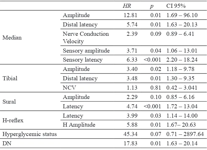
The low amplitude of median and tibial
nerve had significant value in predicting PDN
occurence with hazard ratio 12.81 for median nerve amplitude (95% CI 1.69 – 96.10, p= 0.01) and 3.4 for tibial nerve amplitude (95% CI 1.18 – 9.78, p= 0.02), while sural nerve
with hazard ratio 2.29 was not significant
(95% CI 0.85 – 6.16, p= 0.10). Distal latency of median, tibial, and sural nerve were also
related significantly with PDN occurrence
with hazard ratio 5.74 for median nerve (95% CI 1.63 – 20.13, p= 0.01), 3.48 for tibial nerve (95% CI 1.18 – 9.78, p= 0.02), and 4.74 for sural nerve latency (95% CI 1.72 – 13.04,
p<0.001). Median nerve sensory amplitude
and sensory latency were also significant with
hazard ratio 3.71 (95% CI 1.06 – 13.01, p= 0.04) and 6.33 (95% CI 2.20 – 18.24, p<0.001),
TABLE 1. Hazard ratioof PDN factor
HR p CI 95%
Median
Amplitude 12.81 0.01 1.69 – 96.10 Distal latency 5.74 0.01 1.63 – 20.13 Nerve Conduction
Velocity 2.39 0.09 0.89 – 6.41 Sensory amplitude 3.71 0.04 1.06 – 13.01 Sensory latency 6.33 <0.001 2.20 – 18.24
Tibial
Amplitude 3.40 0.02 1.18 – 9.78 Distal latency 3.48 0.01 1.30 – 9.35 NCV 1.13 0.81 0.42 – 3.041
Sural Amplitude 2.29 0.10 0.85 – 6.16 Latency 4.74 <0.001 1.72 – 13.04
H-reflex Latency 3.99 0.03 1.14 – 14.00 H Amplitude 5.88 0.01 1.67– 20.63 Hyperglycemic status 45.34 0.07 0.71 – 2897.64
DN 17.83 0.01 1.63 – 20.14
the nerve conduction velocityof median nerve (hazard ratio= 2.39) and tibial nerve (hazard
ratio= 1.13) were not significant.
The hazard ratio for H-reflex latency was 3.99 (95% CI 1.14 – 14.00) and for H-reflex
amplitude was 5.88 (95% CI 1.67 – 20.63).
These variables were also significantly related
to PDN occurrence with p value 0.03 and 0.01 respectively. Beside the electrodiagnostic variables, the clinical variables were also measured. The status of DN with hazard ratio 17.83 (95% CI 1.63 – 20.14, p= 0.01) was
significantly related to the occurence of PDN,
while the hyperglycemic status had high
hazard ratio (45.43) but was not significant
(95% CI 0.71 – 2897.64, p= 0.07).
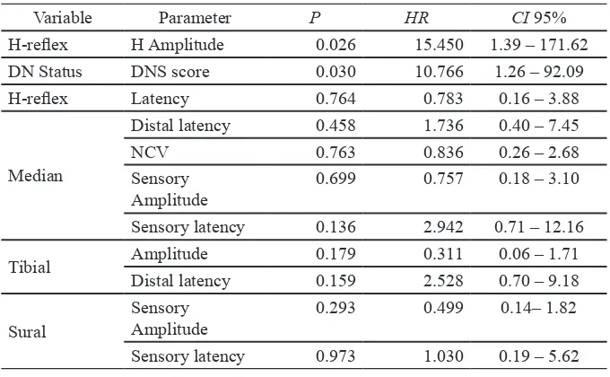
All variables that were significant in
Kaplan Meier analysis were compared using
Cox regression analysis. This analysis showed that only H-reflex amplitude and DN status (measured by DNS score) were significantly
related to PDN occurrence. The hazard ratio
for H-reflex amplitude was 15.450 (95% CI
1.39 – 171.62, p= 0.026), and for DN status was 10.766 (95% CI 1.26 – 92.09, p= 0.030).
Despite of the widened confidence interval, H-reflex amplitude had the highest hazard
ratio among the other predictor variables. This result proved that there was presynaptic inhibition process in PDN that manifested as
low H-reflex amplitude.
DISCUSSION
The study population in this research was patients with abnormality in blood glucose level (DM or IGT). After observation for almost 12 months, 40% of patients had neuropathy and 12% had PDN. Demographic data showed that hyperglycemic and
neuropathic status significantly predict PDN.
This result inherent to mechanisms proposed
TABLE 2. Analysis of PDN Survival Cox regression
Variable Parameter P HR CI 95%
H-reflex H Amplitude 0.026 15.450 1.39 – 171.62 DN Status DNS score 0.030 10.766 1.26 – 92.09 H-reflex Latency 0.764 0.783 0.16 – 3.88
Median
Distal latency 0.458 1.736 0.40 – 7.45
NCV 0.763 0.836 0.26 – 2.68
Sensory
Amplitude 0.699 0.757 0.18 – 3.10 Sensory latency 0.136 2.942 0.71 – 12.16
Tibial Amplitude 0.179 0.311 0.06 – 1.71 Distal latency 0.159 2.528 0.70 – 9.18
Sural Sensory Amplitude 0.293 0.499 0.14– 1.82 Sensory latency 0.973 1.030 0.19 – 5.62
by Fink and Oaklander8 that disruption of nerve
function as a result of glucose accumulation in the nerve include abnormal metabolic product formation and focal tissue ischemia in sensory and autonomic nerves as a result
of endoneural hypoxia. The excess of glucose and oxidative stress, aberrant neurofilament
phosphorylation, disruption of normal nerve growth factors and mitochondrial dysfunction in the dorsal root ganglia, disturb normal function of tissue repair mechanisms.8
This study found that several variables of
NCS were significantly related to PDN.
Long-term changes are likely occur after injury, both peripherally and centrally.9 Because NCS
basically evaluates peripheral nerves, these positive results indicate the likelihood that peripheral nerve abnormality has contribution in PDN development. According to Zhuo,9 the
injury and injury-related areas undergo
long-term plastic changes resulting in significantly
enhanced pain sensation (hyperalgesia) or
misinterpretation of non-noxious stimuli as
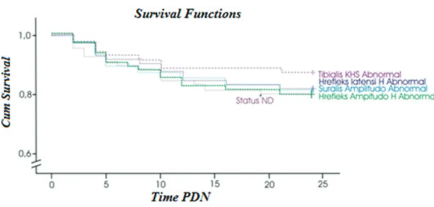
FIGURE 1. Survival diagram of PDN occurence from several predictor variables (the abnormality of tibial nerve conduction velocity, H-reflex latency, sural amplitude, and H-reflex amplitude).
of repetitive application of physiological pain.9
One of the reasons why so many changes happen following a nerve injury is that sensory neurons, and the non-neural cells associated with them, are highly interdependent in generating pain.10 The molecular mechanisms
of pain generation in PDN are multifactorial and not fully understood. The predominantly
involved is distal axon. This is related to the dependence of distal axon upon oxidative metabolism to sustain axonal transport. Disruption in the supply of blood, oxygen,
glucose, ATP, or NADPH will disturb the
transport, which will lead to distal axonal
degeneration.8
Analysis using Kaplan Meier survival in this study showed that H latency and amplitude are related to hazard ratio and the p value.
H-reflex represents activity of peripheral and
central nervous system. Therefore, our result
theory proposed that normal processing of nociceptive and non-nociceptive stimuli
relies on a complex processing which consists
of presynaptic and postsynaptic inhibitory control mechanism in the spinal dorsal horn. Under healthy condition, this inhibition is responsible in keeping the nociceptive and non-nociceptive modalities apart. The loss of this inhibition process has been widely accepted to lead to chronic pain. Blockade of this inhibition mechanism has been shown to cause hyperalgesia and tactile allodynia, two major symptoms of chronic pain. This has been supported by many research showed decrease in inhibition, or ‘‘disinhibition’’, as an important substrate of many chronic pain models.4 This disinhibition will result in increasing neurons excitability and enable excitatory input from non-nociceptive fiber to
nociceptive projection neurons.4
increases in membrane excitability, synaptic efficacy, or a reduced inhibition in dorsal horn
neurons in response to nociceptor input.11
Sensitization of dorsal horn neurons occurs after various types of tissue damage including thermal injury, chemical injury, polyarthritis
or after stimulation of C-fiber afferents.12
After peripheral nerve injury, damaged and
nondamaged A- and C-fibers begin to generate
spontaneous action potentials which will drive central sensitization.11 Repeated C-fiber
afferent stimulation also produces a sequential increase in dorsal horn activity resulting in a prolonged discharge of the cell (wind-up).12
The stimuli that induce wind-up can lead to central sensitization.11
Structural changes in peripheral nerve injury also contribute to altered synaptic function. Peripheral nerve injury leads to
a transganglionic degeneration of C-fiber
terminals in lamina II.11 This will deprive pain transmission neurons in the superficial
dorsal horn of their normal nociceptive input,
and causes patients to experience negative
symptoms.13 There will be increases in the intrinsic axonal growth capacity as part of the
regenerative response of the injured neurons, which will result in sprouting of myelinated A-β fibers from lamina III-IV into lamina I-II and make contact with nociceptive-specific
neurons.11 As a result, stimulation of low
threshold mechanoreceptors abnormally activates pain transmission neurons in lamina II of the spinal cord.13
Diminished synaptic inhibition has also been acknowledged to have a key role in neuropathic pain development.5 Normally,
GABAA receptors in primary afferent terminals of nociceptors will mediate this presynaptic inhibition by causing reduction of transmitter release thus modulating the afferent input from dorsal root ganglion neurons into nociceptive projection neurons
(presynaptic inhibition).4 In PDN this
pre-synaptic inhibition was disturbed, which ultimately caused neuropathic pain. This
disinhibition in the superficial dorsal horn
is caused by the loss of GABAergic and a reduction in glycinergic inhibitory currents. Apoptosis of inhibitory interneurons, caused
by NMDAR-induced cytotoxicity, has been
proposed as the cause of this disinhibition.11
Presynaptic inhibition constitutes an inhibitory mechanism associated with
modulation of monosynaptic reflexes. There-fore, examination of monosynaptic reflex
should be able to detect this pre-synaptic inhibition. One of the proposed methods is by
assessing H-reflex. Our result showed that this
presynaptic inhibition can indeed be assessed
by H-reflex. The H-reflex is evoked by
low-intensity electrical stimulation of the afferent
nerve resulting in monosynaptic excitation of α-motoneurons. Changes in the amount of
presynaptic inhibition acting on Ia afferent terminals has been associated with soleus
H-reflex modulation. Soleus H-reflex has been
shown to be more sensitive to presynaptic inhibition than the mechanically evoked ankle
stretch reflex.6
CONCLUSIONS
This result proved that there was presynaptic inhibition process in PDN that
manifested as low H-reflex amplitude. Our result also showed that depression of H-reflex
amplitude is correlated with the occurrence of
PDN. This indicated that H-reflex amplitude
depression can be used as a marker for presynaptic inhibition in PDN for management of patients with PDN in the future.
ACKNOWLEDGMENTS
REFERENCES
1. Kirby M. Painful diabetic neuropathy-current understanding and management for the primary care team. Br J Diabetes Vasc Dis 2003; 3:138-44. http://dx.doi.org/10.1177/1474651403003002100 1
2. Baron R. Mechanisms of Disease: neuropathic pain--a clinical perspective. Nat Clin Pract Neurol 2006; 2(2):95-106.
http://dx.doi.org/10.1038/ncpneuro0113
3. Aslam A, Singh J, Rajbhandari S. Pathogenesis of painful diabetic neuropathy. Pain Res Treat 2014; 2014.
4. Guo D, Hu J. Spinal presynaptic inhibition in pain control. Neuroscience 2014; 283:95-106.
http://dx.doi.org/10.1016/j. neuroscience.2014.09.032
5. Scholz J, Broom DC, Youn D-H, Mills CD, Kohno T, Suter MR et al. Blocking caspase activity prevents transsynaptic neuronal apoptosis and the loss of inhibition in lamina ii of the dorsal horn after peripheral nerve injury. J Neurosci 2005; 25(32):7317-23.
http://dx.doi.org/10.1523/ JNEUROSCI.1526-05.2005
6. Knikou M. The H-reflex as a probe: pathways and pitfalls. J Neurosci Methods 2008; 171(1):1-12. http://dx.doi.org/10.1016/j.jneumeth.2008.02.012
7. Palmieri RM, Ingersoll CD, Hoffman MA. The hoffmann reflex: methodologic considerations and applications for use in sports medicine and athletic training research. J Athl Train 2004; 39(3):268-77. 8. Fink E, Oaklander A. Diabetic neuropathy. Pain
Manag Rounds 2005; 2:309-14.
9. Zhuo M. Neuronal mechanism for neuropathic pain. Mol Pain 2007; 3:14.
http://dx.doi.org/10.1186/1744-8069-3-14
10. Devor M. Neuropathic pain: what do we do with all these theories? Acta Anaesthesiol Scand 2001; 45(9):1121-7.
http://dx.doi.org/10.1034/j.1399-6576.2001.450912.x
11. Latremoliere A, Woolf CJ. Central sensitization: a generator of pain hypersensitivity by central neural plasticity. J Pain 2009; 10(9):895-926. http://dx.doi.org/10.1016/j.jpain.2009.06.012 12. Coderre TJ, Katz J. Peripheral and central
hyperexcitability: differential signs and symptoms in persistent pain. Behav Brain Sci 1997; 20(3):404-19.
http://dx.doi.org/10.1017/S0140525X97251484 13. Taylor BK. Pathophysiologic mechanisms of
neuropathic pain. Curr Pain Headache Rep 2001; 5(2):151-61.