Difficulty in Controlling Malignant Pleural
Effusion: A Case Report and Literature Review
Ignatius B Prasetya1, Eric D Tenda2
1Department of Internal Medicine, Universitas Indonesia, Cipto Mangunkusumo Hospital, Jakarta, Indonesia 2Division of Respirology and Critical Care, Department of Internal Medicine, Universitas Indonesia,
Cipto Mangunkusumo Hospital, Jakarta, Indonesia
ABSTRACT
Malignant pleural effusion is the second leading cause of exudative pleural effusions, usually recurrent and represents advanced malignant disease. Treatment options were restricted to symptomatic purpose in order to increase functional capacity and quality of life. In this case, a 35-years old woman with history of breast cancer was admitted with worsening dyspnea since 4 months prior. The patient was told that there was fluid in her left lung. She underwent thoracocentesis twice, pleuroscopy and pleurodesis with little success. The treatment option switches to placement of pleural catheter to control the effusion. This option, however, may leave the patient and caregivers with routine task of aspiration of the fluid and with greater risk of infection.
Key words: Malignant pleural effusion
Korespondensi:
ABSTRAK
Efusi pleura maligna merupakan penyebab kedua terbanyak dari efusi pleura eksudatif, biasanya berulang, dan menggambarkan perjalanan keganasan yang telah lanjut. Pilihan terapi terbatas untuk mengurangi gejala, meningkatkan kapasitas fungsional dan kualitas hidup. Dalam kasus ini, seorang wanita berusa 35 tahun dengan riwayat kanker payudara masuk rumah sakit dengan keluhan sesak yang memberat sejak 4 bulan. Pasien diberi tahu bahwa terdapat cairan di paru kirinya. Ia menjalani torakosentesis sebanyak dua kali, pleuroskopi, dan pleurodesis, namun efusi kembali berulang. Akhirnya dilakukan pemasangan kateter pleura untuk mengontrol efusi. Meskipun berhasil, terapi ini dapat membutuhkan aspirasi atau penggantian penampung secara rutin dan lebih berisiko terhadap infeksi.
Kata kunci: Efusi pleura maligna
INTRODUCTION tract as a group accounts for a
dr. Ignatius Bima Prasetya
Email: [email protected]
Indonesian Journal of
CHEST
Critical and Emergency Medicine
Vol. 1, No. 3 July - September 2014
further 25%. Primary tumor sites cannot be identified in around 7-15% of Malignant pleural effusion is pleural effusion
attributed to pleural malignancies. This condition represents the second leading cause of exudative pleural effusions after parapneumonic effusion. However, unlike parapneumonic effusion which has manageable etiology, malignant pleural effusion represents advanced malignancy associated with high morbidity and mortality, precluding the possibility of a curative treatment approach.1 Theoretically all kinds of malignancy can cause malignant pleural effusion but the most common metastatic tumor to the pleura are lung cancer in men and breast cancer in women. Together these two malignancies account for up to 65% of all effusions. Lymphoma, tumors of the genitourinary tract and tumors of the gastrointestinal
malignant pleural effusions.1, 2
CLINICAL CASE
The patient was a 35 years old woman with chief complaint of worsening dyspnea since 4 months prior to admission. She had been diagnosed with breast cancer 3 years prior to admission and since then had undergone a series of surgery, chemotherapy and radiation. The original mass had not been growing further but the patient felt a worsening dyspnea. The dyspnea was especially worsened with strenuous activity. She also felt more comfortable lying on her left side. She complained about dry cough but denied any occurrence of fever. Weight loss was also reported but night sweat was denied. After seeking medical service, the patient was told that there was fluid in her left lung and underwent drainage twice. The fluid, however, quickly re-accumulated soon after each procedure. She was then admitted for pleuroscopy. Physical examination of the patient revealed a decreased breath sound of the left lung. Chest X-ray confirmed the existence of left pleural effusion. No significant abnormalities were detected in the laboratory examination. Pleuroscopy was carried out but no significant lesion can be visualized. Although pleural fluid cytology revealed negative results, due to exclusion of other etiologies, it was concluded that the patient had malignant pleural effusion. Subsequent pleurodesis was then planned for the patient once the fluid production decreased to less than 150 cc/ day. This criteria, however, was not reached after 10 days of observation. Pleurodesis using bleomycin as the sclerosing agent was then carried out since radiologic examination had proved the re-expansion of the left lung. This procedure, however, failed to control the effusion. Placement of pleural catheter was then opted. The patient was discharged with pleural catheter. The patient reported no further dyspnea. Daily fluid production around discharge averaged at 30-50 cc/day.
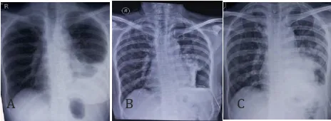
A B C
Figure 1. Chest X-ray of the Patent Before Pleuroscopy (A), Afer Pleuroscopy (B), and Afer Pleurodesis (C)
DISCUSSION
Risk Factors
The primary site of malignancy is the most important predictive factor for survival. Overall survival in general ranges from 3-12 months after diagnosis. Pleural effusion due to metastasis from lung cancer typically has the shortest survival while the longest survival is attributed to metastasis of ovarian cancer. Even a small amount of pleural effusion in patients with non-small cell lung cancer (NSCLC) in whom cytological or histological diagnosis for the effusion using thoracentesis is not feasible confers poor prognosis. Due to this particular reason, the recently revised staging system for NSCLC upstaged the presence of pleural effusion from T4 to M1a.1, 4
Pathophysiology
Pathophysiology of pleural effusion involves increased pleural fluid production due to increased vascular permeability or reduced reabsorption by lymphatic system, usually due to disrupted or occluded drainage channels. Metastatic pleural effusion is typically produced by the second mechanism, occlusion of pleural drainage.4 The most common symptoms of pleural effusion is dyspnea, which occurs in more than 50% of patients, followed by cough, weight loss, and chest pain.1 However, up to 25% of patients may be asymptomatic at diagnosis and pleural effusion was found only after specific physical examination or radiologic signs were found. These signs include reduced breath sound and dull percussion in the involved lung as well as specific pattern seen in chest X-ray (meniscus sign along the lateral chest wall).1,2 Chest X-ray is useful to detect 100 cc of free flowing effusion.4
Diagnosis
Diagnostic thoracocentesis is usually the first diagnostic step in determining pleural effusion characteristics. Analysis of pleural fluid using thoracentesis may help establish the origin of malignant
pleural effusion.1-3 More than 90% of malignant pleural
effusions are exudatives according to Light’s criteria.
The appearance in half of them is hemorrhagic and in 11% is bloody. Transudative malignant pleural effusion usually occurs in mixed condition with other systemic diseases.1 Cytological examination of pleural fluid has traditionally been used as the gold standard
for diagnosis of malignant pleural effusion. This test, however, only yields limited sensitivity of 40-90%.1,4 Furthermore, distinguishing between each individual malignant cell may be difficult as these cells usually have overlapping similar features. Therefore, other procedures, such as immunohistochemistry (IHC) using monoclonal antibodies against tumor markers and chromosomal analysis, complement cytology in the diagnosis of malignant pleural effusion.1 Pleural biopsy is the next step if malignant pleural effusion is still suspected while pleural cytology yields negative result.1, 2 Sensitivity up to 95% can be achieved if the pleural samples are collected using thoracoscopy, while sensitivity of up to 97% can be achieved using combination of cytology and thoracoscopy assisted pleural biopsy.5 The use of tumor marker examinations such as CEA, CA 125, CA 15-3, CA 19-9, Cyfra 21-1 and VEGF levels in the pleural fluid may generally improve specificity but not sensitivity in diagnosing malignant pleural effusions.6,7
Our patient was suspected to have pleural effusion based on clinical signs and symptoms as well as radiologic examinations. Subsequent thoracocentesis confirms this effusion. The pleural fluid analysis showed exudative effusion, consistent with malignant pleural effusion. She also had a history of breast cancer. These facts supported the suspicion that the patient had malignant pleural effusion.
Treatment
The prognosis of patients with malignant pleural effusion is generally poor due to the fact that the existence of pleural effusion indicates advanced stage of the primary tumor. Therefore, the main
principle of malignant pleural effusion’s management
is palliative and not curative. Available therapeutic options for malignant pleural effusions include observation, repeated thoracocentesis, placement of long term indwelling pleural catheter, pleurodesis, pleuroperitoneal shunt and pleurectomy.1,2,4 The best option for each individual patient is determined by symptoms and performance status of the patient, the primary tumor and its response to systemic therapy, and lung re-expansion following pleural fluid evacuation.2
Observation of malignant pleural effusion is generally reserved for asymptomatic patients with good response to systemic chemotherapy. The majority
of these patients, however, will become symptomatic
in due course and require further intervention.1,2 This is also seen in our case where pleural fluid quickly re- accumulated after each thoracocentesis.
Repeated thoracocentesis is ideally recommended for patients with limited survival and slow accumulation of pleural fluid. This procedure provides transient relief of symptoms and avoids hospitalization. However, re-accumulation of pleural fluid can happen in a month in almost 100% of (cough, chest discomfort) and should be limited to 1-1.5 l at a time to prevent re-expansion pulmonary edema.1-3
inflammatory reaction to sclerosing agents. The inflammatory response to the sclerosing agents can be significantly inhibited by corticosteroids. Therefore, steroid treatment before the procedure is associated with increased rate of failure. Recent studies also showed that mesothelial cells play important roles in regulating the pleural coagulation-fibrinolysis balance and may affect the overall success rate of pleurodesis. Success rate of the procedure may also be influenced sclerosant through pleural catheter. The latter method is considered to be less invasive and safer. Smaller clinical trial that stated that although success rate was similar in both group, shorter period of intercostal several agents that have been developed as sclerosing agent for pleurodesis, talc is commonly regarded as the cheapest and most effective option. It may be administered at thoracoscopy as talc poudrage or through an intercostal tube in the form of a suspension termed talc slurry. Studies showed that the efficacy of talc as sclerosing agent in pleurodesis may reach 88-100%. However talc is associated with risk of developing acute respiratory distress syndrome due to unclear mechanism. Other commonly used agents for pleurodesis include tetracycline, bleomycin and doxycycline. Tetracycline was among the first agent used for pleurodesis. Its use, however, declined in recent years due to its inferior efficacy compared to
other agents (success rate 50-92%) and unavailability Other less successful agents for pleurodesis include
Corynebacterium parvum extract, interferons, interleukins (IL-2), cisplatin, cytosine arabinoside and mitoxantrone. Rotation of patients following pleurodesis procedure is no longer recommended but the intercostal drain should be clamped for at procedures. These patients should receive prophylactic radiotherapy to the site of chest drain insertion.2,3 Beside the risk of developing acute respiratory distress syndrome, pleurodesis is associated with other less severe complications. A Cochrane review suggested the frequency of pain and fever in association with talc to be 31% and 26%, respectively.10 The pain after pleurodesis is often so severe that systemic opioids are needed to control it. The risk of empyema may also rise to 4.0% after the procedure.11
Insertion of indwelling pleural catheter (IPC) is a reasonable option for patients with malignant pleural effusion who would like to avoid hospitalization.2,11 Existing randomized trial comparing IPC with pleurodesis showed that patients in the doxycycline arm had shorter hospitalization than patients in the complications are relatively less severe although more frequently found than pleurodesis’ complications. When compared to pleurodesis, IPC also has the advantage of higher recurrence free rate (90-91% patients do not need further procedure). On the other hand, pleurodesis has the advantage of being a finite
treatment while patients with indwelling catheter need day-to-day aspiration to control their effusion.11 Long-term indwelling pleural catheter may lead to
spontaneous pleurodesis in 40−58% of patients.1
Pleuroperitoneal shunt procedure is done by placing unidirectional valves between the pleural and peritoneal cavities via thoracoscopy or a mini- thoracotomy. This valve is pressure-activated, therefore
manual per cutaneous compression of the pump chamber, sometimes over 400 times per day, can be required. Complications of this procedure include shunt
occlusion (found in 12-25% cases), infection and tumor
seeding or implantation into the peritoneal cavity.2,3 Pleurectomy is an effective way of treating malignant pleural effusion. However, this procedure has very high mortality rate (up to 13%). Other complications include empyema, haemorrhage and cardiorespiratory failure. The introduction of video- assisted thoracic surgery (VATS) has shed hope of a safer way of conducting this procedure.2-4
The patient in our case was initially treated with repeated thoracocentesis. However, this approach failed to control the effusion and further drainage technique was carried out. Pleurodesis was decided to be the best option for this patient due to the good functional status. It was initially expected that with successful pleurodesis the patient can have dyspnea- free life without any further special care required. The attempt to conduct pleurodesis, however, resulted in failure to obliterate the pleural space. The aforementioned literature stated that pleurodesis is less likely to be successful in malignant effusion cases than pneumothorax patients due to the thickening of the visceral pleura and stiffening of the lung parenchyma. Apparently this is the case in our patient. Subsequent placement of pleural catheter was then carried out with greater success in controlling the effusion. This option, however, leaves the patient and caregivers with routine task of aspirating the fluid and imposes greater risk of infection. But since it is proven to be able to control symptoms and to increase quality of life, this option can be considered to be the best option for this particular case.
CONCLUSION
Malignant pleural effusion is the second leading
disease. Thoracocentesis should be carried out in every patient with malignant pleural effusion to confirm the diagnosis. The treatment of such effusion is complex and restricted to symptomatic treatment to increase functional capacity and quality of life. Several available treatment options include observation, repeated thoracocentesis, placement of long term indwelling pleural catheter, pleurodesis, pleuroperitoneal shunt and pleurectomy.
REFERENCES
1. Nam HS. Malignant pleural effusion: medical approaches for diagnosis and management. Tuberc Respir Dis (Seoul) 2014 May; 76(5):211-7.
2. Roberts ME, Neville E, Berrisford RG, Antunes G, Ali NJ. Management of a malignant pleural effusion: British Thoracic Society Pleural Disease Guideline 2010. Thorax 2010 Aug; 65 Suppl 2:ii32-40.
3. Vaz MC, Marchi E, Vargas FS. Pleurodesis: technique and indications. J Bras Pneumol 2006 Jul-Aug; 32(4):347-56. 4. Zarogoulidis K, Zarogoulidis P, Darwiche K, Tsakiridis K,
Machairiotis N, Kougioumtzi I, et al. Malignant pleural effusion and algorithm management. J Thorac Dis 2013 Sep; 5 Suppl 4:S413-9.
5. Loddenkemper R. Thoracoscopy--state of the art. Eur Respir J 1998 Jan; 11(1):213-21.
8. Rodriguez-Panadero F, Montes-Worboys A. Mechanisms of pleurodesis. Respiration 2012; 83(2):91-8.
9. Villanueva AG, Gray AW, Jr., Shahian DM, Williamson WA, Beamis
JF, Jr. Efficacy of short term versus long term tube thoracostomy drainage before tetracycline pleurodesis in the treatment of malignant pleural effusions. Thorax 1994 Jan; 49(1):23-5. 10. Shaw P, Agarwal R. Pleurodesis for malignant pleural effusions.
Cochrane Database Syst Rev 2004; (1):CD002916. 11. MacEachern P, Tremblay A. Pleural controversy: pleurodesis
versus indwelling pleural catheters for malignant effusions. Respirology. 2011 Jul; 16(5):747-54.
12. Putnam JB, Jr., Light RW, Rodriguez RM, Ponn R, Olak J, Pollak JS, et al. A randomized comparison of indwelling pleural catheter and doxycycline pleurodesis in the management of malignant pleural effusions. Cancer 1999 Nov 15; 86(10):1992-9. 13. Van Meter ME, McKee KY, Kohlwes RJ. Efficacy and safety of