*Corresponding author. Tel.:#64-9-373-7599 Ext. 8366; fax:#64-9-373-7416.
E-mail address:[email protected] (P.J. Harris)
Ferulic acid is bound to the primary cell walls
of all gymnosperm families
Susan M. Carnachan, Philip J. Harris
*
School of Biological Sciences, The University of Auckland, Private Bag 92019, Auckland, New Zealand
Received 14 July 1999; accepted 26 October 1999
Abstract
Unligni"ed primary cell walls containing ester-linked ferulic acid#uoresce blue in ultraviolet
radiation which changes to green with increased intensity on treatment with ammonium
hydroxide. Using this #uorescence behaviour, we detected ester-linked ferulic acid in the
primary cell walls of all 41 species of gymnosperms we examined. These species were in 17 families representing all four extant classes of gymnosperms. In addition, we obtained cell-wall
preparations containing'95% primary cell walls from nine gymnosperm species in nine
families, representing all four extant classes. These preparations were analysed for ester-linked
monomeric phenolic acids. We found ferulic acid (mostlytrans) (88-1,561lg/g cell walls) in all of
the preparations andp-coumaric acid (mostlytrans) (0}106lg/g cell walls) in all except one of
them. Ferulic acid ester-linked to primary cell walls has previously been found in angiosperms: in the commelinoid monocotyledons and in the dicotyledon order Caryophyllales, both monophyletic groups. From the present results, we postulate that the extant classes of
gymnos-perms are monophyletic and no class is sister to the angiosgymnos-perms. ( 2000 Elsevier Science
Ltd. All rights reserved.
Keywords:Gymnosperms; Gnetales; Primary cell walls; Phenolic acids; Hydroxycinnamic acids; Ferulic acid;p-Coumaric acid; UV Fluorescence microscopy; Biochemical systematics
1. Introduction
The hydroxycinnamic acids ferulic and p-coumaric occur ester-linked to the cell walls of many angiosperm species. These ester-linked phenolic acids were"rst found
in cell-wall preparations from grasses and cereals (family Poaceae) containing a mix-ture of both unligni"ed primary and ligni"ed secondary cell walls (Higuchi et al., 1967; Hartley, 1972). In these early studies, it was assumed that the ferulic acid was ester-linked only to lignin and thus occurred only in the ligni"ed secondary cell walls. However, the presence of ester-linked ferulic acid in primary cell walls was demon-strated in two ways. First, by examining sections by ultraviolet (UV)#uorescence microscopy (Harris and Hartley, 1976). Primary cell walls in these sections#uoresced blue in water (pH 5.4), but this #uorescence changed to green and increased in intensity when the sections were treated with 0.1 M ammonium hydroxide (pH 10.3). Esters of ferulic acid are known to show this change in#uorescence colour with pH (Harris and Hartley, 1976; Fry, 1988). In contrast to the primary cell walls, the ligni"ed secondary cell walls #uoresced blue in water and after treatment with ammonium hydroxide continued to #uoresce blue, but with increased intensity. Second, preparations containing only primary cell walls were isolated from the leaves of perennial ryegrass (Lolium perenne); analyses of these cell walls showed that they contained ferulic acid, together with small amounts ofp-coumaric acid (Harris et al., 1980).
is not easy as they are usually woody plants. However, SaHnchez et al. (1996) obtained cell-wall preparations from the hypocotyls of Pinus pinaster (Pinaceae) which, in contrast to the preparations from gymnosperm leaves, probably contained a high proportion of primary cell walls. SaHnchez et al. (1996) identi"ed ferulic acid as the major monomeric ester-linked hydroxycinnamic acid in these preparations; p-coumaric acid was present in only small amounts.
In the present study we examined the primary cell walls of 41 species of gymno-sperms for the presence of ester-linked ferulic acid using UV#uorescence microscopy. These species were selected so that all 17 gymnosperm families were represented. In addition, we obtained cell-wall preparations containing '95% primary cell walls from nine species of gymnosperms in nine families (representing all four classes). Ester-linked ferulic and p-coumaric acids in these preparations were quanti"ed by capillary GC of their trimethylsilyl derivatives; ester-linkedp-hydroxybenzoic acid, another phenolic acid, was also quanti"ed in the same way.
2. Materials and methods
2.1. Plant material
The sources of plant material are shown in Table 1. Seeds ofWelwitschia mirabilis andPinus radiatawere germinated at 253C in the dark on moist"lter paper. TheW. mirabilis seeds were "rst coated with CaptanTM fungicide and the "lter paper moistened with CaptanTM solution (2 g/l in H
2O). The P. radiata seeds were "rst surface sterilized with sodium hypochlorite solution (1% containing two drops per 100 ml of Tween 20) for 5 min and then soaked in H
2O for 24 h. Cambium (with some di!erentiating xylem and phloem) was collected from the trunk of a tree ofP. radiata by peeling o!and discarding the bark and gently scraping the exposed surface (tissue frozen at!203C).
2.2. Microscopy
Bright-"eld and UV-#uorescence microscopy (Smith and Harris, 1995) were done on fresh transverse sections cut using a razor blade from the organs indicated in Table 1 and on cell-wall preparations obtained from these organs. Staining with the bright-"eld stain toluidine blue O was as described by Harris et al. (1994). Toluidine blue O stains polychromatically: lignin stains green or blue}green; polyanions such as rhamnogalacturonans stain pink or purple (O'Brien and McCully, 1981). Lignin was detected by the red colour reaction given by phloroglucinol}HCl (Harris et al., 1980). Starch was detected using I
2 in KI (Jensen, 1962). Controls were also examined in which the sections or cell walls were mounted in the solvent for the stain or colour reagent. Fluorescence of the cell walls in UV radiation was examined using sections mounted in H
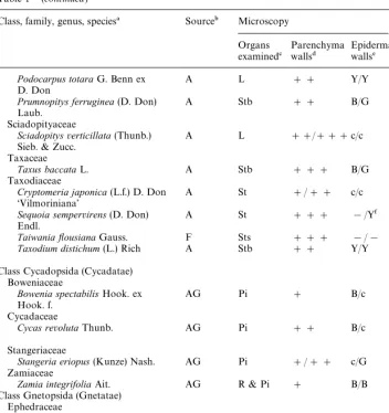
Table 1
Sources of gymnosperms, organs examined, and UV#uorescence of their cell walls
Class, family, genus, species! Source" Microscopy Walls
isolated
Agathis australis(D. Don) Salisb. A L ### Y/Y L#P
Araucaria heterophylla(Salisb.) A Sts ### !/Y NI Franco.
Cephalotaxaceae
Cephalotaxus harringtonia(Forbes) A Stb ### B/Y NI Koch var.drupacea
Cupressaceae
Calocedrus decurrens(Torr.) Florin A Sts ## c/c NI
Chamaecyparis lawsoniana(Murray) A Sts # c/c NI Parl.
Cupressus macrocarpaHartw. ex A Sts ## c/c NI
Gordon
Juniperus confertaParl. A St ### c/c NI
Juniperus squamataBuch.-Ham. Ex A St ### c/c NI D. Don&Blue Star'
Libocedrus plumosa(D. Don) Sarg. A Sts #/## Y/Y NI
Microbiota decussataKomar. A Sts ## !/Y NI
Thuja occidentalisL.&Rheingold' A Sts # c/c NI
Widdringtonia schwarzii(Marloth) F Sts #/## c/c NI Mast.
Phyllocladaceae
Phyllocladus trichomanoides A P ### B/Y NI
D. Don Pinaceae
Abies concolor(Gordon & Glend.) F L ## B/B& NI Lindl. ex Hildebr.
Abies magnixcaA. Murray F L ## B/B& NI
Cedrus atlantica(Endl.) Carr. A L ## Y/Y& NI
Keteleeria evelynianaMast. F St ### c/c NI
Pinus radiataD. Don (open F C ### na C
pollinated clone 268.041) FG ### na NI
Hy24 ### B/B NI
Rd ### B/B NI
Co # c/G NI
Pinus thunbergiiParl. H L # B/!& NI
Pseudotsuga menziesii(Mirb.) F St ### B/! NI
Franco.
Pseudotsuga sinensisDode. F St ### B/B NI
Tsuga heterophylla(Raf.) Sarg. F St ### B/B NI
Podocarpaceae
Dacrycarpus dacrydioides(A. Rich.) A Stb #/## !/! NI Laub.
Dacrydium cupressinumSol. ex A Sts ## !/! NI
Forst.
Lagarostrobos colensoi(Hook.) A Sts ### B/Y Sts
Table 1*(continued)
Class, family, genus, species! Source" Microscopy Walls
isolated
Prumnopitys ferruginea(D. Don) A Stb ## B/G NI
Laub. Sciadopityaceae
Sciadopitysverticillata(Thunb.) A L ##/###c/c NI Sieb. & Zucc.
TaiwaniayousianaGauss. F Sts ### !/! NI
Taxodium distichum(L.) Rich A Stb ## Y/Y L#P#
R Class Cycadopsida (Cycadatae)
Boweniaceae
Bowenia spectabilisHook. ex AG Pi # B/c NI
Hook. f. Cycadaceae
Cycas revolutaThunb. AG Pi ## B/c Pi#R#
P Stangeriaceae
Stangeria eriopus(Kunze) Nash. AG Pi #/## c/G NI Zamiaceae
Zamia integrifoliaAit. AG R & Pi # B/B Pi
Class Gnetopsida (Gnetatae)
Welwitschia mirabilisHook. f. S Hy3 ### B/G NI
!Classi"cation according to Mabberley (1997) and Kramer and Green (1990).
"Plant sources: (H) private garden at Helensville, New Zealand; (F) grounds of Forest Research, Rotorua, New Zealand; (A) grounds of The University of Auckland; (AG) glasshouses at The University of Auckland; (S) seeds from Silverhill Seeds, Silverhill Crescent, Kenilworth, 7700, RSA.
#Organs and tissues examined: (C) cambium with some di!erentiating xylem and phloem; (Hy3) hypocotyl from a 3 day old seedling; (Hy24) hypocotyl from a 24 day old seedling; (FG) female gametophyte; (L) leaf; (P) petiole; (Pi) pinnule/pinna; (R) rachis; (Rd) radicle from a 24 day old seedling; (St) young stem; (Sts) young stem plus scale-like leaves; (Stb) young stem plus bases of leaves; (Co) cotyledon from a 24 day old seedling; (NI) cell walls not isolated.
$Intensity of the green #uorescence of the primary walls of parenchyma cells after treatment with ammonium hydroxide:###"intense#uorescence;##"moderate#uorescence;#"weak# u-orescence.
%Fluorescence colour of walls of epidermal cells, excluding stomatal guard cells, before and after treatment with ammonium hydroxide: (B) blue; (G) green; (Y) yellow; (c)#uorescence of cell walls could not be determined because of intense#uorescence of cell contents;*little or no#uorescence.
2.3. Isolation of cell walls
Cell-wall preparations containing(5% ligni"ed cell walls (estimated microscop-ically after treatment with phloroglucinol}HCl) were obtained from young plant material, collected in September or October, that contained only small proportions of ligni"ed cell walls (Table 1). All procedures were carried out at 43C. Cell-wall isolation and all manipulations of the cell-wall preparations, the phenolic acids and their TMSi derivatives were done in illumination from a halogen re#ector lamp with a UV"lter to avoid UV radiation which causes cis}trans (or Z}E) isomerization (Kahnt, 1967; Hartley and Jones, 1975).
Plant material (1.5}2 g fresh weight) was cut into pieces (3 mm]3 mm) and
hom-ogenized in 35 ml of Mops-KOH bu!er (20 mM, pH 6.8) containing 20 mM sodium metabisulphite using a Polytron blender (4 min). The homogenates were centrifuged (750 g, 5 min) and the pellets divided into three equal batches each of which was further homogenized in fresh bu!er (5 ml) using a Tenbroeck ground-glass homogen-izer. The homogenates were centrifuged (as above), the pellets washed with bu!er three times by centrifugation, resuspended in bu!er, sonicated (for 1 min at full power), and centrifuged. The pellets were washed with H
2O (35 ml) "ve times by centrifugation, and"ltered onto nylon mesh (pore size 11lm). The residue on the mesh was washed with H
2O (750 ml) until the"ltrate was clear, dried using solvent exchange by successively washing with EtOH, MeOH, andn-pentane (50 ml of each), and stored under vacuum over Si gel.
2.4. Removal of contaminating starch
Tris-maleate bu!er (1 ml, 5 mM, pH 6.9) was added to cell-wall preparations (40 mg) contaminated with starch and the starch gelatinized by heating for 5 min at 853C. After cooling to 373C, an equal volume was added of tris-maleate bu!er (15 mM, pH 6.9) containing 2 mM CaCl
2 and porcine pancreatic a-amylase (300 units, Type 1-A, Sigma, St. Louis, MO, USA) and incubated for 1 h at 373C. The suspension was washed with H
2O on to nylon mesh, and further washed with H2O until the "ltrate was clear. The cell-wall preparations were dried and stored as described above. No starch was detected in these preparations.
2.5. Treatment of cell walls with NaOH
The method was based on those used by Ford and Hartley (1988) and Turner et al. (1993). Cell walls (15 mg) were shaken with 1 M NaOH (2 ml) containing 3,4-dimethoxycinnamic acid (10lg/ml) as the internal standard for 20 h at 203C under Ar. The suspension was"ltered (glass micro"bre"lter, type GF/C, Whatman, Maidstone, UK) and the residue washed with H
2O (4]1 ml); the extracts and washings were combined. The"ltrate was adjusted to pH 1.5 with 6 M HCl (370ll), saturated with NaCl, and the phenolic acids extracted by shaking with Et
2O (4]6 ml). The com-bined Et
2O extracts were evaporated in a stream of N2, and dried under vacuum over P
trans-p-coumaric, andtrans-ferulic acids) were also carried through the NaOH treat-ment and extraction procedure as above. The internal standard 3,4-dimethoxycin-namic acid (20lg) was added to the reference phenolic acids before the NaOH treatment.
2.6. Silylation
The residues were silylated by incubating with a mixture of pyridine (100ll) and BSTFA [N,O-bis(trimethylsilyl) tri#uoroacetamide] containing 1% TMCS (trimethyl-chlorosilane) (100ll) for 60 min at 203C under Ar with occasional shaking. Dich-loromethane (0.5 ml for the cell wall samples, 3 ml for the reference phenolic acids) was added before GC. Reference standards (1 mg plus 1 mg of 3,4-dimethoxycinnamic acid) of each of the following were also silyated:trans-ca!eic acid,p -hydroxybenzal-dehyde,trans-sinapic acid, syringaldehyde, syringic acid, vanillic acid, and vanillin. Mixtures ofcis- andtrans-isomers of ca!eic acid, ferulic,p-coumaric, and sinapic acids were obtained by exposing solutions of thetrans-forms (1 mg in 3 ml of MeOH) to UV radiation (maximumk 365 nm) for 8 h. These solutions were evaporated to dryness and silylated.
2.7. GC
The TMSi derivatives of the phenolic acids were separated and quanti"ed on a 30 m]0.32 mm id, 0.25lm"lm thickness DB-1 column (J&W Scienti"c, Folsom,
CA, USA) with a deactivated fused silica retention gap (2 m) in a gas chromatograph "tted with a FID and a dedicated cool on-column capillary inlet. The carrier gas used was He at a column head pressure of 40 kPa; 1ll aliquots were injected. The following oven temperature programme was used: 383C for 30 s after injection; to 1503C at 503C/min; 1503C for 15 min; to 2603C at 53C/min; and 2603C for 5 min. The detector temperature was at 2903C. The phenolic acids in cell-wall extracts were quanti"ed using the internal standard. The cis-isomers of ferulic and p-coumaric acids were quanti"ed using the response factor of their trans-isomer; this has previously been shown to be valid (Turner et al., 1993).
3. Results
3.1. UVyuorescence microscopy
ammonium hydroxide, the#uorescence of ligni"ed cell walls (identi"ed histochemi-cally using phloroglucinol}HCl and toluidine blue O, including the cell walls of xylem tracheary elements, and sclerenchyma"bres) remained blue, but the#uorescence of unligni"ed primary cell walls (including those of phloem and most parenchyma cells) changed to green indicating the presence of ester-linked ferulic acid (Harris and Hartley, 1976). Following treatment with ammonium hydroxide, the intensity of the #uorescence also increased. The intensity of the#uorescence of the primary cell walls varied among species, from intense to only just detectable. An estimate of the intensity of the#uorescence of parenchyma cell walls after treatment with ammonium hydrox-ide is shown in Table 1. Isolated cell walls showed the same#uorescence behaviour as in sections, although the cell types from which the cell walls were derived usually could not be determined. The two histochemical methods we used to identify ligni"ed cell walls gave results that agreed closely. Cell walls that gave a positive colour reaction with phloroglucinol}HCl stained blue/green with toluidine blue O; cell walls that gave no colour reaction with phloroglucinol}HCl stained purple.
In the epidermis, the walls of the stomatal guard cells were always ligni"ed and #uoresced blue before and after treatment with ammonium hydroxide. The walls of the other epidermal cells were ligni"ed in"ve of the species examined:Abies concolor, A. magnixca,Cedrus atlantica,Pinus thunbergii, andSequoia sempervirens. The
#uores-cence of these walls is described in Table 1. The unligni"ed epidermal cell walls of some species, e.g.Ginkgo biloba(Table 1) showed the same blue to green#uorescence behaviour as the walls of other cell types. However, the unligni"ed epidermal walls of the other species showed a variety of #uorescence behaviours, including a yellow #uorescence, either after treatment with ammonium hydroxide, or in some, both before and after treatment, e.g.Libocedrus plumosa. These species with epidermal walls that showed a yellow#uorescence were all in the class Pinopsida.
The cuticles of all the gymnosperms examined#uoresced, and the intensity was increased by treatment with ammonium hydroxide. In some species, e.g. Bowenia spectabilis,Dacrydium cupressinum,G. biloba,Gnetum gnemon, they #uoresced blue, both before and after treatment with ammonium hydroxide; in others, e.g. Agathis australis,Dacrycarpus dacrydioides,L. plumosa, they#uoresced yellow or orange. In others, e.g.Ephedra gerardiana,Sciadopitysverticillata, Widdringtonia schwarzii, they
#uoresced blue when mounted in water which changed to yellow after treatment with ammonium hydroxide. The only exception to the above was the cuticle ofWelwitschia mirabiliswhich when mounted in water#uoresced yellow and changed to blue after treatment with ammonium hydroxide. Unlike the yellow#uorescence of the epider-mal cell walls, the yellow#uorescence of the cuticles was not con"ned to the class Pinopsida.
3.2. Phenolic acids released from the cell walls
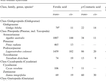
Table 2
Amounts (lg/g) of ferulic,p-coumaric, andp-hydroxybenzoic acids released from the cell walls by treatment with sodium hydroxide
Class, family, genus, species! Ferulic acid p-Coumaric acid p-Hydroxy benzoic acid
trans- cis- trans- cis -Class Ginkgoopsida (Ginkgoatae)
Ginkgoaceae
Ginkgo biloba 79" 11 22 14 *
Class Pinopsida (Pinatae, incl. Taxopsida) Araucariaceae
Agathis australis 1007 144 34 11 17
Pinaceae
Pinus radiata 465 13 * * *
Podocarpaceae
Lagarostrobos colensoi 1459 102 86 20 45
Taxodiaceae
Taxodium distichum 74 19 13 9 *
Class Cycadopsida (Cycadatae) Cycadaceae
Cycas revoluta 106 7 11 * 13
Zamiaceae
Zamia integrifolia 69 19 60 30 31
Class Gnetopsida (Gnetatae) Ephedraceae
Ephedra gerardiana 128 27 32 21 *
Gnetaceae
Gnetum gnemon 264 124 34 21 *
!Classi"cation according to Mabberley (1997) and Kramer and Green (1990). "Averages of determinations on two samples.
to 1561lg/g forLagarostrobos colensoi(Table 2). Smaller amounts ofp-coumaric acid were found. The amounts ofp-coumaric acid found ranged from none detected in the walls ofP. radiatato 106lg/g forL. colensoi. InZ. integrifoliathere was slightly more p-coumaric acid released from the cell walls than ferulic acid. For all species examined, thetrans-forms of both ferulic and p-coumaric acids were more abundant than the cis-forms.p-Hydroxybenzoic acid was found in extracts of the cell walls of only four species:Agathis australis,Cycas revoluta,L. colensoi, and Z. integrifolia.
4. Discussion
4.1. Implications of cell-wall bound ferulic acid for systematics
monocotyledons (Harris and Hartley, 1980; Rudall and Caddick, 1994; Harris et al., 1997; Smith and Harris, 1999) and the dicotyledon order Caryophyllales (Hartley and Harris, 1981), indicating the presence of ester-linked ferulic acid (Harris and Hartley, 1976); the UV#uorescence of epidermal walls was anomalous in some species and is discussed below. In the angiosperms, both the commelinoid monocotyledons and the Caryophyllales have been shown to be monophyletic using nucleotide sequences of therbcL gene (e.g. Chase et al., 1993).
Whether or not extant gymnosperms are monophyletic has been the subject of much recent discussion. In particular, the relationships of the Gnetales have been controversial. Morphological data have indicated that the Gnetales are a sister group to the angiosperms and the gymnosperms are not monophyletic (Doyle, 1998). Molecular data have indicated a variety of relationships for the Gnetales, but support for these has often been weak (Doyle, 1998; Frohlich, 1999). However, a recent study of sequence data from the chloroplastrpoC1 gene showed strong support for gymno-sperms being monophyletic and none of the extant classes being a sister group to the angiosperms (Samigullin et al., 1999). Another recent study, involving sequence data from MADS-box genes, also showed strong support for the Gnetales not being a sister group to the angiosperms (Frohlich, 1999; Winter et al., 1999). Using our data on the occurrence of cell-wall bound ferulic acid as a character, the most parsimonious hypothesis would be to postulate that the gymnosperms are mono-phyletic and none of the extant classes are sister to the angiosperms. Although, as indicated above, some angiosperm families have this character, these families are away from the root of the angiosperms and appear to have acquired this character independently.
4.2. Amounts of hydroxycinnamic acids in isolated cell walls
Our analyses of isolated gymnosperm primary cell walls con"rmed the presence of ferulic andp-coumaric acids. As in angiosperm primary cell walls, both these acids occurred mostly in theirtrans-forms and, with one exception,Zamia integrifolia, there was more ferulic than p-coumaric acid. The amount of ferulic acid (200}460lg/g) found in the cell walls ofPinus pinasterhypocotyls by SaHnchez et al. (1996) was similar to the amount we found in the cell walls ofPinus radiata(478lg/g); although they found small amounts ofp-coumaric acid, we were unable to detect any. However, the amounts of ester-linked ferulic acid we found in gymnosperm primary cell walls (88}1561lg/g) were less than in primary cell walls of angiosperms, although there have been few quantitative studies. Examples of amounts of ferulic acid found in primary cell-wall preparations from angiosperms are as follows: 5700}6300lg/g in mesophyll cell walls ofLolium perenne(Poaceae) (Harris et al., 1980); 14,600lg/g in walls of suspension culture cells of Zea mays (Poaceae) (Grabber et al., 1995); 7217lg/g in parenchyma cell walls ofEleocharis dulcis, a commelinoid monocoty-ledon, (Cyperaceae) (Parr et al. 1996); and 4592 and 6939lg/g in parenchyma cell walls of sugarbeet and beetroot, respectively (Beta vulgaris subsp. vulgaris)
Even the primary cell walls of gymnosperm species with the smallest amounts of ferulic acid showed the blue to green#uorescence behaviour. Thus, this#uorescence was shown even with ferulic acid amounts of 88lg/g (Zamia integrifolia), 90lg/g (Ginkgo biloba) and 93lg/g (Taxodium distichum). Smaller amounts of ester-linked ferulic acid (29.1lg/g) have been reported to occur in the primary cell walls of the dicotyledon carrot (Daucus carota) (Apiaceae) (Parr et al., 1997). However, the cell walls of this species did not show this characteristic#uorescence (Hartley and Harris, 1981). Thus&90lg/g of ferulic acid may be near the lower limit for detection using #uorescence microscopy. Furthermore, it is interesting that the primary cell walls of Zamia integrifoliastill showed the blue to green #uorescence colour change despite containing slightly morep-coumaric than ferulic acid. Esters ofp-coumaric acid do not#uoresce at neutral or acidic pHs but#uoresce blue at alkaline pHs (Fry, 1988). This indicates that the green#uorescence of ferulic esters dominates the wall #uores-cence and masks the#uorescence of thep-coumarate esters.
4.3. Fluorescence of epidermal cell walls
Unlike angiosperms examined in previous surveys (Harris and Hartley, 1980; Hartley and Harris, 1981; Rudall and Caddick, 1994; Harris et al., 1997; Smith and Harris, 1999), the unligni"ed primary walls of epidermal cells in certain gymnosperm species did not show the blue to green change in UV#uorescence following treatment with ammonium hydroxide. In some species, all in the class Pinopsida, these epider-mal cell walls#uoresced yellow, either after, or both before and after, treatment with ammonium hydroxide. This#uorescence may be due, at least in part, to the presence of #avonol glycosides which have been detected, using the histochemical reagent Natursto!reagenz A, in the epidermal cell walls of the leaves of Pinus sylvestris
(Schnitzler et al., 1996) andPicea abies(Hutzler et al., 1998). Strack et al. (1988) also found that cell-wall preparations from leaves of the Pinaceae contained#avonoid glycosides, most commonly astragalin (kaempferol 3-O-glucoside). Although in some gymnosperm species we found that the epidermal cell walls showed little or no #uorescence in UV radiation, the cuticle #uoresced in all the species we examined. Harris and Hartley (1980) and Hartley and Harris (1981) also found that the cuticle #uoresced in all the angiosperm species they examined. This#uorescence is probably due to phenolic components, including hydroxycinnamic acids and#avonoids, known to occur in cuticles (Bacic et al., 1988; Laguna et al., 1999).
4.4. Ester-linkage of hydroxycinnamic acids to polysaccharides
oleracea) and sugarbeet (Chenopodiaceae, Caryophyllales) the predominant non-cellulosic polysaccharides in the primary cell walls are pectic polysaccharides. The ferulic acid is esteri"ed via its carboxyl groups to the C(O)6 hydroxyl of galac-topyranosyl residues in pectic galactans and to the C(O)2 hydroxyl of ara-binofuranosyl residues in pectic arabinans (Ishii and Tobita, 1993; Colquhoun et al., 1994). In the gymnosperm primary cell walls the ferulic andp-coumaric acids may also be linked to the most abundant non-cellulosic polysaccharides which in the few species that have been examined (mostly conifers) are pectic polysaccharides (Edashige et al., 1995; Edashige and Ishii, 1996).
4.5. Ferulic acid dimers
Dimerization of ferulic acid residues linked to di!erent polysaccharides chains results in the cross-linking of these chains. In the present study, we did not analyse for ferulic acid dimers ester-linked to the primary cell walls. However, such dimers have been reported from hypocotyl cell walls inPinus pinaster(SaHnchez et al., 1996). Three dehydrodimers of ferulic acid were found: two 8-8@ dimers and a 8-5@dimer. These, together with other ferulic acid dehydrodimers, have also recently been found in primary cell walls of the Poaceae (Grabber et al., 1995; Ralph et al., 1994),Eleocharis dulcis(Cyperaceae) (Parr et al., 1996), and sugarbeet and beetroot (Chenopodiaceae) (Waldron et al., 1997).
For many years, only one ferulic acid dehydrodimer, the 5-5@dimer, was known, and was "rst found in cell walls of the Poaceae (Hartley and Jones, 1976; Markwalder and Neukom, 1976). This dimer was found by Harris and Hartley (1980) and Hartley and Harris (1981) in their surveys of angiosperm cell walls; they found it in almost all the monocotyledon cell-wall preparations that contained ferulic acid, and in some of the Caryophyllales cell-wall preparations that contained ferulic acid. Thus, ferulic acid dehydrodimers often accompany ferulic acid when it occurs in primary cell walls. It is interesting that SaHnchez et al. (1996) were unable to detect the 5-5@dimer in the hypocotyl cell walls of P. pinaster. De"nitive evidence for the crosslinking of primary cell-wall polysaccharides by ferulic acid dehydrodimers has been obtained for the cell walls of the Poaceae. Here the 5-5@dimer was shown to link glucuronoarabinoxylan molecules by the isolation and characterization of a diferuloyl arabinoxylan hexasaccharide (Ishii, 1991). This cross-linking of cell-wall polysaccharides by diferulate has been shown in the Poaceae to make the primary cell walls mechanically more rigid (Tan et al., 1991) and impede their degradation by a mixture of fungal carbohydrases (Grabber et al., 1998). Similar e!ects of polysaccharide cross-linking may also occur in the primary cell walls of the gymnosperms. This polysaccharide cross-linking may thus protect the cell walls from degradation by enzymes produced by pathogenic microorganisms.
Acknowledgements
from The Agricultural and Marketing Research and Development Trust (New Zea-land), and The University of Auckland. We also thank Dr. J.E. Braggins and Dr. A.G. Rodrigo, The University of Auckland, and Dr. A.G. McDonald, Forest Research, Rotorua, for their helpful advice.
References
Angiosperm Phylogeny Group, 1998. An ordinal classi"cation for the families of#owering plants. Ann. Mo. Bot. Gard. 85, 531}553.
Bacic, A., Harris, P.J., Stone, B.A., 1988. Structure and function of plant cell walls. In: Preiss, J. (Ed.), The Biochemistry of Plants. Academic Press, San Diego, pp. 297}371.
Chase, M.W., Soltis, D.E., Olmstead, R.G., Morgan, D., Les, D.H., Mishler, B.D., Duvall, M.R., Price, R.A., Hills, H.G., Qiu, Y., Kron, K.A., Rettig, J.H., Conti, E., Palmer, J.D., Manhart, J.R., Sytsma, K.J., Michaels, H.J., Kress, W.J., Karol, K.G., Clark, W.D., Hedren, M., Gaut, B.S., Jansen, R.K., Kim, K., Wimpee, C.F., Smith, J.F., Furnier, G.R., Strauss, S.H., Xiang, Q., Plunkett, G.M., Soltis, P.S., Swensen, S.M., Williams, S.E., Gadek, P.A., Quinn, C.J., Eguiarte, L.E., Golenberg, E., Learn jr., G.H., Graham, S.W., Barrett, S.C.H., Dayanandan, S., Albert, V.A., 1993. Phylogenetics of seed plants: an analysis of nucleotide sequences from the plastid generbcL. Ann. Mo. Bot. Gard. 80, 528}580.
Colquhoun, I.J., Ralet, M.-C., Thibault, J.-F., Faulds, C.B., Williamson, G., 1994. Structure identi"cation of feruloylated oligosaccharides from sugar-beet pulp by NMR spectroscopy. Carbohydr. Res. 263, 243}256.
Cronquist, A., 1968. The Evolution and Classi"cation of Flowering Plants. Nelson, London.
Cronquist, A., 1988. The Evolution and Classi"cation of Flowering Plants. The New York Botanical Garden, 2nd Edition. Bronx, New York.
Doyle, J.A., 1998. Phylogeny of vascular plants. Annu. Rev. Ecol. Syst. 29, 567}599.
Edashige, Y., Ishii, T., 1996. Pectic polysaccharides from xylem-di!erentiating zone ofCryptomeria japonica. Phytochemistry 42, 611}616.
Edashige, Y., Ishii, T., Hiroi, T., Fujii, T., 1995. Structural analysis of polysaccharides of primary cell walls from xylem di!erentiating zones ofCryptomeria japonicaD. Don. Holzforschung 49, 197}202. Ford, C.W., Hartley, R.D., 1988. Identi"cation of phenols, phenolic acid dimers, and monosaccharides by
gas-liquid chromatography on a capillary column. J. Chromatogr. 436, 484}489. Frohlich, M.W., 1999. MADS about Gnetales. Proc. Nat. Acad. Sci. USA 96, 8811}8813.
Fry, S.C., 1988. The Growing Plant Cell Wall: Chemical and Metabolic Analysis. Longman, Harlow, Essex, UK.
Grabber, J.H., Hat"eld, R.D., Ralph, J., Zon, J., Amrhein, N., 1995. Ferulate cross-linking in cell walls isolated from maize cell suspensions. Phytochemistry 40, 1077}1082.
Grabber, J.H., Hat"eld, R.D., Ralph, J., 1998. Diferulate cross-links impede the enzymatic degradation of non-ligni"ed maize walls. J. Sci. Food Agric. 77, 193}200.
Harris, P.J., Hartley, R.D., 1976. Detection of bound ferulic acid in cell walls of the Gramineae by ultraviolet#uorescence microscopy. Nature 259, 508}510.
Harris, P.J., Hartley, R.D., 1980. Phenolic constituents of the cell walls of monocotyledons. Biochem. Systems Ecol. 8, 153}160.
Harris, P.J., Hartley, R.D., Lowry, K.H., 1980. Phenolic constituents of mesophyll and non-mesophyll cell walls from leaf laminae ofLolium perenne. J. Sci. Food Agric. 31, 959}962.
Harris, P.J., Webster, J., Weinhandl, J.A., Stone, B.A., 1994. Composition of the walls of pollen grains of the seagrassAmphibolis antarctica. Sex. Plant Reprod. 7, 101}106.
Harris, P.J., Kelderman, M.R., Kendon, M.F., McKenzie, R.J., 1997. Monosaccharide composition of unligni"ed cell walls of monocotyledons in relation to the occurrence of wall-bound ferulic acid. Biochem. Systems. Ecol. 25, 167}179.
Hartley, R.D., Jones, E.C., 1975. E!ect of ultraviolet light on substituted cinnamic acids and the estimation of theircisandtransisomers by gas chromatography. J. Chromatogr. 107, 213}218.
Hartley, R.D., Jones, E.C., 1976. Diferulic acid as a component of cell walls of Lolium multiyorum. Phytochemistry 15, 1157}1160.
Hartley, R.D., Harris, P.J., 1981. Phenolic constituents of the cell walls of dicotyledons. Biochem. Systems Ecol. 9, 189}203.
Higuchi, T., Ito, Y., Kawamura, I., 1967.p-Hydroxyphenylpropane component of grass lignin and role of tyrosine-ammonia lyase in its formation. Phytochemistry 6, 875}881.
Hutzler, P., Fischbach, R., Heller, W., Jungblut, T.P., Reuber, S., Schmitz, R., Veit, M., WeissenboKck, G., Schnitzler, J.-P., 1998. Tissue localization of phenolic compounds in plants by confocal laser scanning microscopy. J. Exp. Bot. 49, 953}963.
Ishii, T., 1991. Isolation and characterization of a diferuloyl arabinoxylan hexasaccharide from bamboo shoot cell-walls. Carbohydr. Res. 219, 15}22.
Ishii, T., Tobita, T., 1993. Structural characterization of feruloyl oligosaccharides from spinach-leaf cell walls. Carbohydr. Res. 248, 179}190.
Jensen, W.A., 1962. Botanical Histochemistry. Freeman, San Francisco.
Kahnt, G., 1967.Trans-cis-equilbrium of hydroxycinnamic acids during irradiation of aqueous solutions at di!erent pH. Phytochemistry 6, 755}758.
Kato, Y., Nevins, D.J., 1986. Isolation and identi"cation ofO-(5-O-feruloyl-a
-L-arabinofuranosyl)-(1P3)-O-b-D-xylopyranosyl-(1P4)-D-xylopyranose as a component ofZeashoot cell-walls. Carbohydr. Res. 137, 71}85.
Kramer, K.U., Green, P.S., 1990. Introduction to Pteridophytes and Gymnosperms. In: Kubitzki, K. (Ed.), The Families and Genera of Vascular Plants. Pteridophytes and Gymnosperms, Vol. 1. Springer, Berlin, pp. 1.
Laguna, L., Casado, C.G., Heredia, A., 1999. Flavonoid biosynthesis in tomato fruit cuticles afterinvivo
incorporation of H3-phenylalanine precursor. Physiol. Plant. 105, 491}498.
Mabberley, D.J., 1997. The Plant-Book, 2nd Edition. Cambridge University Press, Cambridge. Markwalder, H.U., Neukom, H., 1976. Diferulic acid as a possible crosslink in hemicelluloses from wheat
germ. Phytochemistry 15, 836}837.
Mueller-Harvey, I., Hartley, R.D., Harris, P.J., Curzon, E.H., 1986. Linkage ofp-coumaroyl and feruloyl groups to cell wall polysaccharides of barley straw. Carbohydr. Res. 148, 71}85.
O'Brien, T.P., McCully, M.E., 1981. The Study of Plant Structure: Principles and Selected Methods. Termarcarphi, Melbourne.
Parr, A.J., Waldron, K.W., Ng, A., Parker, M.L., 1996. The wall-bound phenolics of Chinese water chestnut (Eleocharis dulcis). J. Sci. Food Agric. 71, 501}507.
Parr, A.J., Ng, A., Waldron, K.W., 1997. Ester-linked phenolic components of carrot cell walls. J. Agric. Food Chem. 45, 2468}2471.
Ralph, J., Helm, R.F., 1993. Lignin/hydroxycinnamic acid/polysaccharide complexes: synthetic models for regiochemical characterization. In: Jung, H.G., Buxton, D.R., Hat"eld, R.D., Ralph, J. (Eds.), Forage Cell Wall Structure and Digestibility. ASA-CSSA-SSSA. Madison, WI, pp. 201}246.
Ralph, J., Quideau, S., Grabber, J.H., Hat"eld, R.D., 1994. Identi"cation and synthesis of new ferulic acid dehydrodimers present in grass cell walls. J. Chem. Soc., Perkin Trans 1, 3485}3498.
Rudall, P.J., Caddick, L.R., 1994. Investigation of the presence of phenolic compounds in monocoty-ledonous cell walls, using UV#uorescence microscopy. Ann. Bot. 74, 483}491.
Samigullin, T.Kh., Martin, W.F., Troitsky, A.V., Antonov, A.S., 1999. Molecular data from the chloroplast rpoCl gene suggest a deep and distinct dichotomy of contemporary spermatophytes into two monophyla: gymnosperms (including Gnetales) and angiosperms. J. Mol. Evol. 49, 310}315.
SaHnchez, M., Pen8a, M.J., Revilla, G., Zarra, I., 1996. Changes in dehydrodiferulic acids and peroxidase activity against ferulic acid associated with cell walls during growth ofPinus pinasterhypocotyl. Plant Physiol. 111, 941}946.
Smith, B.G., Harris, P.J., 1995. Polysaccharide composition of unligni"ed cell walls of pineapple [Ananas comosus(L.) Merr.] fruit. Plant Physiol. 107, 1399}1409.
Smith, B.G., Harris, P.J., 1999. The polysaccharide composition of Poales cell walls: Poaceae are not unique. Biochem. Systems Ecol. 27, 33}53.
Strack, D., Heilemann, J., MoKmken, M., Wray, V., 1988. Cell wall-conjugated phenolics from Coniferae leaves. Phytochemistry 27, 3517}3521.
Tan, K.S., Hoson, T., Masuda, Y., Kamisaka, S., 1991. Correlation between cell wall extensibility and the content of diferulic and ferulic acids in cell walls ofOryza sativacoleoptiles grown under water and in air. Physiol. Plant. 83, 397}403.
Turner, L.B., Mueller-Harvey, I., McAllan, A.B., 1993. Light-induced isomerization and dimerization of cinnamic acid derivatives in cell walls. Phytochemistry 33, 791}796.
Waldron, K.W., Ng, A., Parker, M.L., Parr, A.J., 1997. Ferulic acid dehydrodimers in the cell walls ofBeta vulgarisand their possible role in texture. J. Sci. Food. Agric. 74, 221}228.