253 (2000) 1–15
www.elsevier.nl / locate / jembe
Changes in the structure and ultrastructure of the intestine of
Spadella cephaloptera (Chaetognatha) during feeding and
starvation experiments
*
Yvan Perez , Jean-Paul Casanova, Jacques Mazza
´ ´
UPRES Biodiversite, Laboratoire de Biologie Animale (Plancton), Case 18, Universite de Provence, 3, Place Victor Hugo, 13331 Marseille Cedex 3, France
Received 30 September 1999; received in revised form 15 May 2000; accepted 23 May 2000
Abstract
Ultrastructural changes in the intestinal epithelium of fed and starved specimens of Spadella
cephaloptera are described. Animals were maintained in a circulating natural sea water system and
fed with Artemia salina nauplii. After a period of acclimation, they were individually isolated, deprived of food for 24 h and submitted to controlled feeding experiments. The absorption develop in the intestinal absorptive cells (A-cells) 5 min after the ingestion of prey and consist in the formation of endocytotic vesicles and endosome-like vacuoles. During the following steps up to 10 h, a second type of digestive vacuole containing electron-dense material, and probably corre-sponding to a lysosome-like compartment, appears. Throughout this time, the vacuoles pro-gressively arrange in columns, the youngest at the top and the oldest at the bottom of the A-cells. In addition, large lipid inclusions appear in the apical cytoplasm. The ultrastructural changes of the intestinal secretory cells (S-cells) is less marked, but the number of granules largely diminishes during the first 30 min after the ingestion of prey. In starved specimens, major changes in A-cells occur between the sixth and tenth day of starvation and consist in the increase of endosome-like vacuoles. Lysosome-like vacuoles containing dense material are not observed. At the same time, necrosis features are evident in S-cells. After 30 days of starvation, necrosis features are observed in the totality of the intestinal epithelium and the specimens die few days later. 2000 Elsevier Science B.V. All rights reserved.
Keywords: Spadella cephaloptera; Feeding; Starvation; Intestinal epithelium; Ultrastructure
*Corresponding author. Tel.: 133-4-9110-6758; fax: 133-4-9110-6333.
E-mail address: [email protected] (Y. Perez).
1. Introduction
All chaetognaths are carnivorous marine animals. A number of studies based on gut content analysis and feeding experiments have contributed to the knowledge of diet, digestion time and digestive efficiency (for review, see Feigenbaum and Marris, 1984). In addition, several studies have focused on the structure, ultrastructure and functional morphology of the gut (John, 1933; Dallot, 1970; Welsch and Storch, 1983; Bone et al., 1987; Arnaud et al., 1996; Shinn, 1997; Perez et al., 1999) and reveal that the intestinal epithelium is composed of secretory and absorptive ciliated cells, the latter probably being involved in intracellular digestion of macromolecules (Arnaud et al., 1996; Perez et al., 1999). Although all these studies may help to better understand alimentary behaviour, cytological gut structure and digestive physiology of chaetognaths, they offer no information on the changes occurring in the gut during digestion. The only work correlating nutrition experiments with observations of the gut concerns the benthic species Spadella cephaloptera (Parry, 1944), but the investigations were carried out under a light microscope. Thus, within the framework of our research on the nutrition in chaetognaths, we have endeavoured to follow the gross morphology of the intestine and the ultrastructural changes of its epithelium during the digestive process in the benthic species Spadella cephaloptera.
2. Material and methods
an Olympus CH-2 microscope. Thin sections were stained with uranyl acetate and lead citrate (Reynolds, 1963), before being observed under a Zeiss EM 912 transmission electron microscope.
Characterization of lipids in the intestinal cells was investigated using lysochromes on semi-thin sections mounted on glass slides according to Sire and Vernier (1980), but without H O which is generally used to eliminate the primary effect of blackness due to2 2 osmium fixation, which is absent in this tissue. Two stainings were carried out for 45 min at 608C, either in a Sudan Black B solution saturated in 70% alcohol or in a 1% Nile blue sulphate solution. Conversely, PAS detection in Epon-embedded tissues was carried out to test the presence of Schiff-positive sites in the secretory intestinal cells.
3. Results
3.1. Nutrition experiments
3.1.1. In vivo observations
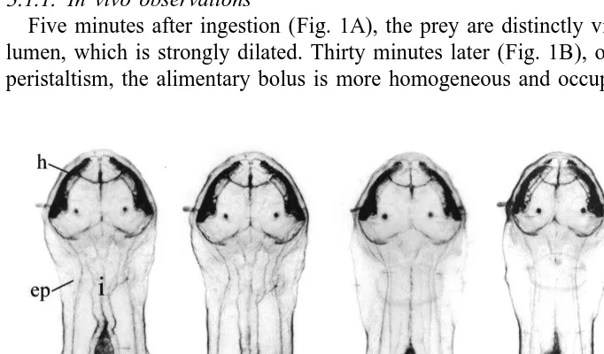
Five minutes after ingestion (Fig. 1A), the prey are distinctly visible in the intestinal lumen, which is strongly dilated. Thirty minutes later (Fig. 1B), owing to the intestinal peristaltism, the alimentary bolus is more homogeneous and occupies the posterior half
of the intestine. One hour later (Fig. 1C), the bolus is very reduced and numerous dark granulations appear inside the intestinal epithelium, which becomes denser. Few changes develop in the following hours, apart from a more and more dense aspect of the intestinal epithelium, achieving a maximum around the fifth hour after ingestion (Fig. 1D). The specimens often swallow water during this time. Twenty-four hours later (Fig. 1E), the intestinal lumen is entirely empty and the epithelium displays a light appearance. However, dark granulations persist in three zones that are always localized in the same parts of the intestine.
3.1.2. Cellular changes in the intestinal epithelium
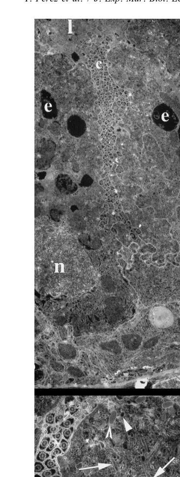
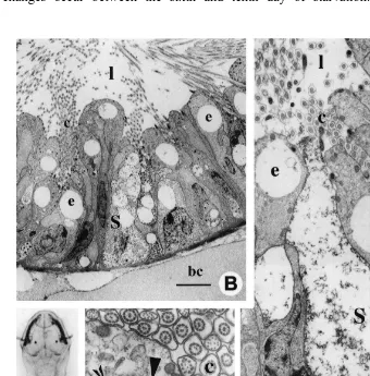
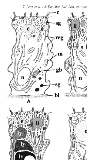
Five minutes after the ingestion of prey, the apical part of the A-cells (Fig. 2A, B) is occupied by a number of coated or uncoated endocytotic vesicles (0.1–0.15 mm in diameter) with an inner layer of dense material arranged against the membrane, a large network of moderately dense tubules, one or several light vacuoles (up to 4 mm in diameter) containing heterogeneous material probably corresponding to the endosome-like compartment and dense secretory granules (0.25–0.5 mm in diameter) frequently gathered against the apical membrane. Cisternae of RER, large mitochondria with tubular cristae, and Golgi bodies surround the nucleus and predominate in the basal half of the cells.
Thirty minutes later (Fig. 3A), the upper third of the A-cells displays the same characteristics. However, large lipid-like inclusions develop. In the rest of the cells, the most outstanding feature consists in the development of irregularly outlined vacuoles containing dense and heterogeneous material and probably corresponding to the lysosome-like compartment.
After 1 h (Fig. 3B) and 2 h (Fig. 3C), endocytosis continues, while the major changes consist in the gradual development of several large and dense lysosome-like vacuoles arranged in columns (Fig. 3B). Some vacuoles display circular light zones while, in the majority, the material becomes very dense (Fig. 3C).
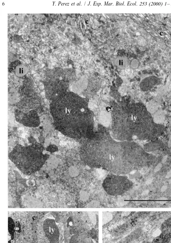
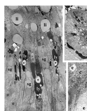
After 10 h (Fig. 4A, B), the changes occurring in the A-cells are less marked. The columnar disposition of the vacuoles is well visible and the material contained inside exhibits distinct aspects according to the localization in the cells, i.e. (Fig. 4B) the most heterogeneous and the least dense material at the top, with the most homogeneous and densest at the bottom. In some specimens, the apical vacuoles contain both light heterogeneous material characteristic of endosome-like vacuoles, and dense rather homogeneous material characteristic of lysosome-like vacuoles (Fig. 4C). Finally, the cells contain voluminous lipid-like inclusions (Fig. 4B) probably resulting from the coalescence of the smaller ones previously observed.
Throughout this time, the development of the secretion products seems less marked. In the A-cells the number of granules only infrequently varies and exocytosis of granules is sometimes observed during the digestive cycle. Contrarily, in the S-cells, the number of granules largely diminishes during the first 30 min, then remains nearly stable, but no exocytosis is observed.
3.2. Starvation experiments
intestinal regions characterized by dense granulations (Fig. 5A) are less marked in comparison with the specimens that are starved for only 1 day (see Fig. 1E). Major changes occur between the sixth and tenth day of starvation. In the A-cells, the
cytoplasm becomes more electron lucent but absorptive and secretory functions continue. The endocytotic vesicles (0.15–0.25mm) and endosomal-like compartment (up to 6mm in diameter) are larger than in fed specimens (Fig. 5B–D). On the other hand, dense bodies and lipid-like inclusions are absent (Fig. 5B, C). In the lower half of the cells (Fig. 5C), the nucleus maintains a normal appearance, with dense peripheral clusters of chromatin and a large nucleolus (Fig. 5C) and the Golgi apparatus always produces dense secretions. In the S-cells (Fig. 5C), features of cell necrosis are evident: burst secretory granules, the contents of which are often released into the gut lumen; degenerative mitochondria; absence of the RER and Golgi apparatus; and a homoge-neously dense nucleus. No change occurs at the intestinal basal lamina level. After the 20th day, both the A-cells and the longitudinal muscles also begin necrosis. Then the animals become moribund, although most of them remain alive for approximately 35 days. Finally, we must note that chaetognaths stop vitellogenesis during starvation because their ovaries do not product eggs after the last laying before the starvation begins. However, the reproductive behaviour is not inhibited.
3.3. Histochemical detection of lipids and polysaccharides
The Sudan Black B strongly stains the light inclusions in the apical region of the A-cells, which thus are lipids (Fig. 6A). With the Nile blue sulphate these inclusions are rosy, which indicates the presence of neutral lipids. Their light ultrastructural aspect, except at their periphery, indicates that the carbon chains are not counterstained by osmium tetroxide and therefore are probably saturated. These inclusions often come into contact with the edges of a number of RER cisternae, which converge towards them and become filled with dense material (Figs. 6B, C). Conversely, the periodic acid–Schiff technique does not demonstrate the presence of neutral mucopolysaccharides in the intestinal epithelium.
4. Discussion
4.1. Interpretation of cellular changes in fed specimens
The observations carried out on living specimens and on cross sections through the gut at different times after ingestion of prey allow us to follow the main changes of the A-cells which represent the only absorptive cell-type in the gut of chaetognaths. The following interpretation arises from our results and also previous data obtained on the functional morphology of the A-cells (Welsch and Storch, 1983; Arnaud et al., 1996; Perez et al., 1999) and on comparable absorptive cells involved in intracellular digestion in several metazoa, such as crustaceans (Arnaud et al., 1984, 1987; Al-Mohanna and Nott, 1986), fishes (Iida and Yamamoto, 1985; Iida et al., 1986), and molluscs (Boucaud-Camou and Yim, 1980), as well as on the knowledge of general endocytosis pathways (Helenius et al., 1983; Steinman et al., 1983; Mellman et al., 1986).
Fig. 6. (A) Photomicrograph of a semi-thin section through the whole intestine after incubation during 45 min at 608C a in Sudan Black solution. Observe that the lipid-like inclusions in A-cells strongly react. Scale bar520 mm. (B, C) Electromicrographs of lipid-like inclusions (li) of A-cells entirely surrounded by numerous cisternae of rough endoplasmic reticulum (arrows). Scale bar55mm.
that probably correspond to endosomes (Fig. 7A). As for the cytoplasmic tubules, most of them probably represent organelles involved in the recycling of plasma membrane and receptors of endocytosis. During the following steps (Fig. 7B) and up to 10 h after ingestion (Fig. 7C), endocytosis continues. The vacuolar apparatus increases with the development of new vacuoles containing dense material, interpreted as lysosomes (Arnaud et al., 1996; Perez et al., 1999), and arising from the transformation of endosomes pushed deeper into the cell. So, it seems that in chaetognaths the endosome does not represent a permanent compartment. Later, the evident stratification of several lysosomes along the upper cell region is related to the time of their formation, the youngest being localized at the top, and the oldest at the bottom of the cell. Thus, and in contrast to other metazoa, the lysosome compartment observed in the A-cells of Spadella
cephaloptera does not consist in a single large vacuole as in the B-cells of crustaceans
In the wild specimens which continuously eat, all the A-cells exhibits several stages of digestion, shown by the superposition of different types of vacuoles (Arnaud et al., 1996). In the specimens maintained under laboratory conditions, a number of A-cells complete their digestive cycle 24 h after the ingestion of prey and probably extrude their contents, especially residual dense bodies, upon termination. This hypothesis is both supported by the cell remnants observed at the basal part of the intestinal epithelium (Arnaud et al., 1996) and dense bodies sometimes released into the gut lumen (personal observation).
The comparison between our observations and those of Parry (1944) is instructive. According to him, the gland cells, i.e. the S-cells, develop a number of disrupted granules 10 min after feeding, which indicates a rapid release of their secretions. However, Parry suggests that these secretions represent digestive enzymes, while their fine structure could rather correspond to muco-substances, the enzymes probably being produced by another type of secretory cell localized in the cephalic region of the gut (Arnaud et al., 1996; Perez et al., 1999). Our results, i.e. the scarcity of the exocytozing granules, could confirm that secretion is mediated and quickly occurs as soon as the prey enters the gut. As for the A-cells, Parry (1944) also describes the progressive development of vacuoles after feeding and the presence of dark granulations, suggesting both absorptive and excretory functions.
During experiments, the A-cells absorb large amounts of lipids. As they are stained in rosy by the Nile blue sulphate, they corresponds to neutral lipids (Sire and Vernier, 1980). Such accumulations were not observed in the A-cells of wild specimens (Arnaud et al., 1996). It is probable that they are caused by the food used for experiments, exclusively consisting of Artemia salina nauplii. Indeed, nauplii contain large amounts of stored lipoproteins which must be absorbed and temporarily stored in A-cells. Finally, the relations between lipid inclusions and numerous cisternae of RER do correspond to lipidic metabolism probably linked to membrane formation and / or biosynthesis of complex lipids. Large amounts of lipid involved in temporary storage and forming voluminous droplets were also in close contact with the RER after experimental feeding in the intestine of fishes (Noaillac-Depeyre and Gas, 1974, 1976).
4.2. Effects of starvation
this time, since most of the specimens die a few days afterwards. The irreversible necrosis of the intestinal epithelium within one month suggests that Spadella
cephalo-ptera is poorly adapted to starvation during mating and reproduction. Comparative data
on starvation in invertebrates are scarce, but in terrestrial isopods the hepatopancreatic cells remain capable of regeneration after several weeks of food deprivation (Storch, 1984).
Intestinal microvilli represent labile structures, the development of which is correlated with nutrition (Hryniewiecka-Szifter and Storch, 1986). During starvation, their gradual reduction can be observed in fishes (Gas and Noaillac-Depeyre, 1976) and crustaceans (Storch and Lehnert-Moritz, 1980; Storch et al., 1982; Hryniewiecka-Szifter and Storch, 1986), which suggests a possible decrease in absorption. In contrast, a short starvation period stimulates absorption in chaetognaths because the size of both the endocytotic vesicles and endosomal compartment increases (Fig. 7D). The increase in the membrane surface involved in absorption is not surprising because chaetognaths are devoid of storage structures. Although absorption is undoubtedly active in starved specimens, the intracellular digestion is less evident because the electron-dense vacuoles considered as the lysosomal compartment are never observed. It is interesting to note that in several
Sagitta species, the A-cells are hypertrophic due to the presence of very large vacuoles
(Dallot, 1970; Bone et al., 1987) which often display a light appearance (personal observation). Although dense vacuoles were not observed, these A-cells are certainly functional because (i) all the organelles involved in the absorptive function are present (personal observations), (ii) the vacuolar fluid pH is about 6.0 (Bone et al., 1987) and corresponds to the acid pH generally observed in lysosomal compartments (Forgac,
1
1989), and (iii) the giant vacuoles contain high concentrations of NH4 and amino-acids (Bone et al., 1987) which certainly arise from intracellular digestion. Thus, it seems that the absence of dense vacuolar material does not reflect the absence of an intracellular digestive function, but rather a low concentration of absorptive products. However, cytochemical tests, such as the detection of acid phosphatase, would be necessary to resolve this problem.
The intestinal absorption is active until the twentieth day of starvation and most specimens remain alive for more than 30 days. Since the chaetognaths lack storage substances, where do they find the nutrients necessary to survive more than 1 month without food ? The intestinal epithelium of chaetognaths display numerous cilia, the beating of which may create a water current in the gut lumen (Parry, 1944). Dissolved organic molecules certainly penetrate into the intestinal lumen via this water and could be internalized by the A-cells via the permanent receptor-mediated endocytosis. The implication of receptors in absorption seems evidenced by the presence of an electron-dense layer closely disposed against the inner side of the vesicle membrane. However, the biochemical nature of the endocytosized substances remains unknown.
Acknowledgements
We are greatly indebted to Deborah Sazer for correcting the text. [SS]
References
Al-Mohanna, S.Y., Nott, J.A., 1986. B-cells and digestion in the hepatopancreas of Penaeus semisulcatus (Crustacea: Decapoda). J. Mar. Biol. Assoc. UK 66, 403–414.
Arnaud, J., Brunet, M., Mazza, J., 1978. Studies on the midgut of Centropages typicus (Copepod, Calanoida). I. Structural and ultrastructural data. Cell Tissue Res. 187, 333–353.
Arnaud, J., Brunet, M., Mazza, J., 1984. Cytochemical detection of phosphatase and arylsulphatase activities in the midgut of Centropages typicus (Copepod, Calanoid). Basic Appl. Histochem. 28, 399–412.
ˆ
Arnaud, J., Brunet, M., Mazza, J., 1987. Role des cellules B de l’intestin moyen chez Centropages typicus (Copepoda, Calanoida). Reprod. Nutr. Dev. 27, 817–827.
Arnaud, J., Brunet, M., Casanova, J.P., Mazza, J., Pascalini, V., 1996. Morphology and ultrastructure of the gut in Spadella cephaloptera (Chaetognatha). J. Morphol. 228, 27–44.
Bone, Q., Brownlee, C., Bryan, G.W., Burt, G.R., Dando, P.R., Liddicoat, M.I., Pulsford, A.L., Ryan, K.P., 1987. On the differences between the two ‘indicator’ species of chaetognaths, Sagitta setosa and S. elegans. J. Mar. Biol. Assoc. UK 67, 545–560.
Boucaud-Camou, E., Yim, M., 1980. Fine structure and function of the digestive cells of Sepia officinalis (Mollusca: Cephalopoda). J. Zool. 191, 89–105.
´ ´
Dallot, S., 1970. L’anatomie du tube digestif dans la phylogenie et la systematique des chaetognathes. Bull. Mus. Natl. Hist. Nat. Paris 42, 549–565.
Duvert, M., Gourdoux, L., Moreau, R., 2000. Cytochemical and physiological studies of the energetic metabolism and osmotrophy in Sagitta (chaetognath). J. Mar. Biol. Assoc. UK (in press).
Feigenbaum, D.L., Marris, R.C., 1984. Feeding in the Chaetognatha. Oceanogr. Mar. Biol. Annu. Rev. 22, 343–392.
Forgac, M., 1989. Structure and function of the vacuolar class of ATP-driven proton pumps. Physiol. Rev. 69, 765–796.
Gas, N., Noaillac-Depeyre, J., 1976. Studies on intestinal epithelium evolution during prolonged fasting. J. Ultrastruct. Res. 56, 137–151.
Helenius, A., Mellman, I., Wall, D., Hubbard, A., 1983. Endosomes. Trends Biochem. 8, 245–250. Hryniewiecka-Szifter, Z., Storch, V., 1986. The influence of starvation and different diets on the hindgut of
Isopoda (Mesidotea entomon, Oniscuc asellus, Porcellio scaber). Protoplasma 134, 53–59.
Iida, H., Yamamoto, T., 1985. Intracellular transport of horseradish peroxidase in the absorptive cells of the gold fish hingut in vitro, with special reference to the cytoplasmic tubules. Cell Tissue Res. 240, 553–560. Iida, H., Shibata, Y., Yamamoto, T., 1986. The endosome-lysosome system in the absorptive cells of the
goldfish hindgut. Cell Tissue Res. 243, 449–452.
John, C.C., 1933. Habits, structure, and development of Spadella cephaloptera. Q. J. Microsc. Sci. 75, 625–696.
Mellman, I., Fuchs, R., Helenius, A., 1986. Acidification of the endocytotic and exocytotic pathways. Annu. Rev. Biochem. 55, 663–700.
Noaillac-Depeyre, J., Gas, N., 1974. Fat absorption by the enterocytes of the carp (Cyprinus carpio L.). Cell Tissue Res. 155, 353–365.
Noaillac-Depeyre, J., Gas, N., 1976. Electron microscopic study on the gut epithelium of the tench (Tinca tinca L.) with respect to its absorptive functions. Tissue Cell 8, 511–530.
Parry, D.A., 1944. Habits, structure and development of Spadella cephaloptera and Sagitta setosa. J. Mar. Biol. Assoc. UK 26, 16–36.
Perez, Y., Arnaud, J., Brunet, M., Casanova, J.-P., Mazza, J., 1999. Morphological study of the gut in Sagitta
Reynolds, E.S., 1963. The use of lead citrate at high pH as an electron-opaque stain in electron microscopy. J. Cell Biol. 17, 208–212.
Sire, M.-F., Vernier, J.-M., 1980. Lipid staining on semithin sections with Sudan black B or Nile blue sulphate. Applications to intestinal fat absorption. Acta Histochem. Cytochem. 13, 193–201.
Steinman, R.M., Mellman, I., Muller, W.A., Cohn, Z.A., 1983. Endocytosis and the recycling of plasma membrane. J. Cell Biol. 96, 1–27.
Shinn, G.L., 1997. Chaetognatha. In: Harrison), F.W. (Ed.), Microscopic Anatomy of Invertebrates. Hemichor-data, Chaetognatha and the Invertebrate Chordates, Vol. 15. Wiley-Liss, New York, pp. 103–220. Storch, V., 1984. The influence of the nutritional stress on the ultrastructure of the hepatopancreas of terrestrial
isopods. Symp. Zool. Soc. Lond. 53, 167–184.
Storch, V., Lehnert-Moritz, K., 1980. The effects of starvation on the hepatopancreas of the isopods Ligia
oceanica. Zool. Anz. 204, 137–146.
Storch, V., Heinrich Janssen, H., Cases, E., 1982. The effects of starvation on the hepatopancreas of the Coconut Crab, Birgus latro (L.) (Crustacea, Decapoda). Zool. Anz. 208, 115–123.