Research Article
Abnormalities in the Male Reproductive System After
Exposure to Diesel and Biodiesel Blend
Elena R. Kisin,
1Naveena Yanamala,
1Mariana T. Farcas,
1Dmitriy W. Gutkin,
2Michael R. Shurin,
2Valerian E. Kagan,
3Aleksandar D. Bugarski,
4and Anna A. Shvedova
1,5*
1
Pathology and Physiology Research Branch, and Exposure Assessment Branch,
HELD, NIOSH, Morgantown, West Virginia
2
Department of Pathology, University of Pittsburgh Medical Center, Pittsburgh,
Pennsylvania
3
Department of Environmental and Occupational Health, University of
Pittsburgh, Pittsburgh, Pennsylvania
4
Office of Mine Safety and Health Research, NIOSH, Pittsburgh,
Pennsylvania
5
Department of Physiology and Pharmacology, West Virginia University,
Morgantown, West Virginia
Altering the fuel source from petroleum-based ultra-low sulfur diesel to biodiesel and its blends is consid-ered by many to be a sustainable choice for controlling exposures to particulate material. As the exhaust of biodiesel/diesel blends is composed of a combination of combustion products of polycyclic aromatic hydrocarbons and fatty acid methyl esters, we hypothesize that 50% biodiesel/diesel blend (BD50) exposure could induce harmful outcomes because of its ability to trigger oxidative damage. Here, adverse effects were compared in murine male reproductive organs after pharyngeal aspira-tion with particles generated by engine fueled with BD50 or neat petroleum diesel (D100). When com-pared with D100, exposure to BD50 significantly altered sperm integrity, including concentration, motility, and morphological abnormalities, as well as increasing testosterone levels in testes during the time course postexposure. Serum level of luteinizing
hormone was significantly depleted only after BD50 exposure. Moreover, we observed that exposure to BD50 significantly increased sperm DNA fragmen-tation and the upregulation of inflammatory cyto-kines in the serum and testes on Day 7 postexposure when compared with D100. Histological evaluation of testes sections from BD50 exposure indicated more noticeable interstitial edema, degenerating spermatocytes, and dystrophic seminiferous tubules with arrested spermatogenesis. Significant differen-ces in the level of oxidative stress assessed by accu-mulation of lipid peroxidation products and depletion of glutathione were detected on exposure to respirable BD50 and D100. Taken together, these results indicate that exposure of mice to inhal-able BD50 caused more pronounced adverse effects on male reproductive function than diesel. Environ. Mol. Mutagen. 00:000–000, 2014. VC2014 Wiley
Periodicals, Inc.
Key words: pulmonary exposure; biodiesel particles; oxidative stress; male reproduction; sperm quality; DNA fragmentation
Grant sponsor: NIOSH; Grant numbers: 2927ZKCY, OH008282. Disclaimer: The findings and conclusions in this report are those of the authors and do not necessarily represent the views of the National Institute for Occupational Safety and Health. Mention of trade names or commer-cial products do not constitute endorsement or recommendation for use.
*Correspondence to: Anna A. Shvedova, Pathology and Physiology Research Branch, and Exposure Assessment Branch, HELD, NIOSH, 1095 Willowdale Rd, Morgantown, WV 26505, USA. E-mail: [email protected]
Received 30 June 2014; provisionally accepted 25 September 2014; and in final form 00 Month 2014
DOI 10.1002/em.21915
Published online 00 Month 2014 in
Wiley Online Library (wileyonlinelibrary.com).
INTRODUCTION
The use of biodiesel (BD) or its blends with petroleum
diesel (D100) is contemplated to be a justifiable approach
to reduce occupational and environmental exposures to
particulate matter (PM). Exposure to diesel exhaust in
humans has been shown to cause a number of adverse
health outcomes, including pulmonary, cardiovascular
dis-eases, and cancer [Tokiwa and Ohnishi, 1986; Watkinson
et al., 1998; Holgate et al., 2003a; Garshick et al., 2004;
Mills et al., 2005, 2007; Rivero et al., 2005; Nemmar
et al., 2007, 2009; Tornqvist et al., 2007; Peretz et al.,
2008; Pronk et al., 2009; Sawyer et al., 2010; Hazari
et al., 2011; Silverman et al., 2012]. Diesel exhaust
par-ticulates (DEPs) have also been reported to cause the
dis-ruption of male reproductive function. Prior studies have
shown that DEP exposure disturbed spermatogenesis,
resulting in reduction of daily sperm production and
motility, increased morphological sperm abnormalities,
and ultrastructural changes in Leydig cells in mice
[Yosh-ida et al., 1999; Yosh[Yosh-ida and Takeda, 2004; Izawa et al.,
2008; Li et al., 2012]. In male rats, the regulation of
tes-ticular function was altered resulting in elevation of
serum testosterone and reduction of luteinizing hormone
(LH) and sperm production after DEP exposure
[Wata-nabe and Oonuki, 1999; Tsukue et al., 2001, 2002; Izawa
et al., 2007; Li et al., 2007, 2009b; Ramdhan et al.,
2009].
On the other hand, most BD fuels are produced by the
transesterification of vegetable oils generating fatty acid
alkyl esters [Knothe et al., 2010]. The major components
of these oils are monounsaturated oleic acid,
polyunsatu-rated linoleic acid, and linolenic acid, which are prone to
oxidation on combustion forming a variety of peroxides
and secondary oxidation products [Song et al., 2000;
Knothe, 2005]. Moreover, exposure to BD was found to
increase the particle-bound volatile organic fraction of
PM and carbonyl emissions [Purcell et al., 1996; Liu
et al., 2009], inducing a rise in cellular toxicity [Liu
et al., 2009]. Recent studies demonstrate that BD or its
blends are more mutagenic and increase formation of
multinucleated cells when compared with ultralow sulfur
diesel fuel (ULSD) [Ackland et al., 2007; Bunger et al.,
2007; Krahl et al., 2009; Kisin et al., 2013]. Brito et al.
[2010] demonstrated that exposure to diesel, neat BD,
and 50% biodiesel/diesel blend (BD50) induces acute
lung inflammation. Recently, we reported that pharyngeal
aspiration or inhalation exposure to BD exhaust causes
pronounced pulmonary damage accompanied by a robust
inflammatory response and severe oxidative stress when
compared with diesel exhaust nanoparticles [Shvedova
et al., 2013; Yanamala et al., 2013].
Diesel engine emissions from the use of diesel, or its
blends with biodiesel, are highly complex mixtures of
aerosols and gases. Most of the particles from these
engine exhausts are of nanoscale and potentially have
hundreds of chemicals absorbed onto their surfaces,
including known and suspected mutagens and
carcino-gens, for example, polycyclic aromatic hydrocarbons
(PAH) and nitrated PAH (nPAH) [Mermelstein et al.,
1981; Ohe, 1984; Rivedal et al., 2003; B
€
unger et al.,
2012]. Recently, it has been shown that human exposure
to PAHs causes alterations in male sperm quality,
includ-ing morphology, concentration, and vitality, as well as
DNA damage, thus affecting male reproductive function
[Gaspari et al., 2003; Meeker et al., 2007; Han et al.,
2011; Jeng et al., 2013].
To
address
whether
exposure
to
respirable
BD
adversely affects spermatogenesis, we compared the
effects of BD50 and neat petroleum diesel in male
C57BL6 mice. We found that pulmonary exposure to
BD50 significantly altered sperm concentration, motility,
and morphology. Moreover, BD50 caused upregulation of
inflammatory cytokines, increase in testicular testosterone,
and reduction of serum LH. Testicular histopathology
revealed interstitial edema, clustering of the dystrophic
seminiferous tubules with arrested spermatogenesis, and
the presence of degenerating spermatocytes. Additionally,
we observed that exposure to inhalable BD50 caused
severe oxidative stress and DNA damage in mouse male
reproductive organs. Here, for the first time, we
demon-strate that exposure to respirable BD50 generates a
pro-nounced toxicity to the male reproductive system when
compared with diesel.
MATERIALS AND METHODS
Animals
Specific pathogen-free adult male C57BL/6 mice (7–8 weeks) were supplied by Jackson Laboratories (Bar Harbor, ME) and weighed 20.061.9 g when used. Animals were housed one mouse per cage receiving filtered high-efficiency particulate air in the Association for Assessment and Accreditation of Laboratory Animal Care (AAALAC) International-accredited National Institute of Occupational and Safety Health (NIOSH) animal facility. All animals were acclimated in the ani-mal facility under controlled temperature and humidity for 1 week prior to use. Beta Chips (Northeastern Products Corp., Warrensburg, NY) were used for bedding and changed weekly. Animals were supplied with water and certified chow 7913 (Harlan Teklad, Indianapolis, IN) ad libi-tum, in accordance with the guidelines and policy set forth by the Insti-tute of Laboratory Animal Resources, National Research Council. All experimental procedures were conducted in accordance with a protocol approved by the NIOSH Institutional Animal Care and Use Committee.
General Experimental Design
To assess reproductive toxicity, mice were treated by repeated pha-ryngeal aspiration with suspensions of BD50 or D100 exhaust particles (15lg/mouse/day of total carbon) in United States Pharmacopeia (USP)
sterile water (Hospira, Lake Forest, IL). The corresponding control mice were administered USP sterile water. Mice were exposed twice a week for two consecutive weeks with a cumulative dose of 60 lg per mouse
deposited cumulative dose of 60 mg of total carbon can be achieved in 88 working weeks at allowable exposure concentration limits defined by MSHA (160 mg/m3 of total carbon). Calculations were performed using the formula published previously by our group [Yanamala et al., 2013]. Animals were weighed and sacrificed on Days 7 and 28 following their last exposure. Blood samples were collected and kept at room tem-perature for 1.5 hr to allow for clotting. The samples were then centri-fuged at 1,700gfor 15 min at 4C, and serum was separated and stored
at 280C until assayed for cytokine responses and LH level. Testes
were removed and weighed. The right testis was used for histologic examination, whereas the left one was processed for testosterone measure-ments, cytokines responses, and biomarkers of oxidative stress. The epidi-dymis were excised and trimmed carefully of excess tissue and fat and immediately weighed. The right epididymis was processed for semen analysis (sperm density, motility, and morphology evaluation), whereas the left epididymis was used for sperm DNA fragmentation screening.
Exhaust/Emission Generation and Diesel Particulate
Matter Samples Collection System
The diesel particulate matter (DPM) samples were collected at the die-sel laboratory at the NIOSH Office of Mine Safety and Health Research. A single batch of neat corn-based fatty acid methyl ester (FAME) BD was acquired from Peter Cremer (Cincinnati, OH, NEXSOLTMBD-100), and a single batch of petroleum-based ULSD fuel was acquired from a local supplier. The BD50 blend was prepared at the site. The fractions of BD and ULSD were determined volumetrically. The samples of BD50 and D100 particulates were collected from the exhaust of a mechanically controlled, naturally aspirated directly injected Isuzu C240 (Isuzu Motors Limited, Tokyo, Japan) diesel engine equipped with a diesel oxidation catalytic converter (DOC, Lubrizol, New Market, ON). The engine was exercised over four steady-state operating conditions [Yanamala et al., 2013]. The exhaust particles originating from the four different loads were collected and combined to perform the study.
A high-volume sampling system was developed to advance the methods of collecting representative samples of diesel particulates. This system allows for collecting nanosized and ultrafine DPM aerosols in liquid media, therefore preserving the sampled aerosol to the highest possible level of physical and chemical characteristics. The method for the DPM sample col-lection system has been described in detail by Yanamala et al. [2013]. The samples collected for each of the fuels were combined and further concen-trated using a rotating evaporator (Eppendorf Vacufuge, Hamburg, Germany). The particle suspensions were sonicated using a Vibra Cell (Sonics & Materials, Newtown, CT) before administering them to animals.
All doses of BD50 and D100 exhaust particles were standardized based on the amount of total carbon analyzed by NIOSH Method 5040 [NMAM, 2003; Birch, 2004] as mentioned previously [Yanamala et al., 2013]. Additionally, the hydrodynamic diameter of BD50 and D100 par-ticulates (216 and 312 nm, respectively) was determined by dynamic light scattering [Yanamala et al., 2013].
Particulate Aspiration
Mouse pharyngeal aspiration was used for particulate administration. Briefly, after anesthesia with a mixture of ketamine (Phoenix Pharma-ceutical Inc, St. Joseph, MO) and xylazine (62.5 and 2.5 mg/kg subcuta-neous in the abdominal area; Phoenix Pharmaceutical Inc, St. Joseph, MO), the mouse was placed on a board in a near vertical position, and the animal’s tongue was extended with lined forceps. A suspension of BD50 or D100 exhaust particles (15lg/mouse/day of total carbon, twice a week, for 2 weeks) was placed posterior in the throat, and the tongue held until the suspension was aspirated into the lungs. All particles were sterilized prior to administration. All mice from the control, BD50-, and D100-treated groups survived this exposure procedure and exhibited no negative behavioral or health outcomes.
Levels of Oxidative Stress Markers
Oxidative damage in the testis following pulmonary exposure to BD50 or D100 was evaluated by the presence of hydroxynonenal-histidine (His) protein adduct and glutathione (GSH) level. HNE-His adducts, lipid peroxidation end products, were measured in tissue homogenates by ELISA using an OxiSelectTMHNE-His adduct kit (Cell Biolabs, San Diego, CA). The quantity of HNE-His adducts in protein samples were determined by comparing its absorbance with that of a known HNE-BSA standard curve.
GSH concentration in testis homogenates was determined using ThioGloTM-1 (Covalent Associates Inc., Corvallis, OR), a maleimide rea-gent, which produces highly fluorescent adducts on its reaction with SHA
groups [Shvedova et al., 2005]. Glutathione content was estimated by an immediate fluorescence response registered on the addition of ThioGloTM -1 to the testis homogenate. A standard curve was established by the addi-tion of GSH (0.04–4.0lM) to 100 mM phosphate buffer (PBS, pH 7.4)
containing 10 lM ThioGloTM-1. The SynergyTM H1 hybrid multimode
microplate reader (BioTek Instruments, Winooski, VT) was used for the assay of fluorescence using excitation at 388 nm and emission at 500 nm. The data obtained were exported and analyzed using Gen5TMdata analysis software (BioTek Instruments).
Epididymal Sperm Concentration and Motility
The total sperm cell counts and motility were assessed as described previously by Polyzos et al. [2009]. The caudal right epididymis was minced into 2 ml prewarmed M16 medium (Sigma-Aldrich, St. Louis, MO) and incubated at 37C in 5% CO2for 5 min to allow sperm to
dis-perse. Sperm concentration and motility were evaluated from the same sperm cell suspension using an improved Neubauer hemocytometer chamber (Hausser Scientific, Horsham, PA). Ten microliter of the warm sperm cell suspension was loaded into the hemocytometer and immedi-ately examined under microscope. About 300–400 cells per slide were tracked for motility assessment by scoring the number of all motile and nonmotile sperm in the same field.
Epididymal Sperm Morphology
Sperm morphology was assessed as described previously by Pereira et al. [1981]. Briefly, 10 ml of Eosin Y (Leica Microsystems, Buffalo Grove, IL) was mixed with 90ml of sperm suspension obtained from the right epididymis as described in the preceding paragraph and incubated for 30 min at room temperature. A drop of sperm suspension (5ml) was smeared on the slide, air dried, and mounted with low-viscosity mount-ing medium. Sperm morphology was examined on bright field micros-copy (Olympus Provis, B&B Microscopes, Pittsburgh, PA), and spermatozoa were classified as follows: sperm cells with normal mor-phology and cells presenting abnormalities in head, neck/mid-piece, and tail. At least 500 sperm cells were recorded for each slide under 1,000 3magnification.
Sperm DNA Fragmentation Screening
Sperm chromatin integrity was assessed by the sperm chromatin structure assay (SCSA). This test provides detection of damaged DNA and altered proteins in sperm nuclei via flow cytometry of acridine orange stained sperm [Evenson and Melamed, 1983]. Cell suspension from the left caudal epididymis was prepared in M16 medium (as described above) and incubated for 5 min at 37C in 5% CO2. After
incubation, the sperm cells were filtered through a 70-mm sterile cell strainer to remove the tissues and other debris and to form a more uni-form cell suspension. Next, the sperm cell suspension was centrifuged for 3 min at 2,000 rcf (37C), the supernatant was removed, and the
suspensions were immediately frozen and stored in liquid nitrogen until shipping to SCSA Diagnostics (Brookings, SD) for evaluation of cells with DNA fragmentation. The DNA fragmentation index value indicates the percent of sperm cells containing DNA damage.
Histopathology of the Testis
Testes were removed and fixed with 10% buffered formaldehyde. The testes were embedded in paraffin and sectioned at a thickness of 5 mm on an HM 320 rotary microtone (Carl Zeiss, Thornwood, NY). Prepared sections were stained with hematoxylin and eosin (H&E), and histopathological evaluation was performed by certified pathologist. Sample identification was coded to ensure unbiased evaluation.
Preparation of Tissue Homogenates
The left testis was homogenized with a tissue tearer (model 985–370, Bio-spec Products, Racine, WI) in cold PBS (pH, 7.4) for 2 min on ice. The homogenate suspensions were aliquoted and frozen at280C until processed.
Measurement of Cytokines and Chemokines
Proinflammatory cytokines, IL-1a, IL-1b, IL-6, and TNF-a, in the serum and testes homogenates from mice exposed to BD50 and D100 exhaust particulates were analyzed using a Bio-Plex system (Bio-Rad, Hercules, CA). The concentrations were calculated using Bio-Plex Man-ager 6.1 software (Bio-Rad) based on standard curves.
Testosterone Level in the Testes
The concentration of testosterone in the testes homogenate was deter-mined by applying the competitive enzyme immunoassay technique using a BlueGene Testosterone ELISA kit (Antibodies-Online, Atlanta, GA). The optical density was determined using a Synergy H1 hybrid multimode microplate reader (BioTek Instruments). Using Gen5TMdata analysis software (BioTek Instruments), the concentration of testosterone in the samples was determined.
Luteinizing Hormone Concentration in the Serum
The concentration of LH was determined using an ELISA kit from BlueGene Biotech (Shanghai, China). The optical density was determined using a Synergy H1 hybrid multimode microplate reader (BioTek Instru-ments, Winooski, VT). Using Gen5TM data analysis software (BioTek Instruments, Winooski, VT), the sample concentrations were calculated. Each sample was run in duplicate.
Protein Assay
Measurements of protein in tissue homogenates from mouse testis were run using a Bio-Rad protein assay kit (Richmond, CA).
Statistical Analysis
Results were compared by one-way ANOVA using all the pairwise multiple comparison procedures (Holm-Sidak method). All results are presented as mean1SEM.Pvalues of less than 0.05 were considered to indicate statistical significant.
RESULTS
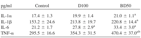
Proinflammatory Cytokine in Serum Following BD50 or
D100 Exposures
The release of cytokines was used as a marker of
proinflammatory responses in the serum of mice exposed
to BD50 and D100 exhaust (Table I). IL-1
a
, IL-1
b
, and
TNF-
a
in the serum were uniquely upregulated only after
exposure to BD50 (Day 7 post-treatment). The elevation
of TNF-1
a
was found to be significantly stronger in
BD50 when compared with that of D100. The level of
IL-6 was found to be significantly elevated after exposure
to either BD50 or D100, with a stronger response from
BD50. Overall, the serum levels of the proinflammatory
cytokines following BD50 exposure were higher when
compared with D100.
Oxidative Stress Markers
The level of oxidative damage in the testes caused by
BD50 or D100 exhaust PM was assessed by lipid
peroxi-dation products and GSH (Fig. 1). Exposure to BD50
resulted in significant 44 and 64% accumulation of lipid
peroxidation products, measured as HNE-His adducts,
over control throughout the time course of 7 and 28 days
post-treatment, respectively (Fig. 1A). Similarly, D100
caused significant 30 and 35% increases of HNE-His
adducts observed on Days 7 and 28 postexposure,
respec-tively. However, accumulation of lipid peroxidation
prod-ucts was significantly greater on exposure to BD50 when
compared with D100 on Day 28 post-treatment. Time
course of GSH depletion in the testes of mice exposed to
BD50 revealed significant 18 and 28% decreases from
control samples at 7 and 28 days postaspiration,
respec-tively (Fig. 1B). No significant changes in the level of
GSH were found after exposure to D100. Additionally,
alterations in the level of GSH were significantly different
on Day 7 postexposure to DB50 and D100.
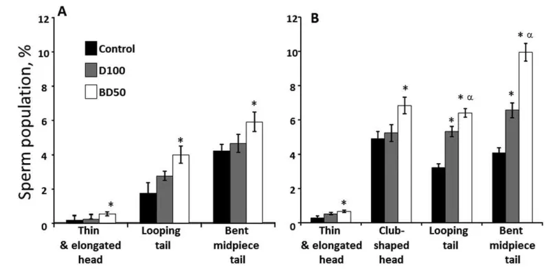
Epididymis Sperm Characteristics
To characterize the impact of BD50 or D100 treatment
on the male reproductive system, epididymis sperm
con-centration (Fig. 2A), sperm motility (Fig. 2B), and
mor-phological abnormalities (Fig. 3) were analyzed on Days
7 and 28 after pharyngeal aspiration.
We found that BD50 exposure induced a significant
72% and 44% decrease in epididymis sperm
concentra-tion in contrast to the control on Days 7 and 28
TABLE I.
Differential Responses in Serum Cytokines on Day 7
Post Repeated Exposure to Diesel (D100) or Biodiesel Blend
(BD50) Exhaust Particulates
pg/ml Control D100 BD50
IL-1a 17.461.3 19.961.4 21.061.1a IL-1b 153.2624.6 213.8619.7 220.8614.4a IL-6 21.261.7 27.862.9a 33.463.0a TNF-a 295.5616.6 354.3631.5 470.4637.0ab
a
P<0.05, vs control. b
postexposure, respectively (Fig. 2A). Exposure to D100
resulted in a significant reduction of sperm concentration
by 24% on Day 28 only.
Similarly, BD50 exhaust significantly diminished sperm
motility by 34% and 19% when compared with the control on
Days 7 and 28 postexposure, respectively (Fig. 2B), whereas
D100 decreased the number of motile cells by 21% when
compared with the control on Day 7 postexposure only.
Sperm morphological examination demonstrated a
sig-nificant 3.4-, 1.7-, and 1.4-fold increase in the number of
abnormal thin/elongated heads, looping, and bent
mid-piece tails versus control on Day 7 post-BD50 exposure,
respectively (Fig. 3A). No significant changes were found
on Day 7 after exposure to D100 exhaust. More
pro-nounced morphological abnormalities were found on Day
28 postexposure in both D100- and BD50-treated mice
(Fig. 3B). Thus, significant 2.2- and 1.4-fold increase in
the number of abnormal thin/elongated and club-shaped
heads, respectively, over control were detected only after
exposure to BD50. Additionally, BD50 induced
signifi-cant twofold and 2.4-fold rises over the control in the
number of looping and bent mid-piece tails, whereas
D100 exposure increased up to 1.65- and 1.6-fold when
compared with control mice, respectively.
Interestingly, effect of BD50 induced significantly
stronger changes in sperm concentration and
morphologi-cal abnormalities only on Day 28 postexposure, whereas
alterations in motility of the sperm were more
pro-nounced throughout the time course of the experiment
when compared with D100.
Testicular Cytokines Following BD50 or D100 Exhaust
Exposures
The release of proinflammatory cytokines in the testes
of mice exposed to BD50 and D100 exhaust are shown in
Fig. 1. The degree of oxidative stress evaluated by HNE-His adduct (A)
and GSH (B) in the testes of C57BL6 mice on Days 7 and 28 after repeated exposure to D100 or BD50 exhaust particles. Mice were exposed via pharyngeal aspiration (15mg/mouse/day of total carbon, twice a week for 2 weeks). Black columns: 7 days after last repeated exposure to BD50
or D100 exhaust particulate; and clear columns: 28 days after last repeated exposure to BD50 or D100 exhaust particulate. Data are pre-sented as percent of control. Means6SE (n510 mice per group). Signif-icantly different from *controls (P<0.05) andaD100 exhaust
particulate-exposed group (P<0.05).
Fig. 2. Concentration (A) and motility (B) of epididymal sperm from
C57BL6 mice on days 7 and 28 after repeated exposure to D100 or BD50 combustion exhaust particles. Mice were exposed to exhaust par-ticles via pharyngeal aspiration (15 mg/mouse/day of total carbon, twice a week for 2 weeks). Black columns: 7 days after last repeated
exposure to BD50 or D100 exhaust particulate; and clear columns: 28 days after last repeated exposure to BD50 or D100 exhaust particulate. Means6SE (n510 mice per group). Significantly different from *controls (P<0.05) and aD100 exhaust particulate-exposed group
Table II. IL-1
a
, IL-1
b
, and TNF-
a
were upregulated after
exposure to BD50 or D100 (Day 7 post-treatment). The
elevation of TNF-1
a
was found to be significantly
stron-ger in BD50 when compared with that of D100. The
lev-els of IL-6 in both groups were not different from the
control.
Testicular Testosterone Level
The testosterone concentration in the testes
homoge-nates was measured after pharyngeal aspiration of BD50
or D100 exhaust particulates on Days 7 and 28
postex-posure (Fig. 4A). A significant 19% augmentation in
tes-ticular testosterone level was found in the BD50 group
on Day 7 postexposure, which progressed further (86%
over control) by Day 28. No significant changes were
found after exposure to D100. Moreover, the elevation
of testosterone concentration was found to be
signifi-cantly stronger in BD50 when compared with that of
D100.
Serum Luteinizing Hormone Concentration
We investigated the serum concentration of LH in
mice on Days 7 and 28 postexposure with BD50 or
D100 exhaust particulate. Serum concentrations of LH
in the BD50 exposure group were significantly decreased
by 21% and 19% on Days 7 and 28 post-treatment,
respectively, when compared with the control group
(Fig. 4B). Exposure to D100 exhaust particulate did not
induce significant depletion of LH in serum when
com-pared with the control group. The results from BD50
exposure were not significantly different when compared
with D100.
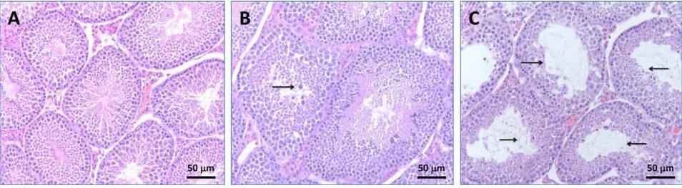
Histological Evaluation
Formalin-fixed, paraffin-wax embedded testes
sec-tions stained with H&E were examined
microscopi-cally. Normal architecture of seminiferous tubules and
orderly spermatogenesis were detected in the testes
from control mice on Days 7 and 28 postexposure.
Additionally,
no
histopathological
changes
were
observed in Leydig or Sertoli cells (Fig. 5A). Sections
of D100 and BD50 groups on Day 7 postexposure
showed no significant pathologic alterations. However,
on Day 28 postexposure, sections of D100 group
showed interstitial edema and occasional dystrophic
seminiferous tubules with arrested spermatogenesis and
the presence of degenerating spermatocytes (Fig. 5B).
These histologic changes were even more prominent in
BD50 group, where dystrophic seminiferous tubules
were
clustering,
especially
in
subcapsular
areas
(Fig. 5C).
Fig. 3. Morphological abnormalities of epididymal sperm from C57BL6
mice on Days 7 and 28 postrepeated exposure to D100 or BD50 combus-tion exhaust particles. (A) Day 7 and (B) Day 28 postexposure to com-bustion exhaust particles. Mice were exposed to exhaust particles via pharyngeal aspiration (15mg/mouse/day of total carbon, twice a week for 2 weeks). Black columns: control mice; gray columns: mice exposed to
D100 exhaust particulate; and clear columns: mice exposed to BD50 exhaust. Data presented as percent of abnormal cells population when compared with total number of sperm. Means6SE (n510 mice per group). Significantly different from *controls (P<0.05) and aD100
exhaust particulate-exposed group (P<0.05).
TABLE II.
Differential Responses in Testicular Cytokines on
Day 7 Post Repeated Exposure to Diesel (D100) or Biodiesel
(BD50) Exhaust Particulates
pg/mg protein Control D100 BD50
IL-1a 0.960.0 1.360.1a 1.3 60.0a IL-1b 8.860.7 13.260.9a 12.3
60.4a IL-6 1.160.1 0.960.1 1.060.0 TNF-a 18.761.2 23.260.9a 25.6
60.6ab
aP<0.05, vs control.
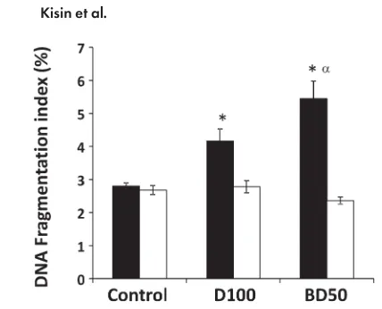
DNA Damage
The sperm chromatin structure abnormalities were
measured in the cells derived from the caudal epididymis
of mice on Days 7 and 28 postexposure to BD50 or D100
using SCSA. The levels of DNA fragmentation were
sig-nificantly increased by 1.5- and 1.9-fold in the samples
from D100- and BD50-exposed mice, respectively, only
on Day 7 post-treatments (Fig. 6). Furthermore, the level
of DNA fragmentation was found to be significantly
stronger in BD50 when compared with that of D100. No
changes in DNA fragmentation were found on Day 28
postexposure in either the BD50 or the D100 samples.
DISCUSSION
Oxidative stress has been implicated in numerous
dis-eases such as cancer, connective tissue disorders, aging,
infection, inflammation, acquired immunodeficiency
syn-drome, and male infertility [Agarwal and Said, 2005;
Benedetti et al., 2012; Guerriero et al., 2014]. Seminal
reactive oxygen species (ROS) were increased in a large
proportion of infertile men, demonstrating that oxidative
stress may be a major cause of male infertility
[Pasqua-lotto et al., 2001; Agarwal and Said, 2005]. Oxidative
stress results from the imbalance between production of
the ROS and the protective effect of the antioxidant
sys-tem responsible for their neutralization and removal. An
excess of ROS causes a pathological reaction resulting in
cell and tissue damage.
Spermatozoa are the most susceptible to the harmful
effects of ROS due to the large amounts of unsaturated
fatty acids that can be oxidized (lipid peroxidation) in
their cell membrane. The lipid peroxidation process leads
to a loss of membrane integrity and an increase in its
per-meability, inactivation of cellular enzymes, and structural
Fig. 4. Testicular testosterone (A) and serum LH (B) levels in
C57BL6 mice on Days 7 and 28 after repeated exposure to D100 or BD50 exhaust particulate. Mice were exposed to exhaust particles via pharyngeal aspiration (15 mg/mouse/day of total carbon, twice a week for 2 weeks). Black columns: 7 days after last repeated exposure to
BD50 or D100 exhaust particulate; and clear columns: 28 days after last repeated exposure to BD50 or D100 exhaust particulate. Data presented as percent of control. Means6SE (n510 mice per group). Significantly different from *controls (P<0.05) and aD100 exhaust
particulate-exposed group (P<0.05).
Fig. 5. Light micrographs of H&E stained sections from testes of mice
28 days after pharyngeal aspiration of D100 or BD50 (cumulative dose 60mg per mouse): (A) control mice, (B) mice exposed to D100, and (C) mice exposed to BD50. Sections of D100 group showed interstitial edema and occasional dystrophic seminiferous tubules with arrested
DNA damage. The overall consequence is reduced sperm
count and activity, decreased motility, abnormal
morphol-ogy, and impaired function [Walczak–Jedrzejowska et al.,
2013].
As biodiesel/diesel blend exhaust is composed of a
combination of PAHs and FAMEs combustion products,
we hypothesized that BD50 exhaust particulate could
induce adverse effects on male reproductive function
because of its ability to cause oxidative damage. As
pre-viously reported, DEPs disrupt male reproductive function
resulting in reduction of daily sperm production and
motility, increased morphological sperm abnormalities,
ultrastructural changes in Leydig cells, elevation of serum
testosterone, and reduction of LH level [Watanabe and
Oonuki, 1999; Yoshida et al., 1999; Tsukue et al., 2001,
2002; Yoshida and Takeda, 2004; Izawa et al., 2007,
2008; Li et al., 2007, 2009b, 2012; Ramdhan et al.,
2009]. Human exposure to PAHs causes modification to
male sperm quality, including morphology, concentration,
and vitality, as well as causing DNA damage in sperm,
thus affecting male reproductive function [Gaspari et al.,
2003; Meeker et al., 2007; Han et al., 2011; Jeng et al.,
2013]. Some PAHs, such as benzo(
a
)pyrene, fluoranthene,
or benzo(
ghi
)perylene (chemically reactive and
nonvola-tile), are emitted at greater levels in biodiesel than diesel
exhaust [Kado et al., 1996] and are well-known endocrine
disrupters [Raychoudhury and Kubinski, 2003].
Nitro-PAHs, such as 4-nitro-3-phenylphenol, have been shown
to induce lipid peroxidation and decrease both superoxide
dismutase and glutathione peroxidase (GSH-Px) activity
[Mi et al., 2010].
Recent publications demonstrate that biodiesel and its
blend (BD50) particles promote more pronounced
cardio-vascular alterations, as well as pulmonary and systemic
inflammation, than diesel [Brito et al., 2010]. Moreover,
Shvedova et al. [2013] and Yanamala et al. [2013]
showed that pharyngeal aspiration or inhalation exposure
to BD exhaust causes pulmonary damage accompanied by
a robust inflammatory responses and severe oxidative
stress. These studies suggest that BD aerosols might
pro-duce greater adverse effects when compared with D100
exposure. Furthermore, very recent publications, including
our own, demonstrate that biodiesel or its blends are
more mutagenic and increase formation of multinucleated
cells when compared with ultralow sulfur diesel fuel
[Ackland et al., 2007; Bunger et al., 2007; Krahl et al.,
2009; Kisin et al., 2013]. These effects are most likely
attributed to the greater levels of PAHs and nPAHs in the
exhaust, which are potent mutagens and carcinogens
[Tokiwa and Ohnishi, 1986]. To the best of our
knowl-edge, the current study is the first to report adverse effects
of biodiesel or its blends exhaust on male reproductive
function.
The major components of BD fuels are
monounsatu-rated oleic, polyunsatumonounsatu-rated linoleic, and linolenic acids
that are prone to oxidation on combustion, causing the
formation of peroxides and a variety of secondary
oxida-tion products [Song et al., 2000; Knothe, 2005]. On the
other hand, reactive metabolites of PAHs can be oxidized,
enter redox cycles, and increase the formation of ROS
[Farmer et al., 2003]. Oxidative stress is known to play a
crucial role in the etiology of defective sperm function
via a mechanism involving the induction of peroxidative
damage to the plasma membrane [Aitken et al., 2014].
An increase in intracellular ROS levels has been shown
to damage tissue and cells. Similarly, in earlier studies
[Shvedova et al., 2013; Yanamala et al., 2013], we found
greater oxidative stress after exposure to BD50 than D100
exhaust. The susceptibility of epididymal spermatozoa to
the existing oxidative stress observed in the testes is not
unexpected due to the fact that spermatozoa are uniquely
rich in polyunsaturated fatty acids [Guerriero et al.,
2014]. The latter are susceptible to free radical attack
generating lipid peroxidation chain reactions that
culmi-nate in electrophilic lipid aldehydes such as 4-HNE or
acrolein. These products of lipid peroxidation are also
capable of triggering ROS generation by sperm
mitochon-dria [Aitken et al., 2012], thus showing that oxidative
stress in spermatozoa is a self-propagating cycle that,
once initiated, will inevitably lead to oxidative damage, a
loss of functionality, and cell death [Aitken et al., 2014].
In the current study, greater accumulation of lipid
peroxi-dation products (HNE-His adduct) and depletion of a
major antioxidant, GSH, were found in the testes of
BD50 exhaust-exposed mice when compared with D100
(Fig. 1). This oxidant/antioxidant imbalance found in the
BD50-exposed mice correlated well with the impairment
of sperm characteristics demonstrated (Figs. 2, 3, and 7).
Fig. 6. DNA damage of epididymal sperm from C57BL6 mice on Days
A pronounced reduction of sperm motility and
amplifica-tion of morphological abnormalities on pulmonary
expo-sure to BD50 exhaust particulates when compared with
D100 was found throughout the postexposure time course.
This is also in agreement with previous studies [Saleh
and Agarwal, 2002; El-Demerdash et al., 2004; Aitken
and Curry, 2011; Aitken et al., 2012], showing that
increased sperm membrane lipid peroxidation inhibits
sperm progress motility, increases the percent of total
sperm abnormalities, and causes a decrease in the
fertiliz-ing potential of sperm (Fig. 7). Experiments involvfertiliz-ing
exposure of mammalian spermatozoa to a variety of ROS
using a xanthine oxidase ROS-generating system
demon-strate the vulnerability of sperm motility to oxidative
stress and identify hydrogen peroxide as the most
cyto-toxic metabolite [Awda et al., 2009; Martinez-Pastor
et al., 2009]. Moreover, it has been shown that H
2O
2inhibits activity of the glucose-6-phosphate
dehydrogen-ase (G6PD) leading to a decredehydrogen-ase in the availability of
NADPH and accumulation of oxidized/reduced
glutathi-one, thus reducing antioxidant defense properties of the
spermatozoa [Griveau et al., 1995]. Furthermore, GSH
deficiency is involved in the instability of the mid-piece
of sperm and the shape of the sperm resulting in defective
motility [Garrido et al., 2004]. In keeping with this, we
found GSH depletion and augmentation of the instability
of the mid-piece and the shape of the sperm in the BD50
exhaust-exposed samples (Figs. 1, 3, and 7).
Exposure to BD50, not D100, exhaust enhanced
testos-terone secretion in testes and decreased LH level in the
serum (Fig. 4). In addition, testicular histopathology
anal-ysis showed more prominent changes in the seminiferous
tubules after exposure to BD50 (Fig. 5). In line with our
findings, previous publications reported an increase in
tes-ticular concentration of testosterone and induction of
degeneration of tubules after exposure to
nanoparticle-rich diesel exhaust [Li et al., 2012]. These results suggest
that nanoparticles affect testosterone biosynthesis and
might have a causal role in steroidogenesis in the testis.
Testosterone is mainly synthesized in the Leydig cells of
the testes, and LH is the major stimulant of testosterone
production [Ewing et al., 1983]. However, a high level of
testosterone suppresses systemic levels of LH by a
nega-tive
feedback
loop
and
inhibits
the
release
of
gonadotropin-releasing
hormone
(GnRH)
and
conse-quently LH [Shibata et al., 2007]. Additionally, IL-1
b
,
detected in the serum of mice (Table I), also plays an
important role in the suppression of GnRH secretion
[Tomaszewska-Zaremba and Herman, 2009] and thus
altered circulatory LH levels in our study.
Apart from the influence on peroxidation of the cell
membrane lipids, ROS can also cause damage to DNA
and may lead to apoptosis through DNA fragmentation
(Fig. 7). Oxidative stress has been correlated with high
frequencies of single and double DNA strand breaks
[Aitken and Krausz, 2001]. Kemal Duru et al. [2000]
reported that exposure of the sperm to artificially
pro-duced ROS increased DNA damage in the form of
modi-fication of all bases, production of base-free sites,
deletion, DNA crosslinks, and chromosomal
rearrange-ments. High levels of ROS mediate the DNA
fragmenta-tion commonly observed in spermatozoa of infertile
males [Aitken et al., 1998; Saleh and Agarwal, 2002].
We found significantly increased degree of DNA
frag-mentation on BD50 exhaust exposure in comparison with
D100 on Day 7 post-treatment, indicating a higher
popu-lation of cells with DNA damage (Figs. 6 and 7). The
absence of DNA damage on Day 28 postexposure could
be due to induction of nucleotide excision repair.
Verhof-stad et al. [2010] exposed male mice to benzo(
a
)pyrene
to study DNA adduct kinetics in sperm and testis. The
maximum adduct level in sperm was observed at about 1
week postexposure with a subsequent decrease in the
fol-lowing 3 weeks. The use of DNA repair-deficient mice
allowed the authors to conclude that DNA damage in
spermatogonia relies on nucleotide excision repair for
maintaining its genetic integrity [Verhofstad et al., 2010].
Despite the often contradictory results from multiple
clinical and experimental studies regarding the effects of
cytokines on sperm quantity and quality, various
cyto-kines are involved in the regulation of gonad and sperm
function [Fraczek et al., 2012]. Moreover, cytokines may
play a crucial role in propagation of oxidative stress in
semen, especially during the inflammatory process (Fig.
7). It was reported that the addition of TNF-
a
, IL-1
a
, or
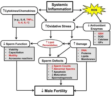
Fig. 7. A schematic representation of several biological effects associated
IL-1
b
to human spermatozoa resulted in an increase of
ROS production by a rise of sperm membrane
peroxida-tion [Buch et al., 1994]. A number of authors observed a
relationship between the increased secretion of cytokines
in the testes or semen and abnormality or fertilizing
abil-ity of sperm [Fraczek et al., 2012]. It was shown that
increased levels of TNF-
a
or IL-1
b
were related to the
quality of semen, such as sperm count, motility, and
mor-phology [Gruschwitz et al., 1996; Sanocka et al., 2003;
Al-Azemi et al., 2010]. In agreement with the previous
studies, we found that exposure to BD50 or D100 induced
upregulation of IL-1
a
, IL-1
b
, and TNF-
a
in the testes of
mice with a stronger effect of BD50 (Table II and Fig.
7). Moreover, among the various inflammatory cytokines,
TNF-
a
is the most potent inducer of apoptosis in
sperma-tozoa [Li et al., 2009a]. Furthermore, we found that
pul-monary exposure to BD50 induced enhanced systemic
inflammation measured by the release of inflammatory
mediators in the serum of mice (Table I) when compared
with D100. Importantly, accumulation of TNF-
a
, IL-1
a
,
and IL-1
b
proinflammatory cytokines was found only in
mice exposed to BD exhaust. Additionally, the level of
IL-6 was greater in B50 samples. These potent
proinflam-matory cytokines, especially IL-1
b
, are likely to play a
central pathophysiological role in the inhibition of the
neuroendocrine reproductive axis [Tomaszewska-Zaremba
and Herman, 2009]. Interestingly, only the IL-1
b
level
was significantly upregulated on Day 28 postexposure in
BD50 samples (data not shown). Most upregulated
cyto-kines found in the current study are similar to those
reported by Yanamala et al. [2013] and clearly indicate
that BD50 particles can potentiate distinct and prolonged
systemic inflammatory responses. Several authors
sug-gested an involvement of sperm apoptosis in the
impair-ment of men’s fertility [Fraczek et al., 2012]. Induction
of sperm apoptosis could be one of the mechanisms by
which proinflammatory cytokines may affect spermatozoa
during male reproductive system inflammation (Fig. 7).
Overall, our data suggest that systemic outcomes of
pulmonary exposure to respirable exhaust PM, leading to
male reproductive toxicity, were more pronounced in
BD50 when compared with D100. The chain of
patholog-ical events was realized through synergized interactions
of oxidative stress and inflammatory response culminating
in the impairment of sperm quality, functions, and DNA
damage (Fig. 7).
AUTHOR CONTRIBUTIONS
E.R.K. designed the study, completed the experiments,
analyzed the data, and prepared the manuscript draft with
important intellectual input from N.Y. and A.A.S. N.Y.
designed the study, completed the experiments, and analyzed
the data. M.T.F. completed the experiments and collected
the data. D.W.G. and M.R.S. performed histopathological
evaluation. A.D.B. collected the diesel/biodiesel exhaust
par-ticles. V.E.K. designed the study. A.A.S. coordinated the
study. All authors approved the final manuscript.
REFERENCES
Ackland ML, Zou LD, Freestone D, de Waasenburg SV, Michalczyk AA. 2007. Diesel exhaust particulate matter induces multinu-cleate cells and zinc transporter-dependent apoptosis in human airway cells. Immunol Cell Biol 85:617–622.
Agarwal A, Said TM. 2005. Oxidative stress, DNA damage and apopto-sis in male infertility: A clinical approach. BJU Int 95:503–507. Agarwal A, Saleh RA. 2002. Role of oxidants in male infertility:
Ration-ale, significance, and treatment. Urol Clin North Am 29:817–827. Aitken RJ, Curry BJ. 2011. Redox regulation of human sperm function: From the physiological control of sperm capacitation to the etiol-ogy of infertility and DNA damage in the germ line. Antioxid Redox Signal 14:367–381.
Aitken RJ, Krausz C. 2001. Oxidative stress, DNA damage and the Y chromosome. Reproduction 122:497–506.
Aitken RJ, Gordon E, Harkiss D, Twigg JP, Milne P, Jennings Z, Irvine DS. 1998. Relative impact of oxidative stress on the functional competence and genomic integrity of human spermatozoa. Biol Reprod 59:1037–1046.
Al-Azemi M, Omu FE, Kehinde EO, Anim JT, Oriowo MA, Omu AE. 2010. Lithium protects against toxic effects of cadmium in the rat testes. J Assist Reprod Genet 27:469–476.
Aitken RJ, Gibb Z, Mitchell LA, Lambourne SR, Connaughton HS, De Iuliis GN. 2012. Sperm motility is lost in vitro as a consequence of mitochondrial free radical production and the generation of electrophilic aldehydes but can be significantly rescued by the presence of nucleophilic thiols. Biol Reprod 87:110.
Aitken RJ, Smith TB, Jobling MS, Baker MA, De Iuliis GN. 2014. Oxida-tive stress and male reproducOxida-tive health. Asian J Androl 16:31–38. Awda BJ, Mackenzie-Bell M, Buhr MM. 2009. Reactive oxygen species
and boar sperm function. Biol Reprod 81:553–561.
Benedetti S, Tagliamonte MC, Catalani S, Primiterra M, Canestrari F, De Stefani S, Palini S, Bulletti C. 2012. Differences in blood and semen oxidative status in fertile and infertile men, and their rela-tionship with sperm quality. Reprod Biomed Online 25:300–306. Birch ME. 2004. Monitoring of diesel particulate exhaust in the
work-place. In: Schlecht PC, O’Connor PF, editors. NIOSH Manual of Analytical Methods (NMAM), 4th ed. Publication No. 2003-154. Cincinnati, OH: U.S. Department of Health and Human Services, Centers for Disease Control and Prevention, National Institute for Occupational Safety and Health, DHHS (NIOSH). Chapter Q, pp 229–259.
Brito JM, Belotti L, Toledo AC, Antonangelo L, Silva FS, Alvim DS, Andre PA, Saldiva PHN, Rivero DHRF. 2010. Acute cardiovas-cular and inflammatory toxicity induced by inhalation of diesel and biodiesel exhaust particles. Toxicol Sci 116:67–78.
Buch JP, Kolon TF, Maulik N, Kreutzer DL, Das DK. 1994. Cytokines stimulate lipid membrane peroxidation of human sperm. Fertil Steril 62:186–188.
Bunger J, Krahl J, Munack A, Ruschel Y, Schroder O, Emmert B, West G, Muller M, Hallier E, Bruning T. 2007. Strong mutagenic effects of diesel engine emissions using vegetable oil as fuel. Arch Toxicol 81:599–603.
B€unger J, Krahl J, Schr€oder O, Schmidt L, Westphal GA. 2012. Poten-tial hazards associated with combustion of bio-derived versus petroleum-derived diesel fuel. Crit Rev Toxicol 42:732–750. El-Demerdash FM, Yousef MI, Kedwany FS, Baghdadi HH. 2004.
Evenson DP, Melamed MR. 1983. Rapid analysis of normal and abnor-mal cell types in human semen and testis biopsies by flow cytometry. J Histochem Cytochem 31:248–253.
Ewing LL, Wing TY, Cochran RC, Kromann N, Zirkin BR. 1983. Effect of luteinizing hormone on Leydig cell structure and testosterone secretion. Endocrinology 112:1763–1769.
Farmer PB, Singh R, Kaur B, Sram RJ, Binkova B, Kalina I, Popov TA, Garte S, Taioli E, Gabelova A, et al. 2003. Molecular epidemiol-ogy studies of carcinogenic environmental pollutants. Effects of polycyclic aromatic hydrocarbons (PAHs) in environmental pol-lution on exogenous and oxidative DNA damage. Mutat Res 544: 397–402.
Fraczek M, Czernikiewicz A, Kurpisz M. 2012. Cytokines and oxidative stress in the germ line. In: Agarwal A, Aitken RJ, Alvarez JG, editors. Oxidative Stress in Applied Basic Research and Clinical Practice. Studies on Men’s Health and Fertility. Springer Scien-ce1Business Media, LLC. New York, NY, pp 179–205. Garrido N, Meseguer M, Alvarez J, Simon C, Pellicer A, Remohı J.
2004. Relationship among standard semen parameters, glutathi-one peroxidase/glutathiglutathi-one reductase activity, and mRNA expres-sion and reduced glutathione content in ejaculated spermatozoa from fertile and infertile men. Fertil Steril 82 (Suppl 3):1059– 1066.
Garshick E, Laden F, Hart JE, Rosner B, Smith TJ, Dockery DW, Speizer FE. 2004. Lung cancer in railroad workers exposed to diesel exhaust. Environ Health Perspect 12:1539–1543.
Gaspari L, Chang SS, Santella RM, Garte S, Pedotti P, Taioli E. 2003. Polycyclic aromatic hydrocarbon-DNA adducts in human sperm as a marker of DNA damage and infertility. Mutat Res 535:155– 160.
Griveau JF, Dumont E, Renard P, Callegari JP, Le Lannou D. 1995. Reactive oxygen species, lipid peroxidation and enzymatic defence systems in human spermatozoa. J Reprod Fertil 103:17– 26.
Gruschwitz MS, Brezinschek R, Brezinschek HP. 1996. Cytokine levels in the seminal plasma of infertile males. J Androl 17:158–163. Guerriero G, Trocchia S, Abdel-Gawad FK, Ciarci GA. 2014. Roles of
reactive oxygen species in the spermatogenesis regulation. Front Endocrinol (Lausanne) 22:56.
Han X, Zhou N, Cui Z, Ma M, Li L, Cai M, Li Y, Lin H, Li Y, Ao L, et al. 2011. Association between urinary polycyclic aromatic hydrocarbon metabolites and sperm DNA damage: A population study in Chongqing, China. Environ Health Perspect 119:652– 657.
Hazari MS, Haykal-Coates N, Winsett DW, Krantz QT, King C, Costa DL, Farraj AK. 2011. TRPA1 and sympathetic activation contrib-ute to increased risk of triggered cardiac arrhythmias in hyperten-sive rats exposed to diesel exhaust. Environ Health Perspect 119: 951–957.
Holgate ST, Sandstrom T, Frew AJ, Stenfors N, Nordenhall C, Salvi S, Blomberg A, Helleday R, Soderberg M. 2003. Health effects of acute exposure to air pollution. I. Healthy and asthmatic subjects exposed to diesel exhaust. Res Rep Health Eff Inst 112:1–30 (discussion 51–67).
Izawa H, Kohara M, Watanabe G, Taya K, Sagai M. 2007. Effects of diesel exhaust particles on the male reproductive system in strains of mice with different aryl hydrocarbon receptor respon-siveness. J Reprod Dev 53:1191–1197.
Izawa H, Kohara M, Aizawa K, Suganuma H, Inakuma T, Watanabe G, Taya K, Sagai M. 2008. Alleviative effects of quercetin and onion on male reproductive toxicity induced by diesel exhaust particles. Biosci Biotechnol Biochem 72:1235–1241.
Jeng HA, Pan CH, Chao MR. 2013. 1-Hydroxypyrene as a biomarker for assessing the effects of exposure to polycyclic aromatic hydrocarbons on semen quality and sperm DNA integrity.
J Environ Sci Health A Tox Hazard Subst Environ Eng 48:152– 158.
Kado NY, Okamoto RA, Kuzmicky PA. 1996. Chemical and bioassay analyses of diesel and biodiesel particulate matter: pilot study. Final report for The U.S. Department of Energy and The Renew-able Energy Report Library by Department of Environmental Toxicology, University of California, Davis.
Kemal Duru N, Morshedi M, Oehninger S. 2000. Effects of hydrogen peroxide on DNA and plasma membrane integrity of human sper-matozoa. Fertil Steril 74:1200–1207.
Kisin E, Shi X, Keane M, Bugarski A, Shvedova AA. 2013. Mutagenec-ity of biodiesel diesel exhaust particles and the effect of engine operating conditions. J Environ Eng Ecol Sci 2:3.
Knothe G. 2005. Dependence of biodiesel fuel properties on the structure of fatty acid alkyl esters. Fuel Process Technol 86:1059–1070. Knothe G, Krahl J, Van Gerpen JH. 2010. The Biodiesel Handbook, 2nd
ed. AOCS Press, Urbana, Il.
Krahl J, Knothe G, Munack A, Ruschel Y, Schroder O, Hallier E, Westphal G, Bunger J. 2009. Comparison of exhaust emissions and their mutagenicity from the combustion of biodiesel, vegeta-ble oil, gas-to-liquid and petrodiesel fuels. Fuel 88:1064–1069. Li C, Taneda S, Suzuki AK, Furuta C, Watanabe G, Taya K. 2007.
Effects of 3-methyl-4-nitrophenol in diesel exhaust particles on the regulation of testicular function in immature male rats. J Androl 28:252–258.
Li MW, Mruk DD, Lee WM, Cheng CY. 2009a. Cytokines and junction restructuring events during spermatogenesis in the testis: An emerg-ing concept of regulation. Cytokine Growth Factor Rev 20:329–338. Li C, Taneda S, Taya K, Watanabe G, Li X, Fujitani Y, Ito Y, Nakajima T, Suzuki AK. 2009b. Effects of inhaled nanoparticle-rich diesel exhaust on regulation of testicular function in adult male rats. Inhal Toxicol 21:803–811.
Li C, Li X, Jigami J, Hasegawa C, Suzuki AK, Zhang Y, Fujitani Y, Nagaoka K, Watanabe G, Taya K. 2012. Effect of nanoparticle-rich diesel exhaust on testosterone biosynthesis in adult male mice. Inhal Toxicol 24:599–608.
Liu YY, Lin TC, Wang YJ, Ho WL. 2009. Carbonyl compounds and toxicity assessments of emissions from a diesel engine running on biodiesels. J Air Waste Manag Assoc 59:163–171.
Martınez-Pastor F, Aisen E, Fernandez-Santos MR, Esteso MC, Maroto-Morales A, Garcıa-Alvarez O, Garde JJ. 2009. Reactive oxygen species generators affect quality parameters and apoptosis markers differently in red deer spermatozoa. Reproduction 137:225–235. Meeker JD, Godfrey-Bailey L, Hauser R. 2007. Relationships between
serum hormone levels and semen quality among men from an infertility clinic. J Androl 28:397–406.
Mermelstein R, Kiriazides DK, Butler M, McCoy EC, Rosenkranz HS. 1981. The extraordinary mutagenicity of nitropyrenes in bacteria. Mutat Res 89:187–196.
Mi Y, Zhang C, Li C, Taneda S, Watanabe G, Suzuki AK, Taya K. 2010. Quercetin attenuates oxidative damage induced by treat-ment of embryonic chicken spermatogonial cells with 4-nitro-3-phenylphenol in diesel exhaust particles. Biosci Biotechnol Bio-chem 74:934–938.
Mills NL, Tornqvist H, Robinson SD, Gonzalez M, Darnley K, MacNee W, Boon NA, Donaldson K, Blomberg A, Sandstrom T, et al. 2005. Diesel exhaust inhalation causes vascular dysfunction and impaired endogenous fibrinolysis. Circulation 112:3930–3936. Mills NL, Tornqvist H, Gonzalez MC, Vink E, Robinson SD, Soderberg
S, Boon NA, Donaldson K, Sandstrom T, Blomberg A, et al. 2007. Ischemic and thrombotic effects of dilute diesel-exhaust inhalation in men with coronary heart disease. N Engl J Med 357:1075–1082.
administered diesel exhaust particles in rats. Am J Physiol Lung Cell Mol Physiol 292:L664–L670.
Nemmar A, Dhanasekaran S, Yasin J, Ba-Omar H, Fahim MA, Kazzam EE, Ali BH. 2009. Evaluation of the direct systemic and cardio-pulmonary effects of diesel particles in spontaneously hyperten-sive rats. Toxicology 262:50–56.
NMAM. 2003. NIOSH method 5040 update. In: Schlecht PC, O’Connor PF, editors. NIOSH Manual of Analytical Methods (NMAM), 4th ed. Third Supplement, Publication No. 2003-154. Cincinnati, OH: U.S. Department of Health and Human Services, Centers for Dis-ease Control and Prevention, National Institute for Occupational Safety and Health, DHHS (NIOSH), pp 94–113.
Ohe T. 1984. Mutagenicity of photochemical reaction products of poly-cyclic aromatic hydrocarbons with nitrite. Sci Total Environ 39: 161–175.
Pasqualotto FF, Sharma RK, Kobayashi H, Nelson DR, Thomas AJ, Agarwal A. 2001. Oxidative stress in normospermic men under-going infertility evaluation. J Androl 22:316–322.
Pereira MA, Sabharwal PS, Gordon L, Wyrobek AJ. 1981. The effect of diesel exhaust on sperm-shape abnormalities in mice. Environ Int 5:459–460.
Peretz AJ, Kaufman D, Trenga CA, Allen J, Carlsten C, Aulet MR, Adar SD, Sullivan JH. 2008. Effects of diesel exhaust inhalation on heart rate variability in human volunteers. Environ Res 107: 178–184.
Polyzos A, Schmid TE, Pi~na-Guzman B, Quintanilla-Vega B, Marchett FI. 2009. Differential sensitivity of male germ cells to main-stream and sidemain-stream tobacco smoke in the mouse. Toxicol Appl Pharmacol 237:298–305.
Pronk A, Coble J, Stewart PA. 2009. Occupational exposure to diesel engine exhaust: A literature review. J Expo Sci Environ Epide-miol 19:443–457.
Purcell DL, McClure BT, McDonald J, Basu HN. 1996. Transient testing of soy methyl ester fuels in an indirect injection, compression ignition engine. J Am Oil Chem Soc 73:381–388.
Ramdhan DH, Ito Y, Yanagiba Y, Yamagishi N, Hayashi Y, Li C, Taneda S, Suzuki AK, Watanabe G, Taya K, et al. 2009. Nano-particle-rich diesel exhaust may disrupt testosterone biosynthesis and metabolism via growth hormone. Toxicol Lett 191:103–108. Raychoudhury SS, Kubinski D. 2003. Polycyclic aromatic
hydrocarbon-induced cytotoxicity in cultured rat Sertoli cells involves differ-ential apoptotic response. Environ Health Perspect 111:33–38. Rivedal E, Myhre O, Sanner T, Eide I. 2003. Supplemental role of the
Ames mutation assay and gap junction intercellular communica-tion in studies of possible carcinogenic compounds from diesel exhaust particles. Arch Toxicol 77:533–542.
Rivero DHRF, Soares SRC, Lorenzi G, Saiki M, Godleski JJ, Antonangelo L, Dolhnikoff M, Saldiva PHN. 2005. Acute cardio-pulmonary alterations induced by fine particulate matter of Sao Paulo, Brazil. Toxicol Sci 85:898–905.
Saleh RA, Agarwal A. 2002. Oxidative stress and male infertility: From research bench to clinical practice. J Androl 23:737–752. Sanocka D, Jedrzejczak P, Szumała-Kaekol A, Fraczek M, Kurpisz M.
2003. Male genital tract inflammation: The role of selected inter-leukins in regulation of pro-oxidant and antioxidant enzymatic substances in seminal plasma. J Androl 24:448–455.
Sawyer K, Mundandhara S, Ghio AJ, Madden MC. 2010. The effects of ambient particulate matter on human alveolar macrophage oxida-tive and inflammatory responses. J Toxicol Environ Health A 73: 41–57.
Shibata M, Friedman RL, Ramaswamy S, Plant TM. 2007. Evidence that down regulation of hypothalamic KiSS-1 expression is involved in the negative feedback action of testosterone to
regu-late luteinising hormone secretion in the adult male rhesus mon-key (Macaca mulatta). J Neuroendocrinol 19:432–438.
Shvedova AA, Kisin ER, Mercer R, Murray AR, Johnson VJ, Potapovich AI, Tyurina YY, Gorelik O, Arepalli S, Schwegler-Berry D, et al. 2005. Unusual inflammatory and fibrogenic pul-monary responses to single-walled carbon nanotubes in mice. Am J Physiol Lung Cell Mol Physiol 289:L698–L708.
Shvedova AA, Yanamala N, Murray AR, Kisin ER, Khaliullin T, Hatfield MK, Tkach AV, Krantz QT, Nash D, King C, et al. 2013. Oxidative stress, inflammatory biomarkers, and toxicity in mouse lung and liver after inhalation exposure to 100% biodiesel or petroleum diesel emissions. J Toxicol Environ Health A 76: 907–921.
Silverman D, Samanic CM, Lubin JH, Blair AE, Stewart PA, Vermeulen R, Coble JB, Rothman N, Schleiff PL, Travis WD, et al. 2012. The diesel exhaust in miners study: A nested case–control study of lung cancer and diesel exhaust. J Natl Cancer Inst 104:855– 868.
Song JH, Fujimoto K, Miyazawa T. 2000. Polyunsaturated (n23) fatty acids susceptible to peroxidation are increased in plasma and tis-sue lipids of rats fed docosahexaenoic acid-containing oils. J Nutr 130:3028–3033.
Tokiwa H, Ohnishi Y. 1986. Mutagenicity and carcinogenicity of nitroarenes and their sources in the environment. Crit Rev Toxicol 17:23–60. Tomaszewska-Zaremba D, Herman A. 2009. The role of immunological
system in the regulation of gonadoliberin and gonadotropin secre-tion. Reprod Biol 9:11–23.
Tornqvist HN, Mills L, Gonzalez M, Miller MR, Robinson SD, Megson IL, MacNee W, Donaldson K, Soderberg S, Newby DE, et al. 2007. Persistent endothelial dysfunction in humans after diesel exhaust inhalation. Am J Respir Crit Care Med 176:395–400. Tsukue N, Toda N, Tsubone H, Sagai M, Jin WZ, Watanabe G, Taya K,
Birumachi J, Suzuki AK. 2001. Diesel exhaust (DE) affects the regulation of testicular function in male Fischer 344 rats. J Toxicol Environ Health A 63:115–126.
Tsukue N, Tsubone H, Suzuki AK. 2002. Diesel exhaust affects the abnormal delivery in pregnant mice and the growth of their young. Inhal Toxicol 14:635–651.
Verhofstad N, van Oostrom CT, van Benthem J, van Schooten FJ, van Steeg H, Godschalk RW. 2010. DNA adduct kinetics in repro-ductive tissues of DNA repair proficient and deficient male mice after oral exposure to benzo(a)pyrene. Environ Mol Mutagen 51: 123–129.
Walczak–Jedrzejowska R, Karol Wolski J, Slowikowska–Hilczer J. 2013. The role of oxidative stress and antioxidants in male fertil-ity. Cent European J Urol 66:60–67.
Watanabe N, Oonuki Y. 1999. Inhalation of diesel engine exhaust affects spermatogenesis in growing male rats. Environ Health Perspect 107:539–544.
Watkinson WP, Campen MJ, Costa DL. 1998. Cardiac arrhythmia induc-tion after exposure to residual oil fly ash particles in a rodent model of pulmonary hypertension. Toxicol Sci 41:209–216. Yanamala N, Hatfield MK, Farcas MT, Schwegler-Berry D, Hummer
JA, Shurin MR, Birch ME, Gutkin DW, Kisin ER, Kagan VE, et al. 2013. Biodiesel versus diesel exposure: Enhanced pulmo-nary inflammation, oxidative stress, and differential morphologi-cal changes in the mouse lung. Toxicol Appl Pharmacol 272: 373–383.
Yoshida S, Sagai M, Oshio S, Umeda T, Ihara T, Sugamata M, Sugawara I, Takeda K. 1999. Exposure to diesel exhaust affects the male reproductive system of mice. Int J Androl 22:307–315. Yoshida S, Takeda K. 2004. The effect of diesel exhaust on murine