Eficient Strategies for Elimination of Phenolic Compounds
During DNA Extraction from Roots of
Pistacia vera
L
.
Saeideh Rajaei1*), Rana Sabagh Farshi2), Maryam Moazzam Jazi2) and Seyed Mahdi Seyedi2)
1) Department of Industrial and Environmental Biotechnology
National Institute of Genetic Engineering and Biotechnology (NIGEB), Iran 2) Department of Agricultural Biotechnology
National Institute of Genetic Engineering and Biotechnology (NIGEB), Iran *) Corresponding author E-mail: [email protected]
Received: November 11, 2015 /Accepted: June 2, 2017
ABSTRACT
Optimization of DNA extraction protocols for plant tissues and including endophytic microorganisms is a critical step of advanced plant-microbe interaction in agricultural studies. Pistachio (Pistacia vera L.) root tissue contains high levels of polyphenols have been known as major extract contaminants and inhibitors of enzymatic activities
during ampliication. The present study aimed
to develop reliable strategies to purify DNA from Pistachio root samples. Inhibiting substances were removed from DNA through a process including
extraction with hot detergent contains SDS-Tris-EDTA, AlNH4(SO4)2.12H2O as chemical coagulating
factor and CTAB-NaCl. Following typically organic
extraction/alcohol precipitation, denaturing agarose electrophoresis performed to purify probable remain
contaminants. The puriied DNA was enough free of
polyphenols based upon loss of color and spectral
quality (260/230>1.6) and eficiently ampliied during
polymerase chain reaction particularly in the present
of GC-clamp primers. This method proved well with
detection of Glomus sp. (arbuscular mycorrhiza fungi) associated with Pistacia vera L. using denaturing gradient gel electrophoresis (DGGE).
Keywords: Arbuscular Mycorrhiza (AM); chemical coagulation; DNA extraction; polyphe-nols; Pistacia vera L.
INTRODUCTION
Preparation of high quality genomic DNA from agricultural plants is a critical stage of most genomic analyses studies toward plant genetic improvement and understanding plant-microbe interaction. Plant roots have been considered as the settlement of soil endophytic microorganisms some of which enhance nutrients availability and plant growth, improve the plant ability to tolerate abiotic (drought, salinity, etc.)
and biotic stress (plant pathogens) nevertheless, most of the endophyte-plant relationships are not well
understood (Hardoim, van Overbeek, & van Elsas, 2008; Porras-Alfaro et al., 2008; Reinhold-Hurek & Hurek, 2011; Bulgarelli, Schlaeppi, Spaepen, Ver Loren van Themaat, & Schulze-Lefert, 2013; Nair & Padmavathy, 2014; Tkacz & Poole, 2015). Direct
isolation of DNA from various part of plant tissues is a preliminary step for studying many associating and symbiosis relationship especially which types are not culturable in experimental culture media (Stewart, 2012).
Pistachio (Pistacia vera L.) is the key of horticultural plant in arid regions of Iran which currently includes 10 percent of non-petroleum export value however, excessive soil salinity as a current major ecological and agronomical problem
has signiicantly reduced productivity of pistachio
trees. Several studies have investigated the role of endophytic microorganisms -for instance arbuscular mycorrhiza (AM)- in protection of plants against salt stress by various mechanisms (Marulanda, Azcón,
& Ruiz-Lozano, 2003; Marulanda, Porcel, Barea, & Azcón, 2007; Wu, Zou, Xia, & Wang, 2007; Wang &
Liu 2001).
In order to study the colonized-mycorrhiza
fungi with Pistachio roots, the irst step was isolation
of DNA from root tissues. AM fungi is being an obligate symbiont that cannot be cultured in the absence of a suitable host therefore, direct extraction of DNA from root tissue and analysis of fungal ribosomal DNA sequence was the reliable way to study AM communities. In this study DNA extraction from Pistachio roots via commercial DNA extraction kits resulted in dark color DNA with low spectral quality
(Table 1). The problem was relevant to dark
brown-colored compounds in root cells, called polyphenols substances that have a similar size and charge with DNA, tending to co-precipitate with extracted DNA,
Cite this as: Rajaei, S., Farshi, R. S., Jazi, M. M., & Seyedi, S. M. (2017). Eficient strategies for elimination of phenolic compounds during DNA extraction from roots of Pistacia vera L. AGRIVITA Journal of Agricultural Science, 39(3), 279–287. http://doi.org/10.17503/agrivita.v39i3.734
interfering with downstream enzymatic applications
(Schrader, Schielke, Ellerbroek, & Johne, 2012; Borse, Joshi, & Chaphalkar, 2011; Healey, Furtado, Cooper, & Henry, 2014). Once the plant cells are
broken apart, polyphenols become exposed to oxygen and reacted with polyphenol oxidases. Polyphenol oxidation products covalently bind to the phosphate backbone of nucleic acids, making them forcefully impossible to be removed (Manoj,
Tushar, & Sushama, 2007; Zhang & Stewart, 2000; Borse, Joshi, & Chaphalkar, 2011). In order to
nucleic acid extraction, selecting very young leaves or cotyledons has been recommended to reduce trouble of polyphenols however, for some studies like gene expression in a certain part of the plant or endophytic investigation, the conditions are not ideal furthermore considering age and type of the plant tissue (roots, leaves or stems), the content of polyphenol compounds/secondary metabolite (as DNA contaminants) will be various. In that case, the purity of extracted DNA is out of the power of commercial DNA extraction kits which are usually mentioned in the user instruction booklet as troubleshooting. Under these circumstances, it should be developed a particular strategy or improve available isolation protocols for elimination of polyphenol compounds from DNA. Common procedures involve using antioxidants (Ascorbic acid) and certain polymers (polyvinylpyrrolidone
(PVP) and polyvinylpolypyrrolidone (PVPP) for
removing phenolic compounds in leaf tissues
(Peterson, Boehm, & Stack, 1997; Porebski, Bailey, & Baum, 1997; Khanuja, Shasany, Darokar, & Kumar, 1999; Carrier et al., 2011; Sahu, Thangaraj, & Kathiresan, 2012). PVPP-40 has addressed to
remove polyphenols along with high concentration of NaCl, resulting in polyphenol-free DNA. Ascorbic acid was also being used for isolation of nucleic
acids in polyphenol rich plants (Bielski, 1982). In
the current study with regard to high concentration of polyphenol compounds in the root of Pistachio, it was suggested a reliable DNA extraction and
puriication method to eliminate polyphenol so
that being well-suited for sensitive downstream reactions.
MATERIALS AND METHODS
Preparing Soil Samples
Pistachio (Pistacia vera L.) roots were collected from Pistachio Orchard in Rafsanjan (Kerman province, south-eastern of Iran). After
removing soil particles, roots were sterilized by 10 % sodium hypochlorite for 10 min and grinded in the liquid nitrogen.
DNA Extraction and Puriication
Two grams of grinded root was mixed with 4 ml of pre-warmed extraction buffer (65 °C) included 100 mM Tris-HCl (pH 8.5-9), 25mM sodium EDTA (pH 8.5), 2 % SDS and 50-mM AlNH4(SO4)2.12H2O
(adjusted pH to 8.5 with 1 M NaOH) and incubated for 2 hours at 65 °C, inverting every 15 minutes through incubation. Then, the mixture was centrifuged at 1500 xg for 5 minutes and 2 ml of pre-warmed (37 °C) 5M NaCl-5% CTAB (Cetyl trimethyl
ammonium bromide) was added to the supernatant
and incubated for 10 minutes at 65 °C. The
temperature was necessary to assure high yields of
DNA, due to lower solubility of CTAB salts bellow 50 °C (Abu Almakarem, Heilman, Conger, Shtarkman, & Rogers, 2012). Incubated mixture was extracted
two times with equal volume of chloroform-isoamyl
alcohol (24:1), followed by centrifugation at 2500 xg, for 15 minutes at room temperature. Upper
phase was carefully recovered and precipitated with 0.6 volumes of isopropanol and incubated at
-20 °C for at least 2 hours (or one overnight). The
DNA was precipitated by centrifugation at 26000 xg
for 15 minutes at 4 °C. The DNA pellet was washed
using a washing solution (7 vol absolute ethanol, 2
vol ddH2O and 1 vol ammonium acetate 3M) and
centrifuged at 26000 xg for 15 minutes. DNA pellet was diluted in sterile ddH2O and puriied by loading
in 2 % agarose gel containing 1X Tris-acetate-EDTA (TAE) and equal volume formamide. Following
electrophoresis and staining with GelRed, the bands containing the large molecular weight DNA were excised then transferred to a sterile tube and agarose solubilizing buffer was added (agarose Gel DNA Extraction Kit (Roche) regarding to the kit instructions). Melted agarose mixed by biding buffer and precipitated by washing buffer. By discarding solution via centrifuge at 16000 xg for 30 seconds, DNA pellet resolved in sterile water.
For extracting DNA using plant DNA extraction kit, 100 mg grinded root mixed with lysis and protein precipitation buffers. Lysate was centrifuged to
remove residual debris. The clear supernatant
Polymerase Chain Reaction and DGGE Fingerprinting
The ampliication potential of the extracted
DNA has evaluated using PCR with universal primers including, 18S rDNA universal primers for plant
(F:5-GTACAAAGGGCAGGGACGTA-3 and R:5-GGAAGGCTGAGGCAATAACA-3 (Rajaei, Niknam, Seyedi, Ebrahimzadeh, & Razavi, 2009)) and 18SrDNA primers for AM fungi (NS31-GC: 5-GC(2)
C(3)G(4)CG(2)C(4)G(3)CG(1)G(3)CG(1)G(4)
CACG(1)G(4)TTGGAGGGCAGTCTGGTGCC(1)-3 and Glo1: 5-GCCTGCTTTAAACACTCTA-3 (Liang et al., 2008)). PCR ampliications were performed
using Bio-Rad thermal cycler as following; 2
minutes at 94 °C for initial denaturation, 30 cycles with denaturation for 45 seconds at 94 °C, annealing for 45 seconds at 60 °C, and extension for 45 minutes at 72 °C. A inal extension step at 72 °C for 15 minutes was conducted to allow
complete extension. PCR products were visualized by running the agarose electrophoresis. Denaturing
gradient gel electrophoresis was performed for 25
µl of NS31-GC and Glo1 PCR products on a Bio-Rad DCode system (Bio-Bio-Rad, Mississauga, Ont.) described by Lawrence et al. (2004).
Hybridization
Extracted DNAs were denatured and then
spotted onto a Hybond nylon membrane (Roche). The membranes were hybridized with DNA
probes under the high-stringency prehybridization,
hybridization and washing conditions at 65 °C. The
probes were labeled with the digoxigenin (DIG), and detected using the DIG DNA Labeling and Detection Kit (Roche), according to the manufacturer’s instructions.
RESULTS AND DISCUSSION
The irst step of DNA extraction was to break
up the tissues and cells to access DNA. Woody Pistachio roots seemed tougher and hearty during the bead beating and they were ground using liquid nitrogen and a mortar and pestle instead. Following breaking the cells via grinding in the liquid nitrogen, products of polyphenols oxidation which had a high
afinity for the nucleic acid, covalently bound to
DNA in the extract mixture; adding the alcohol gave a brown color and viscous feature to the extract
mixture and inally precipitated DNA. Polyphenols
are most common PCR inhibitors which require the
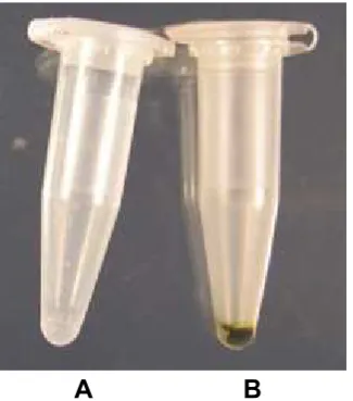
extensive clean-up steps to be used in ampliication process (Moazzam Jazi, Rajaei, & Seyedi, 2015). In the present study, AlNH4(SO4)2.12H2O as a major component for phenolic compounds precipitation was added to the extraction buffer in order to chemical coagulation of phenolic compounds during lysis step. Fig. 1 shows the color of extracted and precipitated DNA using current protocol (A) and
DNA extraction kit (B). The clear DNA was obtained
using chemical coagulation method (Fig. 1A), while DNA was dark brown when the extraction was conducted using plant tissue extraction kit (Fig. 1B).
A
B
Direct application of solid AlNH4(SO4)2.12H2O
in the extraction buffer (inal concentration of 50
mM) remarkably improved the quality and quantity
of extracted DNA (Table 1). The quantity of isolated DNA with this modiication was approximately
twice as that of obtained using 100mM solution
of AlNH4(SO4)2.12H2O (Fig. 2) which could be attributed to the chaotropic effect and DNA loss
in higher concentrations of AlNH4(SO4)2.12H2O
(Braid, Daniels, & Kitts, 2003; Bakken & Rostegård, 2006; Gadkar & Filion, 2013). Addition of solid AlNH4(SO4)2.12H2O directly to the lysis buffer also
led to pH decline to 4; therefore, following addition of AlNH4(SO4)2.12H2O adjusting pH to 9 was indispensable.
Agarose gel pattern of root genomic DNA
(stained with Ethidium bromide) obtained by proposed method has been shown in Fig. 3.In this protocol, chloroform and isoamyl alcohol (24:1, v/v) was used for the denaturation of contaminating proteins. Phenol is a very hazardous chemical which usually used for removing proteins (Liao
et al., 2004). Phenol-based method intensiied
production of brown color in DNA pellet (Chang,
Puryear, & Cairney, 1993; Moazzam Jazi, Rajaei, & Seyedi, 2015), therefore it was important to ignore
the application of phenol.
The quality and quantity of the extracted
DNA was determined using a Nano Drop
spectrophotometer and shown in Table 1. The yield of extracted-puriied DNA using proposed protocol varied from 48.6 to 293.4 µg µl-1. Since root Table 1. The quality and quantity of extracted DNA from root of Pistacia vera L. (using the Nanodrop)
Method Root sample DNA (μg/μl) a 260/280 260/230
Present PR1 88.2± 0.19 1.31± 0.025 1.65±0.045
PR2 163.2± 0.17 1.56± 0.020 1.55± 0.038
PR3 48.6± 0.27 1.44± 0.042 1.80± 0.026
PR4 293.4± 0.35 1.62± 0.086 1.64± 0.074
PR5 49.8± 0.20 1.73± 0.044 1.57± 0.069
DNA extraction kits PR1 44.2± 0.13 1.62± 0.087 0.10± 0.016b
PR2 80.5± 0.10 1.83± 0.039
-PR3 12.7± 0.05 1.53± 0.011 0.25± 0.021
PR4 179.1± 0.07 1.75± 0.045 0.02± 0.040
PR5 26.5± 0.09 1.81± 0.020 0.15± 0.036
Remarks: a Mean± standard error (n=3); b Dilution ratio1:50
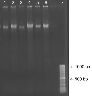
Fig. 2. Agarose electrophoresis of extracted DNA from roots of Pistacia vera L. under 100 (1, 3 and 5) and
50 mM (2, 4 and 6) concentration of AlNH4(SO4)2.12H2O in extraction buffer ← 1000 pb
← 500 bp
samples were prepared from mature trees in the
ield, they were very woody and tough containing very small quantities of DNA because of ligniied cells dominance in wood. The present protocol
could successfully extract DNA from samples however, the aim of this study was not focusing on the yield of DNA, in fact the main objective was removing polyphenolic compounds which precipitated concomitant with DNA through nucleic acid extraction. Polyphenolic contamination of DNA was determined by A260/A230 ratio, the ratio closed to 2 or > 2 showed a very low or no contamination
in DNA (Kasem, Rice, & Henry, 2008; Rodrigues et al., 2007). The A260/A230 ratio of extracted DNAs by introduced method varied between
1.55-1.80 whereas the ratio for extracted DNAs using
commercial kit was very low between 0.02-0.25,
indicating the presence of organic contaminants
(Table 1). Healey, Furtado, Cooper, & Henry (2014)
tried to raise the quality of extracted DNA from
recalcitrant plant species (Corymbia and Coffea) by
adding β-mercaptoethanol to a CTAB based method and the centrifugation step after 65 °C incubation. In
this investigation the high concentration of phenolic compounds accumulated in pistachio roots was
eliminated using AlNH4(SO4)2.12H2O during DNA
extraction. Similarly, Braid, Daniels, & Kitts (2003) reported that adding AlNH4(SO4)2.12H2O to the
DNA extraction buffer signiicantly declined the
concentration of humic inhibitors with a minimal loss in the quantity of soil DNA.
Prepared DNA by the present method and
DNA extraction kit were ampliied using a standard
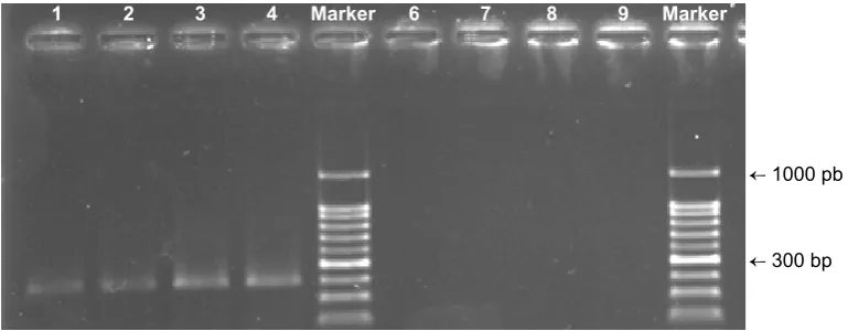
PCR protocol. Fig. 4 shows agarose electrophoresis of PCR products includes 18S rDNA fragments.
Ampliication of the 18S rDNA ribosomal subunit of
plants was not possible in the presence of extracted DNA template via DNA extraction kit while the sharp PCR bands were gained when the DNA had been extracted using the chemical coagulating method.
Fig. 4. Agarose electrophoresis of PCR products (300 bp) extracted DNA as template and 18S primers for plant. PCR products resulted from isolated DNA using the coagulating extracted method and
plant-tissue extraction kit were illustrated in the lanes of 1-4 and lanes of 6-9, respectively
← 1000 pb
← 300 bp 1 2 3 4 Marker 6 7 8 9 Marker
Fig. 3. Agarose electrophoresis of extracted DNA from roots of Pistacia vera L. (1-8) using plant-tissue DNA extraction kit
12 kb
1 kb DNA
Polyphenols
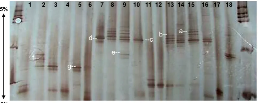
According to the DGGE image in Fig. 5,
ingerprints were obtained by separation of PCR
products (which were produced in the present of NS31-GC and Glo1 primers, Fig. 6) on denaturing
gradient gel (gradient range of 35-55 %). PCR–
DGGE produced high number of distinct and sharp bands, demonstrating the current method appears
to be an eficient protocol for studying biodiversity of
AM fungi which colonized pistachio trees. Fungi were characterized from excised DGGE bands, which
mainly belonged to the Glomus genus according
to the basic local alignment search tool (BLAST) (https://www.ncbi.nlm.nih.gov) (Fig. 5).
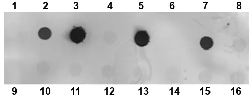
Extracted DNAs from pistachio roots were hybridized using an oligonucleotide probe complementary to a highly conserved sequence in the region between NS31 and Glo1 with dot blot
technique. Location of spotted DNA onto a Hybond
nylon membrane was clear without any pollution; no
signiicant cross-hybridization was observed (Fig. 7).
1 2 3 4 5 6 7 8 9 10 11 12 13 14 15 16 17 18
Marker Marker
35% 55%
a-- b----c
d--
e--
f--
g--Fig. 6. Silver-stained band pattern of DGGE analysis for 18S rDNA fragments of arbuscular mycorrhiza
were ampliied in PCR-DGGE using pistachio roots DNA as the template and NS31-GC and Glo1
primers. Each lane belonged to the individual root. DGGE gel composed of 6% acrylamide in a
denaturing gradient, form55 to 35%. a, b, c, d, e, f and g bands were cloned and sequenced. a: uncultured Glomus (KT033907), b: uncultured Xylariales (KT033908), c: uncultured Glomus (KT033909), d: uncultured Glomus (KT033910), e: uncultured Glomus (KT033911), f: uncultured Glomus (KT033912), g: uncultured Glomus (KT033913)
Fig. 5. Agarose electrophoresis of PCR products (280 pb in lane 1-8) ampliied using NS31-GC and Glo1
primers (280 bp) speciic of arbuscular mycorrhiza and extracted DNA from pistachio roots as template
500 pb →
200 bp →
Proposed post puriication step, denaturing
agarose electrophoresis using formamide, appropriately removed other residual-PCR
inhibitors. Considering beneicial denaturing activity
of formamide through agarose electrophoresis, residual-PCR inhibitors were detached from DNA. Moreover, PCR inhibitor including polyphenols traveled faster through the gel compared to DNA according to Fig. 2. Newman, Feminella, & Liles
(2010) embedded the extracted genomic DNA in agarose plugs and incubated in a formamide-NaCl solution to remove contaminants, however, in the present study electrophoresis of DNA was more time consuming than incubation. Depending
upon concentration of AlNH4(SO4)2.12H2O in lysis
buffer, the co-puriication of PCR inhibitors were
reduced; nonetheless, upper concentrations of
AlNH4(SO4)2.12H2O (above 50mM) noticeably decreased amount of DNA yield (Fig. 2). Recovery of agarose gel-embedded DNA also leveled up quality of DNA in polyphenolics-rich samples. Combination of two steps promoted the quality of highly contaminated DNA. Lack of smears and the appearance of sharp bands indicated that DNA degradation or shearing had not taken place (Fig. 2).
CONCLUSION
Acquiring the high quality DNA from plant tissues is the prerequisite key of plant microbe
interaction studies. As the biochemical proiles of
plant tissues and species considerably vary, it is almost impossible to rely on a universal isolation
protocol/kit. The present study provided a reliable
and simple technique for isolation of intact and high quality DNA from polyphenolic-rich pistachio roots. Inhibiting substances were eliminated from DNA through processes, including the chemical coagulating and denaturing agarose electrophoresis
purifying. Based upon the color of puriied DNA and
260/230 ratio>1.5, DNA was polyphenols-free while
the 260/230 ratio of prepared DNA using commercial extraction kits was nearly zero. Regarding to the results, extracted DNA from the studied procedure
was quite appropriate for PCR ampliication and
hybridization as well. Extracted DNA was too proper for studying biodiversity of plant endophytes, particularly the mycorrhiza fungi that cannot be cultured in the routine laboratory media (without host). Furthermore, using the current protocol and subsequent molecular biology techniques, Glomus sp. was reported as the most important symbiont of pistachio root. Overall, the research proposed that the current procedure can be considered for extracting DNA from other plants containing high levels of polyphenol.
ACKNOWLEDGEMENT
This research was supported by National
Institute of Genetic Engineering and Biotechnology of Iran.
REFERENCES
Abu Almakarem, A. S., Heilman, K. L., Conger, H. L., Shtarkman, Y. M., & Rogers, S. O. (2012).
Extraction of DNA from plant and fungus tissues in situ. BMC Research Notes, 5, 266.
http://doi.org/10.1186/1756-0500-5-266 Bakken, L. R., F & Rostegård, Å. (2006). Nucleic
acid extraction from soil. In P. Nannipieri &
K. Smalla (Eds.), Nucleic acids and proteins in soil – Soil biology vol.8 (pp. 49-73). Berlin:
Springer.
Bielski, B. H. J. (1982). Chemistry of ascorbic acid radicals. In P. A. Seib & B. M. Tolbert (Eds.),
Ascorbic acid: Chemistry, metabolism, and uses (pp. 81-100). Washington, USA: American Chemical Society. http://doi.
org/10.1021/ba-1982-0200.ch004
1 2 3 4 5 6 7 8
9 10 11 12 13 14 15 16
Fig. 7. DNA-DNA hybridization of extracted DNA from Pistacia vera L. roots using synthetic probe NS31/
Borse, T., Joshi, P., & Chaphalkar, S. (2011).
Biochemical role of ascorbic acid during the extraction of nucleic acids in polyphenol rich medicinal plant tissues. Journal of Plant Molecular Biology and Biotechnology,
2(2), 1–7. Retrieved from https://www.re
Removal of PCR inhibitors from soil DNA by
chemical locculation. Journal of Microbio-logical Methods, 52(3), 389–393. http://doi.
org/10.1016/S0167-7012(02)002 10-5 Bulgarelli, D., Schlaeppi, K., Spaepen, S., Ver
Loren van Themaat, E., & Schulze-Lefert,
P. (2013). Structure and functions of the bacterial microbiota of plants. Annual Review of Plant Biology, 64, 807–838. http://
doi.org/10.1146/annurev-arplant-050312-
120106
Carrier, G., Santoni, S., Rodier-Goud, M., Canaguier,
A., de Kochko, A., Dubreuil-Tranchant, C., … le Cunff, L. (2011). An eficient and rapid
protocol for plant nuclear DNA preparation suitable for next generation sequencing methods. American Journal of Botany, 98(1),
13–15. http://doi.org/10.3732/ajb.1000371 Chang, S., Puryear, J., & Cairney, J. (1993). A
simple and eficient method for isolating
RNA from pine trees. Plant Molecular
Biology Reporter, 11(2), 113–116. http://doi.
org/10.1007/BF02670468
Gadkar, V. J., & Filion, M. (2013). Quantitative
real-time polymerase chain reaction for tracking microbial gene expression in complex environmental matrices. Current Issues in Molecular Biology, 15, 45–58. Retrieved from
http://www.caister.com/cimb/v/v15/45.pdf Hardoim, P. R., van Overbeek, L. S., & van Elsas, J. D.
(2008). Properties of bacterial endophytes and their proposed role in plant growth.
Trends in Microbiology, 16(10), 463–471.
http://doi.org/10.1016/j.tim.2008.07.008
Healey, A., Furtado, A., Cooper, T., & Henry, R.
J. (2014). Protocol: A simple method for extracting next-generation sequencing quality genomic DNA from recalcitrant plant species. Plant Methods, 10, 21. http://doi. org/10.1186/1746-4811-10-21
Kasem, S., Rice, N., & Henry, R. J. (2008). DNA extraction from plant tissue. In R. J. Henry
(Ed.), Plant genotyping II: SNP technology (pp.
219-271). Oxfordshire, UK: CAB International.
Khanuja, S. P. S., Shasany, A. K., Darokar, M. P.,
& Kumar, S. (1999). Rapid isolation of
DNA from dry and fresh samples of plants producing large amounts of secondary metabolites and essential oils. Plant Molecular Biology Reporter, 17(1), 1–7.
http://doi.org/10.1023/A:1007528101452
Lawrence, J. R., Chenier, M. R., Roy, R., Beaumier, D., Fortin, N., Swerhone, G. D., .... Greer, C. W. (2004). Microscale and molecular assessment of impacts of nickel, nutrients, and oxygen level on structure and
function of river bioilm communities.
Applied and Environmental Microbiology, 70(7), 4326-4339. http://doi.org/10.1128/
AEM.70.7.4326-4339.2004
Liang, Z., Drijber, R. A., Lee, D. J., Dwiekat, I. M.,
Harris, S. D., & Wedin, D. A. (2008). A
DGGE-cloning method to characterize arbuscular mycorrhizal community structure in soil. Soil Biology and Biochemistry, 40(4), 956–966.
http://doi.org/10.1016/j.soilbio.2007.11.016
Liao, Z., Chen, M., Guo, L., Gong, Y., Tang, F., Sun, X., & Tang, K. (2004). Rapid isolation of
high-quality total RNA from taxus and ginkgo.
Preparative Biochemistry & Biotechnology,
34(3), 209–214.
http://doi.org/10.1081/PB-200026790
Manoj, K., Tushar, B., & Sushama, C. (2007). Iso
-lation and puriication of genomic DNA from
black plum (Eugenia jambolana Lam.) for analytical applications. International Journal of Biotechnology & Biochemistry, 3, 49-55.
Retrieved from https://www.thefreelibrary.
com/Isolation+and+puriication+of+genom - ic+DNA+from+Black+Plum+%28Euge-nia...-a0172131886
Marulanda, A., Azcón, R., & Ruiz-Lozano, J. M. (2003).
Contribution of six arbuscular mycorrhizal fungal isolates to water uptake by Lactuca sativa plants under drought stress. Physiologia Plantarum, 119(4), 526–533. http://doi.org/10.
1046/j.1399-3054.2003.00196.x
Marulanda, A., Porcel, R., Barea, J. M., & Azcón, R.
Glomus Species. Microbial Ecology, 54(3),
543–552. http://doi.org/10.1007/s00248-00 7-9237-y
Moazzam Jazi, M., Rajaei, S., & Seyedi, S. M. (2015). Isolation of high quality RNA from
pistachio (Pistacia vera L.) and other woody plants high in secondary metabolites.
Physiology and Molecular Biology of Plants,
21(4), 597–603. http://doi.org/10.1007/s122
98-015-0319-x
Nair, D. N., & Padmavathy, S. (2014). Impact of
endophytic microorganisms on plants, environment and humans. The Scientiic
World Journal, 2014, 1–11. http://doi.org/10.
1155/2014/250693
Newman, M. M., Feminella, J. W., & Liles, M. R. (2010). Puriication of genomic DNA
extracted from environmental sources for use in a polymerase chain reaction. Cold Spring Harbor Protocols, 2010(2), 1–16.
http://doi.org/10.1101/pdb.prot5383
Peterson, D. G., Boehm, K. S., & Stack, S. M. (1997). Isolation of milligram quantities of
nuclear DNA from tomato (Lycopersicon esculentum), A plant containing high levels of polyphenolic compounds. Plant Molecular Biology Reporter, 15(2), 148–153. http://
doi.org/10.1007/Bf02812265
Porebski, S., Bailey, L. G., & Baum, B. R. (1997). Modiication of a CTAB DNA extraction
protocol for plants containing high polysaccharide and polyphenol components.
Plant Molecular Biology Reporter, 15(1),
8–15. http://doi.org/10.1007/BF02772108 Porras-Alfaro, A., Herrera, J., Sinsabaugh, R. L.,
Odenbach, K. J., Lowrey, T., & Natvig, D.
O. (2008). Novel root fungal consortium associated with a dominant desert grass.
Applied and Environmental Microbiology,
74(9), 2805–2813. http://doi.org/10.1128/A
EM.02769-07
Rajaei, S. M., Niknam, V., Seyedi, S. M., Ebrahimzadeh, H., & Razavi, K. (2009).
Contractile roots are the most sensitive organ in Crocus sativus to salt stress.
Biologia Plantarum, 53, 523. http://doi.org/
10.1007/s10535-009-0095-y
Reinhold-Hurek, B., & Hurek, T. (2011). Living
inside plants: Bacterial endophytes. Current Opinion in Plant Biology, 14(4), 435–443.
http://doi.org/10.1016/j.pbi.2011.04.004
Rock, C., Alum, A., & Abbaszadegan, M. (2010). PCR
inhibitor levels in concentrates of biosolid samples predicted by a new method based on excitation-emission matrix spectroscopy.
Applied and Environmental Microbiology,
76(24), 8102–8109. http://doi.org/10.1128/
AEM.02339-09
Rodrigues, S. M., Soares, V. L., de Oliveira, T. M., Gesteira, A. S., Otoni, W. C., & Costa, M. G. (2007). Isolation and puriication
of RNA from tissues rich in polyphenols, polysaccharides, and pigments of annatto (Bixa orellana L.). Molecular Biotechnology,
37(3), 220–224. http://doi.org/10.1007/s120
33-007-0070-9
Sahu, S. K., Thangaraj, M., & Kathiresan, K. (2012).
DNA extraction protocol for plants with high levels of secondary metabolites and polysaccharides without using liquid nitrogen and phenol. ISRN Molecular Biology, 2012,
1–6. http://doi.org/10.5402/2012/205049 Schrader, C., Schielke, A., Ellerbroek, L., & Johne,
R. (2012). PCR inhibitors - occurrence, properties and removal. Journal of Applied Microbiology, 113(5), 1014–1026. http://doi.
org/10.1111/j.1365-2672.2012.05384.x
Stewart, E. J. (2012). Growing unculturable bacteria.
Journal of Bacteriology, 194(16), 4151–
4160. http://doi.org/10.1128/JB.00345-12 Tkacz, A., & Poole, P. (2015). Role of root
microbiota in plant productivity. Journal of Experimental Botany, 66(8), 2167–2175.
http://doi.org/10.1093/jxb/erv157
Wang, F., & Liu, R. (2001). A preliminary survey
of arbuscular mycorrhizal fungi in saline-alkaline soil of the Yellow River Delta. Chinese Biodiversity, 9(4), 389-392. Retrieved from
http://europepmc.org/abstra ct/cba/354910 Wu, Q.-S., Zou, Y.-N., Xia, R.-X., & Wang, M.-Y.
(2007). Five Glomus species affect water relations of Citrus tangerine during drought stress. Botanical Studies, 48, 147–154.
Retrieved from https://ejournal.sinica.edu. tw/bbas/content/2007/2/Bot482-03.pdf
Zhang, J., & Stewart, J. M. (2000). Economical and
rapid method for extracting cotton genomic DNA. The Journal of Cotton Science, 4,
193–201. Retrieved from https://www.