Majalah Ortodontik Vol. 8 No. 1, Juni 2012 ISSN 1411-7843 Hal. 33-37
Orthodontic camouflage treatment of skeletal class III malocclusion
(Case report)
Arya Brahmanta *, Pambudi Rahardjo **
* Department of Orthodontics Faculty of Dentistry University of Hang Tuah Surabaya ** Department of Orthodontics Faculty of Dentistry University of Airlangga
Abstract
Background: Class III malooclusion can be defined as a skeletal facial deformity characterized by a forward mandibular position with respect to the cranial base and or the
maxilla. There are three main treatment options for skeletal class III malocclusion : growth
modification, orthodontic camouflage and orthognathic surgery. Purpose: This article presented a case of an adult patient with skeletal class III malocclusion treated with
orthodontic camouflage treatment. Case: A female patient, 21 year old with concave facial profile complaining for the difficulty of occlusion due to anterior crossbite and openbite.
Case Management : Extraction of the poor conditioned mandibular first molars to gain
space for anterior segment retraction, placement of lingual arch bar to prevent anchorage
loss and class III intermaxillary elastics for dentoalveolar compensation by proclining
maxillary incisor and retroclining mandibular incisors (orthodontic camouflage).
Conclusion: The results of this treatment indicated that orthodontic camouflage can be
considered an effective therapy for corection of skeletal class III malocclusion.
Key words : class III malocclusion, orthodontic camouflage, tooth extraction, intermaxillary elastic.
Correspondence : Arya Brahmanta, Bagian Ortodonti Fakultas Kedokteran Gigi Universitas Hang Tuah. Jl. Arif Rahman Hakim 150 Surabaya. Telp. (031) 5912191.
Email: [email protected]
INTRODUCTION
Class III malocclusion, using the Angle classification system, are characterized by a mesial position of the mandibulardental arch and malposition of the incisors with a anterior
crossbite 1,2. Class III malocclusion can be classified into 2 types: pseudo class III and true
Class III malocclusion. According to Gianelly, pseudo Class III involves anterior crossbite in maximum intercuspation but in centric relation there is Class I malocclusion. Pseudo class III may be caused by interferes with mandibular closure, displacing it anteriorly. This problem is solved after removal of the interference. The true class III involves a skeletal class
III pattern in centric relation 3. As mentioned by Bench, the true class III malocclusion shows
a genetic trend toward extreme upward and backward condylar growth, anterior crossbite,
open bite and dolichofacial pattern 4.
The true class III malocclusion may also be called skeletal due to the involvement of
skeletal structure, caused by maxillary retrusion, mandibular protrusion or a combination of both. The skeletal class III malocclusion is characterized by mandibular prognatism, maxillary deficiency or both. Clinically these patients exhibit a concave facial profile, a retrusive nasomaxillary area and a prominent lower third face. The lower lip is often
protruded relative to the upper lip 5.
Mandibular prognathism is an anomaly of development, especially of a hereditary nature, but in which such diverse factors as endocrine, occlusal, parafunctional or habits
sometime intervene 1.
The frequency of class III malocclusion varies from author to author and with the population studied. In majority of published studies, this appears to be less frequent type of malocclusion, with prevalence under 10% 1. Class III malocclusion is far more prevalent in Asia than in the west 2. The incidnece of class III malocclusion is 2.3 – 13 % among Japanese, 9,4 – 19 % among Koreans and 12,8 % among Chinese. In contrast, the prevalence of class III malocclusion in the United States is only about 1 % of the total population and
only 5 % of orthodontic patients 6.
The choice of treatment of skeletal class III maloccluison depends on the
diagnosis,facial pattern, age, patient compliance and the severity of the malocclusion 5. There
dentolaveolar compensation (orthodontic camouflage) and orthognathic surgery. Growth modification should be commenced before the pubertal growth spurt, after this spurt only the
latter two options are possible 6.
cases certain extraction are necessary as a camouflage method 7. Camouflage is a therapeutic
process that most of the time, through extraction and orthodontic treatment, masks the skeletal discrepancies instead of correcting them. Therefore a dentoalveolar compensation is
made without correcting the basal dysplasia 1.
Surgical correction of class III malocclusion can be achieved by mandibular setback, maxillary advancement, or a combination of both procedures. The main skeletal changes after surgery were setback of the mandibular dentoalveolus and the uprighting of the lower incisors, whereas the skeletal base of both jaws did not show any significant changes. The setback of the dental alveolus in the mandibular anterior region contributed to the decreased
ANB angle 6.
Skeletal class III maloccclusion case is often complicated and difficult to treat. Cases or chapters related to skeletal class III malocclusion are often less discussed in orthodontic textbooks and diagnosed to treat surgically. Moreover multidisciplinary treatment approaches are avoided by patients.
The purpose of this article is to deliver a case of an adult patient with skeletal class III malocclusion, treated with extraction and intermaxillary elastic (orthodontic camouflage).
CASE REPORT
A patient a 21 year old woman, presented a skeletal class III malocclusion with anterior open bite came to the orthodontic specialist clinic Airlangga university dental
hospital. She complaining about the difficulty of occlusion due to anterior crossbite and
( Figure 1 a,b,c). Over jet – 4 mm and over bite - 2 mm ( Figure 1 e). Occlusal contact recognized as Angle class III malocclusion ( Figure 1 d,f ). The upper maxillary left and right first molar, lower mandibular left first molar were in the poorer condition and mandibular right molar were missing (Figure 1 d,f).
( a ) ( b ) ( c )
( d ) ( e ) ( f )
Figure 1. facial photographs (a) front side, (b) front side- smile, (c) left side, Intra oral
photographs right side (d), front side (e), left side (f)
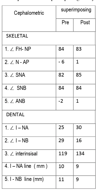
Cephalometric analysis showed a skeletal class III jaw base relationship SNA 82º; SNB 84º (prognathic mandible); ANB – 2 º ( class III). The facial profile was concave FH-NP 84º; NAP -6 º; The upper and lower incisor were labially inclined I -NA line 10 mm; I- NA angle 25 º ; I- NB line 11 mm ; I- NB angle 29 º ; Interinsisal angle 119º. The mandibular plane angle 41 º and the gonial angle was large 140 º.
CASE MANAGEMENT
A patient diagnosed with Angle Class III malocclusion with anterior open bite and skeletal class III jaw base relationship.
The treatment objectives were extraction of the left mandibular first molar 36 and the bilateral maxillary first molar 16,26 because it is in poorer condition. Placement of pre – adjusted braket (Roth) with 0.018 inch slot on upper and lower arch, followed by placement of lingual arch as an anchorage. Correction of the mandibular incisor by retraction use elastik class III. Retention using hawley retainer in both jaws.
Before starting orthodontic treatment, the patient received periodontal treatment.
Periodontal treatment involved oral hygiene instruction and scalling. The upper left first incisor was refered to endodontist because of poor condition caries. Bilateral mandibular lower first molar were extracted to gain space for retraction.
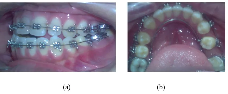
Firstly levelling with 0.012 inch round NiTi archwire was initiated. After leveling with a 0.016 inch NiTi arch wire, the lingual arch inserted onto the mandibular lower arch as an anchorage (Figure 2 a,b). After leveling with 0.016 inch NiTi arch wire in lower arch, retraction of the mandibular second premolar with closed coil spring, after that retraction first premolar and retraction mandibular canines with closed coil spring. Mandibular anterior teeth retraction was started by vertikal loop mechanic. A class III intermaxillary elastic was applied from the upper molar region to the mandibular anterior region for retraction with sliding mechanic (Figure 3 a, b, c).
(a) (b)
(a) (b) (c)
Figure 3. Intermaxillary class III Elastic (a) right side, (b) front side, (c) left side
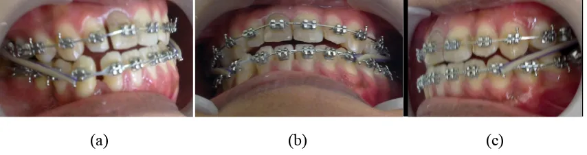
Treatment progress showed that upper and lower canine relation ship becomes Angle class I classification and anterior cross bite – open bite corrected.
(a) (b) (c)
Figure 4. Treatment progress (a) right side, (b) front side, (c) left side
The results of this treatment showed space in the upper and lower dentition were closed. The lower incisor lingually inclined and severe anterior crossbite were corrected. Acceptable occlusion achieved and the overjet and overbite come to normal. The caninus relation were Class I on the both sides (Figure 5 a, b, c). Facial photographs showed overall facial balance was improved. The lips becomes less tension on closure (Figure 5 d,e,f). No signs or symptoms of temporomandibular dysfunction after treatment.
(a) (b) (c)
(d) (e) (f)
Figure 5. facial photographs (a) front side, (b) front side- smile, (c) left side, Intra oral
photographs right side (d), front side (e), left side (f)
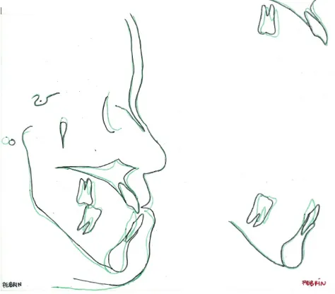
Cephalometric superimposing analysis showed a normal SNA 85º; SNB 84º; ANB 1 º . The facial profile was becoming straight FH-NP 83º; NAP 1 º. The upper and lower incisor have been corected I -NA line 9 mm; 30 º ; I- NB line 11 mm; 16 º ; Inter Insisal 134º . Comparing the superimposing pretreatment and posttreatment cephalometric tracings
showed maxillary and mandibular incisor crown had moved posteriorly (Figure 6).
Table 1. Cephalometric superimposing analysis
During retention with Hawley retainer acceptable occlusion and facial profile also maintained, indicating a stability of the occlusion (Figure 7).
Figure 7. Retention phase with Hawley retainer
Cephalometric superimposing
Pre Post
SKELETAL
1. ∠ FH- NP 84 83
2. ∠ N - AP -‐ 6 1
3. ∠ SNA 82 85
4. ∠ SNB 84 84
5. ∠ ANB -‐2 1
DENTAL
1. ∠ I – NA 25 30
2. ∠ I – NB 29 16
3. ∠ interinsisal 119 134
4. I – NA line ( mm ) 10 9
DISCUSION
Most of skeletal class III malocclusion cases are planned to be treated surgically and still considered high risk procedures, require of skillful operator and team work hence often being avoided either by orthodontists or patients. Therefore non surgical treatment or camouflage considered the most favorable choice. Extraction of lower posterior teeth followed by anterior retraction of lower anterior teeth combined with class III intermaxillary
elastic would be the best treatment plan for skeletal class III malocclusion 8,9.
It is commonly believed that successful camouflage treatment for class III malocclusion can be achieved by proclination of maxillary incisors, retrusion of mandibular incisors, and downward and backward rotation of mandible. Thus, the retraction of the lower incisors and rotation of the mandible were crucial for crossbite correction. The bodily movement of the roots was important in preventing over retroclination of the lower incisors. In order to do that, lingual root torque should be applied to the lower incisors during
treatment 6.
According to Chipman, extraction of posterior lower teeth is indicated in order to
facilitate the distal movement of the anterior teeth. For this case we choose to extract the first
molar lower arch because there are in the poor condition, very decayed. By extract the first
molar, we can get enough space for anteror retraction due to correction anterior cross bite 7.
In class III malocclusion maxillary anterior teeth are protrusive and facially inclined
and the maxillary dental arch takes a V shape in the anterior aspect. If there was a missing
teeth or ectopic canines and palatal eruption of lateral incisors, could cause maxillary
deficiency and crowding of the teeth. The mandibular anterior teeth may be upright in
relation to the basal supporting bone of the mandible, or they may be inclined lingually. In
generally, this malocclusion showed a reversed horizontal overlap in incisor area 10.
Orthodontic camouflage is built around the idea of displacing the teeth relative to the
jaws to compensate for a jaw discrepancy. The goals of camouflage are to obtain satisfactory
dental and facial esthetics. The method was developed as extraction treatment, followed by
orthodontic treatment. Patients with severe crowding would not be a good candidate for
camouflage treatment. Because in a patients with severe crowding, the extraction spaces will
be required to achieve proper alignment of the incisors. So that the extraction spaces
wouldn’t be avaible for controlled anteroposterior displacement 10.
The strategy to camouflage a Class III malocclusion usually involves proclination of the maxillary incisors and retroclination of the mandibular incisors to improve the dental occlusion.The use of Class III elastics provided forward movement of the maxillary molars, decreasing the incisors proclination. Class III intermaxillary elastic also contributed to the correction of the overbite. This can be explained by the use of Class III elastics, there was
counterclockwise rotation of the occlusal plane. Lin and Gu reported similar results and
found that the relative extrusion of the mandibular incisors in relation to the maxillary molars during Class III traction of elastics seemed to contribute to the counterclockwise rotation of the occlusal plane. An increase in the interincisal angle usually means less proclination of the
incisors 11.
At the end of treatment, the teeth were aligned, with better interdigitation and class I canine relationship was obtained. The retraction of the mandibular occured without loss anchorage in the posterior segment because the use of lingual arch bar. The patient main
complaint about the difficulty of occlusion due to anterior crossbite ,openbite and the facial
profile was improved by the treatment from a concave to a straight.
REFERENCE
1. Pinho C, Torrent U, Pinto C. Orthodontic camouflage in the case of a skeletal class III
malocclusion. World Journal of Orthodontics. 2004;5(3). pp: 213-23
2. Graber TM, Vanarsdall RL. Orthodontics: Current Principles and techniques, 3rd .
St.Louis. Mosby.2005
3. Gianelly AA, Bednar J, Cociani S, Giancotti F, Maino G, Ritcher O. Bidimensional
technique: theory and practice. Islandia NY: GAC International.2000. pp 128 – 141
4. Bench RW, Gugino CF, Hilgers JJ. Bioprogresive therapy part 8: Bioprogressive
mixed dentition treatment. J Clin Orthod 1978 (12) pp: 279 -98
5. Figueiredo MA, Siqueira DF, Bommarito S, Scanavini MA. Orthodontic
compensation in skeletal class III malocclusion. World Journal of Orthodontics 2007;8 (4) pp: 385 – 96
6. Rabie Ar- Bakr M, Wong RWK,Min GU. Treatment in borderline class III
malocclusion : orthodontics camouflage (extraction) versus orthognathic suregry. The Open Dentistry Journal. 2008 (2) pp 38-48
7. Mora DR, Oberti G, Ealo M, Baccetti T. Camouflage of moderate class III
malocclusion with extraction of lower second molars and mandibular cervical headgear. Progress in orthodontics 2007;8 (2) pp: 300-7
8. Proffit WR, Contemporary Orthodontics. St.Louis ,Mosby 2007; 607- 609
9. Faerovig E, Zachrisson BU. Effects of mandibular incisor extraction on anterior
occlusion in adults with class III malocclusion and reduced overbite. Am J Orthod Dentofacial Orthop.1999;155(2): 113-24.
10.Marcella B, Anggani HS.Consideration before orthodontic camouflage treatment in
skeletal Clas III malocclusion. Padjadjaran Journal of Dentistry 2008;20(1) pp: 23-33
11.Burns NR, Musich DR, Martin C, Razmus T, Gunel E, Ngan P. Class III camouflage
treatment : What are the limits ?. Am J Orthod Dentofacial Orthop