Flying PublisheR
#
7
2012
Kitchener, Hashem, Wahba,
Khalaf, Zarif, Mansoor
Critical Care
in
Neurology
Flying Publisher Guide to
the
More books at www.FlyingPublisher.com
Nabil Kitchener Saher Hashem Mervat Wahba Magdy Khalaf Bassem Zarif Simin Mansoor
The Flying Publisher Guide to
Critical Care in Neurology
2012 Edition
Correspondence:
Disclaimer
Neurocritical care is an ever-changing field. The publishers and author of The Flying Publisher Guide to Critical Care in
Neurology have made every effort to provide information that is accurate and complete as of the date of publication. However, in view of the rapid changes occurring in medical science, as well as the possibility of human error, this site may contain technical inaccuracies, typographical or other errors. It is the
responsibility of the reading physician who must rely on experience and knowledge about the patient to determine the best treatment and care pathway. The information contained herein is provided “as is”, without warranty of any kind. The contributors to this book, including Flying Publisher & Kamps, disclaim responsibility for any errors or omissions or for results obtained from the use of information contained herein.
This work is protected by copyright both as a whole and in part. Copy editing: Nilly Nagy and Rob Camp
© 2012 by Flying Publisher & Kamps / Design: Attilio Baghino
|
55
Prologue
Neurointensive care is a relatively new field that has developed as a subspecialty of critical care and neurology. The goal of neurointensive care, and the neurointensivist, is to treat and prevent primary and secondary brain (or other nervous system) injury. Inherent in this goal are the monitoring tools unique to the neurointensive care unit, including the most basic but perhaps the most important tool, the neurologic examination. In the era of the subspecialty of critical care neurology, the
neurologist is working now as an aggressive interventionalist who manages life-threatening disorders of the nervous system. The neurointensivist’s role is to help follow the neurologic status and treat the patient while integrating his/her knowledge of other organ systems and expertise in critical care, to provide the most comprehensive care possible for the patient.
Critical care neurology is practiced in emergency rooms, in consultations in general medical and surgical intensive care units, in intermediary care units such as stroke units, and in specialized neurointensive care units where patients are frequently on life-support systems involving ventilators, intravascular lines, and monitoring and treatment devices. Data has shown that care provided by clinicians specializing in neurologic injury, and within dedicated neurointensive care units, improves patient functional outcome, and reduces hospital mortality, length of stay and resource utilization.
This book emphasizes the clinical and practical aspects of management in the neurointensive care unit.
This book is written, mainly, for the neurologist working in, or directing, a specialized neurointensive care unit
nurses, and therapists all working together towards improved neurologic recovery.
We hope this book can provide a new addition to the emerging literature of critical care neurology, and heighten the
recognition by general medical and surgical intensivists of the importance and complexities of nervous system dysfunction in critically ill and injured patients.
The Editors
7
|
Editors
Nabil Kitchener, MD, PhD Professor of Neurology, GOTHI, Egypt
President of Egyptian Cerebro-Cardio-Vascular Association (ECCVA) and Board Director of World Stroke Organization (WSO)
www.ECCVA.com
Saher Hashem, MD Professor and Chairman of Neurology and Neurocritical Department
Cairo University, Egypt
Mervat Wahba, MD, FCCP Assistant Professor of Neurology Department of Neurology University of Tennessee Health Sciences Center, UTHSC, USA
Authors
Magdy Khalaf, MD Consultant Neurologist and Chairman of Neurocritical Care Unit
GOTHI, Egypt
Bassem Zarif, MD Lecturer of Cardiology
National Heart Institute, GOTHI, Egypt
|
9Table of Contents
1. Assessment of Patients in Neurological Emergency... 13
History ... 15
Physical Exam ... 16
1. Mental status ... 16
2. Cranial nerve (CN) exam ... 16
3. Motor exam ... 18
4. Reflexes ... 18
5. Sensory exam ... 18
6. Coordination and balance ... 18
7. Neuroanatomical localization ... 19
Conclusions ... 20
2. How to Approach an Unconscious Patient ... 21
Diagnosis ... 23
Basic assessments ... 24
General Care of the Comatose Patient ... 27
Permanent Vegetative State ... 27
Diagnosis ... 29
Management ... 29
Locked-in Syndrome ... 30
Brain Death ... 31
3. Documentation and Scores ... 32
Scoring and Documentation ... 34
4. Brain Injuries ... 39
Types of Brain Injuries ... 39
Primary brain injuries ... 39
Secondary brain injuries ... 42
Management of Special Issues ... 44
Traumatic brain injury ... 44
Acute stroke ... 45
Status epilepticus (SE) ... 46
Neuromuscular emergencies ... 47
Management of subarachnoid hemorrhage ... 50
5. Basic Hemodynamic Monitoring of Neurocritical Patients ... 53
6. Neurocritical Monitoring ... 59
Neuro-Specific Monitoring ... 59
Clinical Assessment ... 60
The Glasgow Coma Scale ... 60
Pupillary response ... 60
Invasive Monitoring ... 61
Measuring ICP ... 62
Indications for ICP monitoring ... 62
Intracranial Pressure Waveforms and Analysis... 63
Jugular Venous Oximetry (SjvO2) ... 67
Brain Tissue Oximetry ... 69
Noninvasive Monitoring ... 71
Continuous measures of CBF by Transcranial Doppler.. 71
Near Infrared Spectroscopy ... 73
Electrophysiological Monitoring ... 73
Application of the EEG in the ICU: ... 76
Multimodal Monitoring ... 77
Conclusions ... 77
7. Cerebral Edema ... 79
Types of Cerebral Edema ... 81
|
118. General Neurological Treatment Strategies ... 84
Swallowing Disturbances ... 85
Respiratory Management in Neurocritical Care ... 86
Infection Control in Neurocritical Care ... 89
Pain Relief and Sedation ... 90
Bedside approach to the agitated patient ... 91
Role of Rehabilitation ... 92
Diagnostic Findings in Cerebral Death ... 93
Conclusion ... 96
9. Medical Diseases and Metabolic Encephalopathies ... 97
Examination of the Encephalopathic Patient ... 98
General Pathophysiology ... 99
Hepatic Encephalopathy ... 99
Complications of hepatic encephalopathy (HE) ... 100
Treatment of hepatic encephalopathy ... 102
Renal Encephalopathies ... 102
Fluid and Electrolyte Imbalance ... 104
Osmolarity disorders ... 104
Encephalopathy in Diabetic Patients ... 106
Hypoxic Ischemic Encephalopathy (HIE) ... 107
Septic Encephalopathy ... 108
Drug-induced Encephalopathies ... 108
Assessment of Patients in Neurological Emergency
|
131.
Assessment of Patients in
Neurological Emergency
Nabil Kitchener, Saher Hashem
Care in specialized intensive care units (ICUs) is generally of higher quality than in general care units. Neurocritical care focuses on the care of critically ill patients with primary or secondary neurosurgical and neurological problems and was initially developed to manage postoperative neurosurgical patients. It expanded thereafter to the management of patients with traumatic brain injury (TBI), intracranial hemorrhage and complications of subarachnoid hemorrhage; including
vasospasm, elevated intracranial pressure (ICP) and the cardiopulmonary complications of brain injury.
Neurocritical care units have developed to coordinate the management of critically ill neurological patients in a single specialized unit, which includes many clinical domains. Care is provided by a multidisciplinary team trained to recognize and deal with the unique aspects of the neurological disease processes, as several treatable neurological disorders are characterized by imminent risk of severe and irreversible neurological injury or death if treatment is delayed.
1) Impaired level of consciousness.
2) Progressive respiratory impairment or the need for mechanical ventilation in a neurological patient. 3) Status epilepticus or prolonged seizures.
4) Clinical or Computed Tomographic (CT) evidence of raised Intracranial Pressure (ICP), whatever the cause (space occupying lesion, cerebral edema or hemorrhagic
conversion of a cerebral infarct, intracerebral hemorrhage, etc.)
5) Need for monitoring (for example, level of consciousness, ICP, continuous electroencephalography (cEEG)), and 6) Need for specific treatments (Baldwin 2010) (e.g.,
neurosurgery, intravenous or arterial thrombolysis). In the Neurocritical Care unit, patients with primary
neurological diseases such as myasthenia gravis, Guillain-Barré syndrome, status epilepticus, and stroke have a better outcome than those patients with secondary neurological diseases. So, we can conclude that these specialized units have greater
experience in the anticipation, early recognition, and management of potentially fatal complications.
Early identification of patients at risk of life threatening neurological illness in order to manage them properly and to prevent further deterioration is the role of general assessment of new patients in a neurological emergency. History taking and a rapid neurological assessment of a specific patient in specialized neurocritical care units helps answer the question ‘how sick is this patient?’.
The neurologic screening examination in the emergency settings focuses primarily on identifying acute, potentially life-threatening processes, and secondarily on identifying disorders that require other opinions, of other specialists.
Assessment of Patients in Neurological Emergency
|
15stroke, anticonvulsants for nonconvulsive and subtle generalized status epilepticus, and plasmapheresis for Guillain-Barré, etc.
It is obvious that interventions can be time-sensitive and can significantly reduce morbidity and mortality.
A comprehensive neurologic screening assessment can be accomplished within minutes if performed in an organized and systematic manner (Goldberg 1987). Neurologic screening assessment includes six major components of the neurologic exam, namely:
1) Mental status 2) Cranial nerve exam 3) Motor exam 4) Reflexes 5) Sensory exam
6) Evaluation of coordination and balance.
Based on the chief findings of the screening assessment, further evaluation or investigations can be then decided upon.
History
A careful history is the first step to successful diagnosis, and then intervention. Careful history provides clues to the pathology of the patient’s condition, and may help direct the diagnostic workup. For example, an alert patient with a headache associated with neck pain that started after a car accident might help direct the examination and radiographic imaging to focus on cervical spine injury or neck vessels (carotid or vertebral artery) dissection, while the same patient not in a car accident may direct your attention to a spontaneous subarachnoid hemorrhage.
Physical Exam
1. Mental statusUsually we start neurologic examination by assessing the patient’s mental status (Strub 2000).
A full mental status exam is not necessary in the patient who is conscious, awake, oriented, and conversant; on the contrary it must be fully investigated in patients with altered mental status.
Sometimes, we can find no change in mental status; at that point careful consideration should be given to concerns of family.
A systematic approach to the assessment of mental status is helpful in detecting acute as well as any chronic disease, such as delirious state in a demented patient (Lewis 1995). The CAM (confusion assessment method) score was developed to assist in diagnosing delirium in different contexts. CAM assesses four components: acute onset, inattention, disorganized thinking or an altered level of consciousness with a fluctuating course. A ‘Mini-Mental Status’ test can also be used but usually is reserved for patients with suspected cognitive dysfunction as it evaluates five domains – orientation, registration, attention, recall, and language (Strub 2000).
2. Cranial nerve (CN) exam
Cranial Nerves II - VIII function testing are of utmost value in the neurologic assessment in an emergency setting (Monkhouse 2006).
Assessment of Patients in Neurological Emergency
|
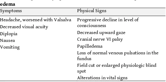
17Assessment of the optic disc, retinal arteries, and retinal veins can be done by a fundus exam, to discover papilledema, flame hemorrhages or sheathing.
Cranial Nerves III, IV, VI – CN III innervates the extraocular muscles for primarily adduction and vertical gaze. CN III function is tested in conjunction with IV, which aids in internal depression via the superior oblique, and VI, which controls abduction via the lateral rectus. Extraocular muscle function is tested for diplopia, which requires binocular vision and thus will resolve when one eye is occluded. Marked nystagmus on lateral gaze or any nystagmus on vertical gaze is abnormal; vertical nystagmus is seen in brainstem lesions or intoxication, while pendular nystagmus is generally a congenital condition.
The pupillary light reflex is mediated via the parasympathetic nerve fibers running on the outside of CN III. In the swinging flashlight test a light is shone from one eye to the other; when the light is shone directly into a normal eye, both eyes constrict via the direct and the consensual light response.
Pupillary size must be documented. Asymmetry in pupils of less than 1 mm is not significant. Significant difference in pupil size suggests nerve compression due to aneurysms or due to cerebral herniation, in patients with altered mental status.
Bilateral pupillary dilation is seen with prolonged anoxia or due to drugs (anticholinergics), while bilateral pupillary constriction is seen with pontine hemorrhage or as the result of drugs (e.g., opiates, clonidine).
Cranial Nerve V – Individual branch testing of the trigeminal nerve is unnecessary, as central nervous system lesions affecting CN V usually involve all three branches.
Cranial Nerve VII – The facial nerve innervates motor function to the face, and sensory function to the ear canal, as well as to the anterior two-thirds of the tongue. Central lesions cause contralateral weakness of the face muscles below the eyes.
When vestibular nerve defects are suspected, patients are assessed for nystagmus, via a past-pointing test and a positive response to the Nylen-Barany maneuver.
3. Motor exam
Motor system assessment focuses on detecting asymmetric strength deficits, which may indicate an acute CNS lesion. Testing motor power can be difficult or impossible in the uncooperative patient. It is not mandatory to test different muscle groups but instead just test for the presence of a “drift”. In diseased patients, the hand and arm on the affected side will slowly drift or pronate when they try to hold their arm out horizontally, palms up with eyes closed.
4. Reflexes
For rapid assessment of reflexes, major deep tendon reflexes and the plantar reflex must be elicited. Major deep tendon reflexes include the patellar reflex, the Achilles reflex, the biceps reflex, and the triceps reflex. Response can be graded from 0 (no reflex) to 4+ (hyperreflexia). Asymmetrical reflexes are the most important as they are considered pathologic. Many reflexes indicate upper motor neuron disease; the most commonly elicited is Babinski’s reflex.
5. Sensory exam
For rapid assessment of the sensory system, pain and light touch sensations should be done. Testing for other sensory modalities is reserved for patients with suspected neuropathies or for further evaluation of sensory complaints.
6. Coordination and balance
Coordination depends on functional integration of the
Assessment of Patients in Neurological Emergency
|
19By the end of the examination, you should reach a clinical diagnosis, which includes answers to the two critical questions, what is the lesion? and where is the lesion?
7. Neuroanatomical localization
Some knowledge of neuroanatomy is essential for correct localization. The first step in localizing neurological lesions should be to determine if it is a central (upper motor neuron) lesion (i.e., in the brain or spinal cord) versus a peripheral (lower motor neuron) lesion (i.e., nerve or muscle).
The hallmark of upper motor neuron lesions is hyperreflexia with or without increased muscle tone. Central (upper motor neuron) lesions are localized to:
Brain
– Cortical brain (frontal, temporal, parietal, or occipital lobes)
– Subcortical brain structures (corona radiata, internal
capsule, basal ganglia, or thalamus)
– Brainstem (medulla, pons, or midbrain)
– Cerebellum
Spinal cord
– Cervicomedullary junction
– Cervical
– Thoracic
– Upper lumbar
The hallmark of a lower motor neuron (LMN) lesion is decreased muscle tone, leading to flaccidity and hyporeflexia. Peripheral LMN lesions are localized to:
– Anterior horn cells
– Nerve root(s)
– Plexus
– Peripheral nerve
– Neuromuscular junction
Conclusions
1. The neurological screening examination provides the clinician with the necessary data to make management decisions.
How to Approach an Unconscious Patient
|
212.
How to Approach an Unconscious
Patient
Magdy Khalaf, Nabil Kitchener
Coma (from the Greek κώμα [ko̞ma], meaning deep sleep) is a state of unconsciousness lasting more than 6 hours, in which a person cannot be awakened, fails to respond normally to painful stimuli, light or sound, lacks a normal sleep-wake cycle and does not initiate voluntary actions.
All unconscious patients should have neurological
examinations to help determine the site and nature of the lesion, to monitor progress, and to determine prognosis. Neurological examination is most useful in the well-oxygenated,
response, the airway clearance, the pattern of breathing and circulation integrity, etc.
Special consideration must be given to neurological causes which may lead to unconsciousness like status epilepticus (either convulsive or non-convulsive), locked-in state, persistent vegetative state and lastly brain stem death. Any disturbances of thermoregulation must be measured.
Coma may result from a variety of conditions including intoxication, metabolic abnormalities, central nervous system diseases, acute neurologic injuries such as stroke, hypoxia or traumatic injuries including head trauma caused by falls or vehicle collisions. Looking for the pathogenesis of coma, two important neurological components must function perfectly that maintain consciousness. The first is the gray matter covering the outer layer of the brain and the other is a structure located in the brainstem called the reticular activating system (RAS or ARAS), a more primitive structure that is in close connection with the reticular formation (RF), a critical anatomical structure needed for maintenance of arousal. It is necessary to investigate the integrity of the bilateral cerebral cortices and the reticular activating system (RAS), as a rule. Unilateral hemispheric lesions do not produce stupor and coma unless they are of a mass sufficient to compress either the contralateral hemisphere or the brain stem (Bateman 2001). Metabolic disorders impair
consciousness by diffuse effects on both the reticular formation and the cerebral cortex. Coma is rarely a permanent state although less than 10% of patients survive coma without significant disability (Bateman 2001); for ICU patients with persistent coma, the outcome is grim.
Maneuvers to be established with an unconscious patient include cardiopulmonary resuscitation, laboratory
investigations, a radiological examination to recognize brain edema, as well as any skull, cervical, spinal, chest, and multiple traumas. Intracranial pressure and neurophysiological
How to Approach an Unconscious Patient
|
23Diagnosis
In the initial assessment of coma, it is common to judge by spontaneous actions, response to vocal stimuli and response to painful stimuli; this is known as the AVPU (alert, vocal stimuli, painful stimuli, unconscious) scale. The most common scales used for rapid assessment are:
1. The Glasgow Coma Scale (GCS), which aims to record the conscious state of a person, in initial as well as continuing assessment. When a patient is assessed and the resulting score is either 14 (original scale) or 15 (the more widely used modified or revised scale), this means ‘normal’; while if a patient is unable to voluntarily open their eyes, does not have a sleep-wake cycle, is unresponsive in spite of strong sensory (painful) or verbal stimuli and who generally scores between 3 to 8 on the Glasgow Coma Scale, (s)he is considered to be in coma.
2. Pediatric Glasgow Coma Scale: The Pediatric Glasgow Coma Scale (PGCS) is the equivalent of the Glasgow Coma Scale used to assess the mental state of adult patients. As with the GCS, the PGCS comprises three tests: eye, verbal and motor responses. The three values separately as well as their sum are considered (Holmes 2005). The lowest possible PGCS is 3 (deep coma or death) whilst the highest is 15 (fully awake and aware) (Holmes 2005).
Diagnosis of coma is simple; but determining the cause of the underlying pathology may prove to be challenging. As in those with deep unconsciousness, there is a risk of asphyxiation as control over the muscles in the face and throat is diminished, so those in a coma are typically assessed for airway management, nasopharyngeal airway or endotracheal intubation to safeguard the airway (Formisano 2004).
Following the previous assessment patients with impaired consciousness can be classified according to their degree of consciousness disturbance into lethargic, stuporous or comatose.
and attentive but slow to respond, unable to adequately perform simple concentration tasks such as counting from 20 to 1, or reciting the months in reverse.
Stupor means incomplete arousal to painful stimuli, little or no response to verbal commands, the patient may obey commands temporarily when aroused by noxious stimuli but more often only by pain.
Coma is the absence of verbal or complex motor responses to any stimulus (Stevens 2006).
Basic assessments
1. Pupillary functions may be normal if the lesion is rostral to the midbrain, while if the injury is diffuse, e.g., global cerebral anoxia or ischemia, the pupillary abnormality is bilateral. Pupil size is important as midposition (2-5 mm) fixed or irregular pupils imply a focal midbrain lesion; pinpoint reactive pupils occur in global hypoxic ischemic insult with pontine damage, or poisoning with opiates and cholinergic active materials; and bilateral fixed and dilated pupils can reflect central herniation or global hypoxic ischemic or poisoning with barbiturates, scopolamine, and atropine. Unilateral dilated pupil suggests compression of the third cranial nerve and midbrain, which necessitates an immediate search for a potentially correctable abnormality to avoid irreversible injury. In case of post-cardiac arrest coma, if pupils remain nonreactive for more than 6-8 hours after resuscitation, the prognosis for neurological recovery is generally guarded (Stevens 2006).
How to Approach an Unconscious Patient
|
253. Ocular reflexes: assessment of the brainstem and cortical functions happen through special reflex tests such as the oculocephalic reflex test (Doll’s eyes test), oculovestibular reflex test (cold caloric test), nasal tickle, corneal reflex, and the gag reflex.
4. Vital signs: temperature (rectal is most accurate), blood pressure, heart rate (pulse), respiratory rate, and oxygen saturation (Inouye 2006). It is mandatory to evaluate these basic vital signs quickly and efficiently to gain insight into a patient’s metabolism, fluid status, heart function, vascular integrity, and tissue oxygenation status.
5. The respiratory pattern (breathing rhythm) is significant and should be noted in a comatose patient. Certain stereotypical patterns of breathing have been identified including Cheyne-Stokes respiratory pattern, where the patient’s breathing is described as alternating episodes of hyperventilation and apnea, a dangerous pattern often seen in pending herniation, extensive cortical lesions, or brainstem damage. Apneustic breathing is characterized by sudden pauses of inspiration and is due to pontine lesion. Ataxic (Biot’s) breathing is an irregular chaotic pattern and is due to a lesion of the medulla. The first priority in managing a comatose patient is to stabilize the vital functions, following the ABC rule (Airway, Breathing, and Circulation). Once a person in a coma is stable, assessment of the underlying cause must be done, including imaging (CT scan, CT
angiography, magnetic resonance imaging (MRI), magnetic resonance angiography (MRA) and magnetic resonance venography (MRV) if needed ) and special studies, e.g., EEG and transcranial Doppler.
and potassium; chloride levels are rarely measured except for arterial blood gases (Bateman 2001). Once a patient is stable and no longer in immediate danger, the medical staff should start parallel work, first investigating the patient to find out any underlying pathology of his presenting illness, second, managing the presenting illness symptoms. Infections must be prevented and a balanced nutrition provided. The nursing staff, to guard against pressure ulcers, may move the patient every 2–3 hours from side to side and, depending on the state of consciousness, sometimes to a chair. Physical therapy may also be used to prevent contractures and orthopedic deformities that would limit recovery for those patients who emerge from coma (Wijdicks 2002).
How to Approach an Unconscious Patient
|
27General Care of the Comatose Patient
1. Airway protection: adequate oxygenation, ventilation and prevention of aspiration are the most important goals; most patients will require endotracheal intubation and frequent orotracheal suctioning.
2. Intravenous hydration: stuporous patients should receive nothing by mouth; use only isotonic fluids in these patients to avoid increasing the size of the cerebral edema or increased intracranial pressure (ICP).
3. Nutrition: enteral feeds via a small bore nasogastric tube. 4. Skin: the patient must be turned every 1-2 hours to prevent pressure sores; an inflatable or foam mattress and protective heel pads may also be beneficial.
5. Eyes: prevent corneal abrasion by taping the eyelids shut or by applying a lubricant.
6. Bowel care: constipation and gastric stress ulcers should be avoided.
7. Bladder care: indwelling urinary catheters are a common source of infection and should be used judiciously; intermittent catheterization every 6 hours when possible.
8. Joint mobility: passive range of motion daily exercises to prevent contractures.
9. Deep venous thrombosis prophylaxis: subcutaneous
anticoagulants and external pneumatic compression stockings or both (Upchurch 1995).
To complete this important critical situation, we will discuss two other categories, the permanent vegetative state and locked-in syndrome.
Permanent Vegetative State
set of criteria defining and ensuring diagnosis of PVS in infants under 3 months old, apart from anencephalics.
There are three major categories of disease in adults and children that result in PVS, upon which the outcome of PVS depends:
A. In acute traumatic and nontraumatic brain injury, PVS usually evolves within 1 month of injury from a state of eyes-closed coma to a state of wakefulness without awareness with sleep-wake cycles and preserved brainstem functions.
B. Some degenerative and metabolic disorders of the brain (i.e., late stage of dementia of Alzheimer type, end stage of Parkinson disease, and motor neuron disease, pontine myelinolysis, severe uncorrected hypoglycaemic coma) inevitably progress toward an irreversible vegetative state. Patients who are severely impaired but retain some degree of awareness may lapse briefly into a vegetative state from the effects of medication, infection, superimposed illnesses, or decreased fluid and nutritional intake. Such a temporary encephalopathy must be corrected before labeling that patient with the diagnosis of PVS.
Consciousness recovery is unlikely if the vegetative state persists for several months.
How to Approach an Unconscious Patient
|
29Diagnosis
The vegetative state is diagnosed, according to its definition, as being persistent at least for one month. Based upon class II evidence and consensus that reflects a high degree of clinical certainty, the following criteria is standard concerning PVS:
– PVS can be judged to be permanent, at 12 months after
traumatic brain injury in adults and children. Special attention to signs of awareness should be devoted to children during the first year after traumatic injury.
– PVS can be judged to be permanent if it lasts more than 3
months, in case of nontraumatic brain injury in both adults and children.
– The chance for recovery, after these periods, is exceedingly
low and recovery is almost always to a severe disability.
Management
When a patient has been diagnosed as being in a PVS by a physician skilled in neurological assessment and diagnosis, physicians have the responsibility of discussing with the family or surrogates the probability of the patient’s attaining the various stages of recovery or remaining in a PVS:
– Patients in PVS should receive appropriate medical, nursing,
or home care to maintain their personal dignity and hygiene.
– Physicians and the family/surrogates must determine
appropriate levels of treatment relative to the
administration or withdrawal of 1) medications and other commonly ordered treatments; 2) supplemental oxygen and use of antibiotics; 3) complex organ-sustaining treatments such as dialysis; 4) administration of blood products; and 5) artificial hydration and nutrition.
self and their environment, reproducible voluntary behavioral responses to visual and auditory stimuli, and interaction with others. Recovery of function occurs when a patient becomes mobile and is able to communicate, learn, and perform adaptive skills and self care and participate in recreational or vocational activities. Using these parameters, recovery of function can be defined with the Glasgow Outcome Scale.
The life span of adults and children in a PVS proves to be reduced; for most PVS patients, life expectancy ranges from 2 to 5 years and survival beyond 10 years is unusual. Once PVS is considered permanent, a “Do not resuscitate (DNR)” order is appropriate which includes no ventilatory or cardiopulmonary resuscitation. The decision to implement a DNR order, however, may be made earlier in the course of the patient’s illness if there is an advanced directive or agreement by the appropriate surrogate of the patient and the physicians responsible for the care of the patient (Plum 2007).
Locked-in Syndrome
How to Approach an Unconscious Patient
|
31the upper brain (Leon Carrion 2002). Possible causes of locked-in syndrome include: traumatic brain injury, diseases of the circulatory system, overdose of certain drugs, various causes which lead to damage to the nerve cells, particularly destruction of the myelin sheath, e.g., central pontine myelinolysis
secondary to rapid correction of hyponatremia and basilar artery (ischemic or hemorrhagic) stroke.
There is neither a standard treatment for locked-in syndrome, nor is there a cure, but stimulation of muscle reflexes with electrodes (NMES) has been known to help patients regain some muscle function. Assistive computer interface technologies in combination with eye tracking may be used to help patients communicate. Direct brain stimulation research developed a technique that allows locked-in patients to communicate via sniffing (Leon Carrion 2002). It is extremely rare for any significant motor function to return and the majority of locked-in syndrome patients do not regalocked-in motor control, but devices are available to help patients communicate. 90% die within the first four months after onset. However, some patients continue to live for much longer periods of time (Bateman 2001).
Brain Death
After exclusion of the previous syndromes, and in the absence of brain stem reflexes, brain death in deeply comatose patients should be established through the following criteria:
1. Irreversible coma
2. Absence of brain stem reflexes
3.
Documentation and Scores
Nabil Kitchener
Medical documentation is important for communication among health care professionals, for research, legal defense, and reimbursement. Neurological scoring systems are used to assess the severity of illness in patients with neurological emergencies, and can be used to monitor the clinical course, to document complications of therapy and to help identify prognostic factors. Two types of scoring systems are commonly used: neurological scoring systems, to quantify neurological deficits, like the Glasgow Coma Scale or the Mini-Mental Status Examination; and functional scoring systems to characterize patients’ abilities to perform activities of daily living, used to quantify the functional outcome with or without therapy, like the Canadian Neurological Score, the NIH stroke scale, the modified Rankin scale, the expanded disability status scale, the Richmond agitation sedation score and the confusion assessment method.
Documentation and Scores
|
33monitoring and care is no longer necessary (Egol 1999).
However, a number of patients who are successfully discharged from intensive care subsequently die during their hospital admissions. This may indicate premature discharge from the ICU or suboptimal management in the ICU or the general ward (Campbell 2008). As trends move towards earlier ICU discharge, it becomes increasingly important to be able to identify those patients at high risk of subsequent clinical deterioration, who might benefit from longer ICU stays or from transfers to intermediate care units. A strategy to reduce premature discharges in patients at high risk of in-hospital death could result in a 39% reduction in post-ICU death in these patients (Daly 2001). It can be concluded that reliable baseline and follow-up assessment is crucial to document any improvement or deterioration in neurological status of patients admitted to a neurocritical care unit. Interrater differences may be significant, so the need for standardized neurological scales and scores comes into play.
Scales seek to quantify different aspects of function within the framework of the World Health Organization hierarchy of impairment, disability, and handicap (Thuriaux 1995). Since the introduction of the Mathew scale in 1972 (Mathew 1972), there has been a steadily increasing number of scales that seek to quantify neurological impairment. These scales involve scoring different modalities of neurological function and then sum the scores to provide an index for neurological status. These scales were developed for a variety of reasons, including monitoring neurological status for improvement or deterioration (Cote 1986) and predicting final outcome in a defined group of patients (Brott 1989). The primary purpose for these scales in
Scoring and Documentation
Each neurocritical care unit should adopt a special scoring and documentation system, to be used to assess and document baseline patient neurological status and status at time of discharge. These include:
– Vital Signs: BP, temp, pulse, respiration, oximetry
– Pupils: size and reaction to light
– Eye movement: gaze, vergence, individual extraocular
movement and nystagmus
– Mental status: LOC, orientation and speech
– Motor functions: state, power, tone, deep reflexes and
pathological reflexes
– Coordination: gate, upper and lower limbs, if applicable
Documentation and Scores
|
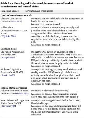
35Table 3.1 – Neurological Scales used for assessment of level of consciousness and mental status
Name and Source Strengths and Weaknesses
Level-of-consciousness scale
Glasgow Coma Scale
(Teasdale 1974, 1979) Strength: Simple, valid, reliable, for assessment of level-of-consciousness. Weaknesses: none observed.
Full Outline
Unresponsiveness – FOUR Score
(Wijdicks 2005)
Strength: The FOUR score is easy to apply and provides more neurological details than the Glasgow scale. This scale is able to detect conditions such locked-in syndrome and the vegetative state, which are not detected by the GCS.
Strength: CAM-ICU is an adaptation of the Confusion Assessment Method (CAM), which was adapted to be a delirium assessment tool for use in ICU patients (e.g., critically ill patients on and off the ventilator who are largely unable to talk). Weaknesses: none observed.
Richmond Agitation Sedation Scale (RASS) (Sessler 2002)
Strength: RASS is logical, easy to administer, and readily recalled. RASS has high reliability and validity in medical and surgical, ventilated and non-ventilated, and sedated and non-sedated adult ICU patients.
Weaknesses: none observed
Mental status screening
Folstein Mini-Mental State
Examination (Folstein 1975) Strength: Widely used for screening. Weaknesses: Several functions with summed score. May mis-classify patients with aphasia. Neurobehavioral Cognition
Status Exam (NCSE) (Kiernan 1987)
Strength: Predicts gain in Barthel Index scores. Unrelated to age.
Table 3.2 – Neurological Scales used for assessment of stroke deficits
Name and Source Strengths Weaknesses
Measures of disability/activities of daily living (ADL)
Barthel Index (Mahoney 1965, Wade 1988)
Widely used for stroke. Excellent validity and reliability.
Low sensitivity for high-level functioning
Functional Independence Measure (FIM) (Granger 1987)
Widely used for stroke. Measures mobility, ADL, cognition, functional communication.
"Ceiling" and "floor" effects
Stroke deficit scales
NIH Stroke Scale
(Brott 1989) Brief, reliable, can be administered by non-neurologists
Low sensitivity
Canadian Neurological Scale (Cote 1986)
Brief, valid, reliable Some useful measures omitted
Assessment of motor function
Fugl-Meyer
(Fugl-Meyer 1975) Extensively evaluated measure. Good validity and reliability for assessing sensorimotor function and balance
Considered too complex and time-consuming by many
Motor Assessment
Scale (Poole 1988) Good, brief assessment of movement and physical mobility
Reliability assessed only in stable patients. Sensitivity not tested
Motricity Index
(Collin 1990) Brief assessment of motor function of arm, leg, and trunk
Valid, brief, reliable test of
Documentation and Scores
|
37Name and Source Strengths Weaknesses
Assessment of speech and language functions
Boston Diagnostic
Long time to administer; half of patients cannot be classified
Porch Index of
Long time to administer. Special training required to administer. Inadequate sampling of language other than one word and single sentences
Western aphasia Battery (Kertesz 1982)
Widely used,
comprehensive Long time to administer. "Aphasia quotients" and "taxonomy" of aphasia not well validated
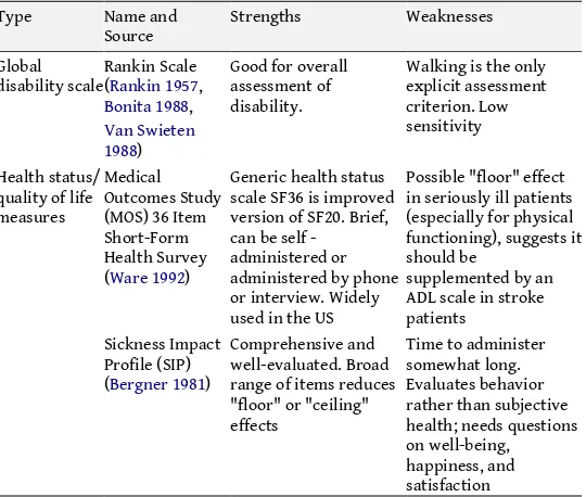
Table 3.3 – Neurological Scales used for assessment of health status and global disabilities
Type Name and
Source Strengths Weaknesses Global
disability scale Rankin Scale (Rankin 1957, Bonita 1988,
Walking is the only explicit assessment scale SF36 is improved version of SF20. Brief, can be self - in seriously ill patients (especially for physical functioning), suggests it should be
Delirium
Delirium is a disturbance of consciousness characterized by acute onset and fluctuating course of inattention accompanied by either a change in cognition or a perceptual disturbance, so that a patient’s ability to receive, process, store, and recall information is impaired.
Delirium, a medical emergency, develops rapidly over a short period of time, is usually reversible, and is a direct consequence of a medical condition or a brain insult. Many delirious ICU patients have recently been comatose, indicating a fluctuation of mental status. Comatose patients often, but not always, progress through a period of delirium before recovering to their baseline mental status.
ICU delirium is a predictor of increased mortality, length of stay, time on ventilator, costs, re-intubation, long-term cognitive impairment, and discharge to long-term care facility; it
necessitates special attention, assessment and management. Delirium assessment is actually an important part of the overall assessment of consciousness.
Delirium includes three subtypes: hyperactive, hypoactive and mixed. Hyperactive delirium is characterized by agitation, restlessness, and attempts to remove tubes and lines. Hypoactive delirium is characterized by withdrawal, flat affect, apathy, lethargy, and decreased responsiveness. Mixed delirium is characterized by fluctuation between the hypoactive and hyperactive. In ICU patients mixed and hypoactive are the most common, and are often undiagnosed if routine monitoring is not implemented. Few ICU patients (less than 5%) experience purely hyperactive delirium.
Brain Injuries
|
394.
Brain Injuries
Magdy Khalaf, Nabil Kitchener
Neurocritical care focuses on critically ill patients with primary or secondary neurological problems. Initially neurocritical care was developed to manage postoperative neurosurgical patients; it then expanded to the management of patients with traumatic brain injury (TBI), intracranial hemorrhage and complications of subarachnoid hemorrhage, including vasospasm, elevated intracranial pressure (ICP) and the cardiopulmonary complications of brain injury (Bamford 1992). The striking improvements noted in many studies suggest that high-quality neurocritical care with the delivery of targeted therapeutic interventions does have an impact, not only on survival, but importantly also on the quality of survival.
Types of Brain Injuries
Primary brain injuriesIschemic brain injury: either global, which includes cardiac arrest or anoxia, or regional ischemic brain injury, which includes vasospasm, compression of blood vessels or stroke. Stroke can be classified into ischemic and hemorrhagic strokes.
thrombotic stroke accounts for 77% while ischemic embolic stroke constitutes the remainder.
Hemorrhagic strokes constitute 10-20% of all strokes, and can be further classified into two types, the intracerebral
hemorrhage that constitutes up to 75% and the subarachnoid hemorrhage that makes up the other 25%.
Acute ischemic stroke is the third leading cause of death in industrialized countries and the most frequent cause of
permanent disability in adults worldwide, so understanding the pathogenesis of ischemic stroke is mandatory. Despite great strides in understanding the pathophysiology of cerebral ischemia, therapeutic options remain limited. Only recombinant tissue plasminogen activator (rTPA) for thrombolysis is
currently approved for use in the management of acute ischemic stroke.
However, its use is limited by its short therapeutic window (3-4.5 hours), complications from the risk of hemorrhage, and the potential damage from reperfusion injury. Effective stroke management requires recanalization of the occluded blood vessels. However, reperfusion can cause neurovascular injury, leading to cerebral edema, brain hemorrhage, and neuronal death by apoptosis or necrosis (Hajjar 2011).
Brain Injuries
|
41encephalitis predominantly in immunocompetent hosts include herpes simplex virus 1 (HSV-1) and 2 (HSV-2), human herpes virus 6 (HHV-6) and 7 (HHV-7), and Epstein-Barr virus (EBV). Cytomegalovirus (CMV) and varicella-zoster virus (VZV) may in some situations cause encephalitis in immunocompetent patients, but more commonly they produce an opportunistic infection in immunocompromised individuals, such as those with HIV infection, organ transplant recipients, or other patients using immunosuppressive drugs. HSV-1 is the most common cause of severe sporadic viral encephalitis in the United States; diagnosis has been become more familiar due to the availability of cerebrospinal fluid (CSF) polymerase chain reaction (PCR) analysis techniques that allow for rapid, specific, and sensitive diagnoses. The use of CSF PCR instead of brain biopsy as the diagnostic standard for HSV encephalitis has expanded awareness of mild or atypical cases of HSV encephalitis. Adult encephalitis is caused by 2 viral serotypes, HSV-1 and HSV-2. Patients with greater than 100 DNA copies/µL HSV in CSF are more likely than those with fewer copies to have a reduced level of consciousness, more significant abnormal findings on
neuroimaging, a longer duration of illness, higher mortality, and more sequelae (Domingues 1997). EBV is almost never cultured from CSF during infection, and serological testing is
inconclusive, so CSF PCR diagnosis is mandatory.
Semiquantitative PCR analysis of EBV DNA suggests that copy numbers are significantly higher in patients with active EBV infection. HHV-6 and -7 can cause exanthema subitum, and appear to be associated with febrile convulsions, even in the absence of signs of exanthema subitum. Almost all children (>90%) with exanthema subitum have HHV-6 or HHV-7 DNA in CSF. Inflammatory primary brain damage like meningitis and encephalitis come from pyogenic infections that reach the intracranial structures in one of two ways - either by
congenital sinus tracts). In a good number of cases, infection is iatrogenic, being introduced in the course of cerebral or spinal surgery, during the placement of a ventriculoperitoneal shunt or rarely through a lumbar puncture needle. Nowadays, nosocomial infections are as frequent as the non-hospital acquired variety. The reason for altered sensorium in meningitis is postulated to be the spillage of inflammatory cells to the adjacent brain parenchyma and the resultant brain edema (Levin 1998).
Compressive brain injury: e.g., tumors and cerebral brain edema are considered as important causes for impairment of the level of consciousness. During tumor growth, cerebral tissues adjacent to the tumor and nearby venules are compressed, which results in elevation of capillary pressure, particularly in the cerebral white matter, and there is a change in cerebral blood flow and consequently intracranial pressure. At that stage the tumor begins to displace tissue, which eventually leads to displacement of tissue at a distance from the tumor, resulting in false localizing signs such as transtentorial herniations,
paradoxical corticospinal signs of Kernohan and Woltman, third and sixth nerve palsies and secondary hydrocephalus, originally described in tumor patients.
Secondary brain injuries
Secondary brain injuries include renal coma, hepatic coma, salt and water imbalance, disturbance of glucose metabolism, other endocrinal causes of coma, disturbances of calcium and magnesium metabolism, drug intoxication and other material intoxication, not only drug toxicity, hypertensive and metabolic encephalopathies, sleep apnea syndromes and other ventilator disturbances. Mechanisms of secondary brain injury include hypoxia, hypoperfusion, reperfusion injury with free radical formations, release of excitatory amino acids and harmful mediators from injured cells, electrolyte and acid base changes from systemic or regional ischemia (Semplicini 2003).
Brain Injuries
|
43through the maintenance of oxygen delivery via the following parameters:
1. Assuring systemic oxygen transport and adequate oxygenation, maintaining hemoglobin level (at approximately 10 g/dl or more) and cardiac output. 2. Assuring optimal mean arterial pressure (MAP). Many
insults are associated with hypertension, which may be a physiologic compensation, so excessive lowering of blood pressure may induce secondary ischemia. In general, systolic pressure should be treated when more than 200 mmHg or MAP when more than 125 mmHg. Cautious reduction in mean arterial pressure by only 25% is recommended (Adams 2007).
3. Avoiding prophylactic or routine hyperventilation - a decrease in extracellular brain pH may produce
vasoconstriction in responsive vessels and reduce CBF to already ischemic zones. This applies to patients with head trauma in whom routine hyperventilation is no longer considered desirable; brief hyperventilation may be lifesaving in the patient with herniation, pending the institution of other methods to lower elevated ICP. 4. Assuring euvolemia. Hypervolemia may also be helpful
when vasoconstriction is suspected, as in the setting of subarachnoid hemorrhage.
5. Consideration should be given to administering
intravenous lidocaine 1.5 mg/kg or intravenous thiopental (5 mg/kg) to blunt the rise in ICP associated with
intubation.
6. Nimodipine should be instituted immediately in patients with SAH and is advocated by some in patients with subarachnoid bleeding after head trauma. Nimodipine probably improves outcome by decreasing calcium-mediated neuronal toxicity.
patient is hypoglycemic; hypotonic solutions should also be avoided.
8. Assessing and treating coagulation defects.
9. Sedation and/or neuromuscular blockade after intubation may be required to control harmful agitation.
10. If seizure occurrs, it should be aggressively treated. 11. Titration of the ICP and cerebral perfusion pressure.
Management of Special Issues
Traumatic brain injuryOutcome after traumatic brain injury depends upon the initial severity of the injury, age, the extent of any subsequent complications, and how these are managed. Much of the early management of traumatic brain injury falls upon emergency room staff, primary care and ambulance services prior to hospital admission. Most patients who attend hospital after a traumatic brain injury do not develop life-threatening complications in the acute stage. However, in a small but important subgroup, the outcome is made worse by failure to detect promptly and deal adequately with complications. General rules:
1. A traumatic brain injury should be discussed with neurosurgery when
a. a CT scan in a general hospital shows a recent intracranial lesion
b. a patient fulfills the criteria for CT scanning but this cannot be done within an appropriate period c. whatever the result of the CT scan, the patient has
clinical features that suggest that specialist
neurological assessment, monitoring, or management are appropriate. Reasons include:
i. Persistent coma (GCS <9, no eye opening) after initial resuscitation
Brain Injuries
|
45iii. Deterioration in level of consciousness after admission (a sustained decrease of one point in the motor or verbal GCS subscores, or 2 points on the eye opening subscale of the GCS)
iv. Persistent focal neurological signs v. A seizure without full recovery vi. Compound depressed fracture
vii. Suspected or definite penetrating injury viii. A CSF leak or other sign of base of skull fracture 2. Keep sodium >140 mmol/L. A fall in serum sodium produces
an osmotic gradient across the blood–brain barrier, and aggravates cerebral edema.
3. Avoid hyperglycemia (treat blood glucose >11 mmol/L). Hyperglycemia increases cerebral lactic acidosis, which may aggravate ischemic brain injury.
4. Feed via an orogastric tube. Gastric motility agents can be given as required.
5. Use TED stockings; avoid low-dose heparin.
6. Apply 15–30° head-up tilt with head kept in neutral position; this may improve CPP.
7. No parenteral hypotonic fluid must be given.
Acute stroke
The World Stroke Organization declared a public health emergency on World Stroke Day (WSO 2010). There are 15 million people who have a stroke each year. According to the World Health Organization, stroke is the second leading cause of death for people above the age of 60, and the fifth leading cause in people aged 15 to 59. Stroke also happens to children, including newborns. Each year, nearly six million people die from stroke. In fact, stroke is responsible for more deaths annually than those attributed to AIDS, tuberculosis and malaria put together. Stroke is also the leading cause of long-term disability irrespective of age, gender, ethnicity or country.
negative perception is shared by the general public, who often has a poor understanding of the early symptoms and significance of a stroke.
Yet within the last few years there have been many important developments in the approach to awareness and caring for stroke patients, for both the acute management and secondary prevention. Clinical research and interest in stroke has increased greatly in the last few years. Each minute of brain ischemia causes the destruction of 1.9 million neurons, 14 billion synapses, and 7.5 miles of myelinated nerves (Hand 2006). Ischemic stroke is characterized by one or more focal neurological deficits corresponding to the ischemic brain regions. It requires an immediate decision regarding
thrombolytic therapy (tissue plasminogen activator, TPA, in the dosage of 0.9 mg/kg, 10% as a bolus over 1 minute and infuse the remaining 90% over the next hour).
Wise control of hypertension is essential, control of
hyperglycemia and fever is protective against more destruction of neurons (Mistri 2006).
Status epilepticus (SE)
Status epilepticus is defined as more than 30 minutes of
continuous seizure activity or recurrent seizure activity without an intervening period of consciousness (Manno 2003).
In one survey, only 10% of patients who develop seizures in a medical ICU will develop SE. The most common causes of SE are noncompliance with or withdrawal of antiepileptic medications, cerebrovascular disease and alcohol withdrawal.
non-Brain Injuries
|
47convulsive generalized status epilepticus, refractory status epilepticus and myoclonic status epilepticus. Also, seizures that persist for longer than 5-10 minutes should be treated urgently because of the risk of permanent neurological injury and because seizures become refractory to therapy the longer they persist (Stasiukyniene 2009).
General measures for management are shown in Table 4.1. Intravenous drug therapy for convulsive seizures in the ICU are as follows:
1. Lorazepam: 0.10 mg/kg up to 2 mg/min or diazepam 0.15 mg/kg; if seizure continues, give
2. Fosphenytoin: 20 mg/kg up to 150 mg/min or phenytoin 20 mg/kg up to 50 mg/min; if seizure continues, one of the
following medications may be used but these require intubation and mechanical ventilation:
– phenobarbital 20 mg/kg up to 50 mg/min
– propofol 3-5 mg/kg load then 1-15 mg/kg/hr
– midazolam 0.2 mg/kg load then 0.05-2 mg/kg/hr
– pentobarbital 5-15 mg/kg load, then 0.5-10 mg/kg/hr
Neuromuscular emergencies
Neuromuscular emergencies are composed of a group of severe life-threatening neuromuscular diseases such as myasthenic crises, cholinergic crises, critical illness myopathy and critical illness polyneuropathy.
Respiratory paralysis occurs in a small percentage of patients with acute neuromuscular disease and accounts for less than 1% of admissions to general intensive care units. Its development may be insidious so that patients with acute neuromuscular disease should have their vital capacity monitored. Orotracheal intubation and ventilatory support should be instituted
Table 4.1 – General measures for management of Status Epilepticus* 1 (0–10 minutes)
Assess cardiorespiratory function Secure airway and resuscitate Administer oxygen
2 (0–60 minutes)
Institute regular monitoring Emergency antiepileptic drug therapy Set up intravenous lines
Emergency investigations
Administer glucose (50 ml of 50% solution) and/or intravenous
thiamine (250 mg) as high potency intravenous Pabrinex where appropriate Treat acidosis if severe
3 (0–60/90 minutes) Establish etiology
Identify and treat medical complications Pressor therapy where appropriate 4 (30–90 minutes)
Transfer to intensive care
Establish intensive care and EEG monitoring Initiate seizure and EEG monitoring
Initiate intracranial pressure monitoring where appropriate Initiate long term, maintenance, antiepileptic therapy
These four stages should be followed chronologically; the first and second within 10 minutes, and stage 4 (transfer to intensive care unit) in most settings within 60–90 minutes of presentation.
*Derived from Shorvon 1994
After assessment of these important conditions, and once the respiratory consequences of progressive neuromuscular weakness are established, the following requirements for management of critical neuromuscular diseases in ICU should be fulfilled:
1. Continuous monitoring of oxygen saturation to stay above 95%; pacemaker to be considered if heart rate variability is abnormal.
Brain Injuries
|
493. Management of inability to swallow through frequent suction, head positioning to allow use of a nasogastric, an orogastric or a Guedel tube.
4. Assessment of cardiac output, e.g., in myositis and arrhythmias (autonomic fiber involvement in GBS), heparinization for prevention of deep venous thrombosis, care for decubital ulcers.
5. Indications for intubation and artificial ventilation in neuromuscular critical cases: If oxygen saturation is below 90% (below 85% if more chronic), exhaustive respiratory work, forced vital capacity falling below 15 ml/kg and recurrent minor aspiration, avoid use of muscle relaxants. If artificial ventilation is likely to be required for more than approx. seven days, a tracheostomy should be created and is more comfortable for the patient than continued orotracheal intubation. Nutrition should be provided early via a nasogastric tube. Strenuous efforts should be made to reduce the incidence of nosocomial infection. Patients with neuropathy should be monitored for autonomic
dysfunction causing cardiac arrhythmia or fluctuating blood pressure. Deep vein thrombosis should be avoided by regular passive limb movements and low-dose
subcutaneous heparin.
6. Use assisted ventilation with IMV mode with low PEEP of 3 cm H20 except in pneumonia, atelectasis and use as few
sedatives as possible to monitor neurologic findings (Murray 2002). Critical illness polyneuropathy and myopathy are considered conditions associated with inflammatory injury to major organs involving peripheral nerves and skeletal muscles, and may add considerable value to the morbidity and mortality of the ICU stays. 7. If systolic pressure remains below 90 mmHg after adequate
volume replacement, begin dopamine infusion to maintain systolic pressure above 90 mmHg; if dopamine is
urine output exceeding 250 ml/hour for 2 hours, start a vasopressin infusion at a dose of 0.5-1.0 U/hour for adults, titrate infusion to maintain urine output at 100-200 ml/hour. Send tracheal aspirate, urine and blood for routine and fungal culture (Shoemaker 2000).
Metabolic disturbances such as hypokalemia or
hypermagnesemia should always be looked for and corrected first. In Guillain–Barré syndrome we recommend intravenous immunoglobulin as being equally effective to plasma exchange, safer, and more convenient. In myasthenia gravis we
recommend intravenous immunoglobulin followed by
thymectomy or, where thymectomy is inappropriate or has been unsuccessful, intravenous immunoglobulin combined with azathioprine and steroids. In polymyositis and dermatomyositis, steroids are the mainstay of treatment but intravenous
immunoglobulin is also effective.
Management of subarachnoid hemorrhage
Subarachnoid hemorrhage (SAH) is a complex medical and surgical event. Among its multiple etiologies, one of the most common relates to bleeding from a cerebral aneurysm. The optimal management of this life-threatening condition relies on a systematic and organized approach leading to the correct diagnosis and timely referral to a capable neurosurgeon. The following is a brief summary of steps that should be initiated when SAH is suspected, and the role of a medical neurocritical care facility.
A CT scan should be obtained immediately after the diagnosis is suspected. If the CT scan is positive, lumbar puncture is
Brain Injuries
|
51relates a history typical of SAH, a cerebral CT arteriogram should be performed despite a negative CT scan. Up to 15% of CT scans obtained within 48 hours of SAH will be negative.
Once the diagnosis is confirmed with a CT scan, a neurosurgeon who can treat the patient should be contacted immediately. Delays in transfer may prove fatal because of the potential for aneurismal rebleeding prior to intervention. It is often best to allow the interventionist or surgeon who will be caring for the patient to arrange for the diagnostic arteriogram to be performed at the institution where the patient will undergo intervention or surgery to repair the aneurysm. Arteriography performed by institutions infrequently treating SAH may be technically inadequate and require repetition upon transfer to the interventionist.
Blood pressure must be closely monitored and controlled following SAH. Hypertension will increase the chance of catastrophic rebleeding. Blood pressure control should be initiated immediately upon diagnosis of SAH.
Medical preoperative management includes prophylactic anticonvulsants, calcium channel blockade, corticosteroids, and antihypertensives as needed. We do not initiate antifibrinolytic therapy unless surgery is not considered within 48 hours of the initial SAH.
Medications that can be initiated prior to transfer to interventionist or neurosurgeon include:
– dexamethasone, 4 mg IV six hourly
– nimodipine, 60 mg orally four hourly
– phenytoin, 10 mg/kg IV load, then 100 mg orally/IV three
times daily
A frequent source of diagnostic difficulty for the
Basic Hemodynamic Monitoring of Neurocritical Patients
|
535.
Basic Hemodynamic Monitoring of
Neurocritical Patients
Bassem Zarif, Magdy Khalaf, Nabil Kitchener
The importance of basic hemodynamic monitoring of
neurocritical patients comes from the goal of maintaining brain autoregulation. Brain autoregulation and other biological signals are the variables to be monitored by using biomedical sensors. Complications that may occur in neurocritical patients (e.g., sepsis, dehydration, post-cardiac arrest status) make hemodynamic monitoring of greater importance.
The goals of hemodynamic monitoring in neurocritical care units are to assess the magnitude of physiological derangements in critically ill patients and to institute measures to correct the imbalance.
The following steps should be taken to reach these goals. Although our review of data may be helpful, the attending physician should decide what to use and when to use it. Menu to work with for proper patient management:
1. Pulse oximeter (SpO2) is regarded as one of the most
important advances in critical care monitoring. SpO2
oxyhemoglobin and deoxyhemoglobin and the
characteristics of pulsatile blood can thus be determined. SpO2 is accurate to within ± 2% for saturations >70%. SpO2 is
widely used in monitoring patients who have a variety of neurological conditions (Adams 1997), and calculations made from the processed signals provide estimates of the tissue or venous and arterial blood and provide an estimate of the amount of oxygenated hemoglobin and the percent saturation of hemoglobin by oxygen SaO2, which is not the
same as the PaO2 (partial pressure of oxygen) in the blood
(Adams 1997). The PaO2 and SaO2 measurements of
oxygenation are related through the oxyhemoglobin dissociation curve. Importantly, SpO2 is a measure of
arterial oxygenation saturation, not arterial oxygen tension (PaO2). Given the characteristics of the oxygen dissociation
curve, large fluctuations in PaO2 can occur despite minimal
changes in SpO2. In addition to its inability to measure PaO2,
SpO2 provides no measure of ventilation or acid-base status.
Therefore, it cannot be used to determine pH or arterial carbon dioxide tension. Significant increases in arterial carbon dioxide can occur with normal readings in SpO2.
Although useful for arterial oxygen saturation, SpO2 should
not be assumed to provide information about ventilation. Studies have shown that to assure a saturation of 60 torr (8.0 kPa), an SpO2 of 92% should be maintained in patients
with light skin, whereas 94% saturation may be needed in patients with dark skin. Oxygenation is considered
adequate if the arterial oxygen saturation is above 95%. The majority of these patients are placed on positive end expiratory pressure (PEEP) at 5cm H2O (Curley 1990).
Also, for patients with manifestations consistent with hypoxemia (e.g., tachycardia, hypotension, anxiety, and agitation) there is a time delay in pulse oximeter to express fluctuation in SpO2. Again in hypothermia, low CO2, and
Basic Hemodynamic Monitoring of Neurocritical Patients
|
55all increase bias, imprecision, and response time for hypoxic episodes, so we proceed to the next step.
2. Arterial blood gas analysis is widely available in hospitals and offers direct measurements of many critical
parameters (pH, PaO2, PaCO2). Arterial blood gas analysis is
among the most precise measurements of oxygen tension and pressure that will reflect tissue oxygenation (García 2011).
3. Non-invasive automated blood pressure devices are frequently used to obtain non-invasive, intermittent blood pressure measurements. Measurements of systolic and diastolic pressure to calculate the mean arterial pressure (MAP) is mandatory to calculate the cerebral perfusion pressure. These devices are less accurate in critically ill patients as well as in those with secondary brain injury. These less accurate readings can distract the attention of the caregiver. The evaluation of blood pressure will be significantly affected by the use of vasopressors. Therefore, the numeric reading may reflect vasoconstriction in spite of decreasing perfusion with adequate blood pressure. 4. Invasive blood pressure monitoring for continuous
monitoring and recording of the arterial pressure via an arterial catheter is preferable to the use of an automated blood pressure device in hemodynamically unstable patients (García 2011). The radial artery is most commonly cannulated because of its superficial location, relative ease of cannulation, and in most patients, adequate collateral flow from the ulnar artery. Other potential sites for percutaneous arterial cannulation include the femoral, brachial, axillary, ulnar, dorsalis pedis, and posterior tibial arteries. Possible complications of intra-arterial
monitoring include hematoma, neurologic injury, arterial embolization, limb ischemia, infection, and inadvertent intra-arterial injection of drugs. Intra-arterial catheters are not placed in extremities with potential vascular