124
Ethanolic Extract of
Moringa oleifera
L. Increases Sensitivity
of WiDr Colon Cancer Cell Line Towards 5-Fluorouracil
Kholid Alfan Nur, Herwandhani Putri, Fany Mutia Cahyani, Aulia Katarina, Ratna
Asmah Susidarti, Edy Meiyanto*
Cancer Chemoprevention Research Center, Faculty of Pharmacy Universitas Gadjah Mada
Jalan Sekip Utara Sleman Yogyakarta 555281 (Telp. 0274 6492662 Fax. 543120) *Corresponding author email : meiyan_e@ugm.ac.id
http//ccrc.farmasi.ugm.ac.id Abstract
For more than four decades, combination chemotherapy (co-chemotherapy) has been employed as a means to increase the effectiveness of chemotherapy regiments. The aim of our research is to investigate the activity of Moringa oleifera L. (tanaman kelor) ethanolic extract (MEE) as a co-chemotherapy agent with 5-fluorouracil (5-FU) on WiDr colon cancer cell line. Evaluation of MEE potency as a co-chemotherapy agent with 5-FU was based on cytotoxic activity based on percent cell viability via MTT assay, and based on apoptosis observation via the double staining method using acrydin orange – ethidium bromide (AE) as the staining reagent.Cytotoxicity evaluation of single treatment using concentrations of 5, 20, 50, 100,125, and 250 µg/ml of MEE reduced cell viability 24 hours post-treatment. 5, 50, and 250 µg/ml of MEE was chosen as the combination concentrations with 1000 µM 5-FU. MTT assay 24 hours and 48 hours post-combination treatment showed significant cell viability reduction in comparison to those of single treatments. Apoptosis observation using the double staining method shows the presence of apoptotic cells 48 hours post combination treatment. MEE is a potential co-chemotherapy agent by increasing the sensitivity of WiDr colon cancer cell line towards 5-FU.
Key words: co-chemotherapy, 5-fluorouracil, Moringa oleifera L., colon cancer
INTRODUCTION
Of the many cancer therapies, chemotherapy remains the conventional and main method to prescribe. Chemotherapy involves the use of synthetic chemical agents to suppress cancer cell proliferation or to destroy cancer cells. 5-Fluorouracil (5-FU) is currently dubbed as the main chemotherapy agent for the treatment of colon cancer. However, 5-FU is also notorious for its adverse side effects and is prone to cause resistance on cancer cells (Kodach et al, 2006). To solve this problem, combination chemotherapy (co-chemotherapy) is a viable option to delve in to.
Co-chemotherapy is an effort to reduce the side effects of chemotherapy by introducing additional anticancer agent(s) within the therapy regiment(s). When the interaction between the two agents is synergistic, co-chemotherapy can increase the potency of the chemotherapy agent to cancer cell proliferation (Brenner, 2002).
Moringa oleifera L. is a plant known to have anticancer activity.
Many research have reported the anticancer activity of Moringa oleifera L. This plant contains a unique combination of molecules called isothiocyanates and glucocynolates. It has been reported that isothiocyanates have chemopreventive properties by modifying the metabolic processes of carcinogenesis through inhibition of phase 1 metabolic enzymes and/or induction of phase 2 metabolic enzymes (Hetch, 2009). Benzyl isothiocyanate, an analogue of isothiocyanate, has shown apoptotic activity on ovarian cancer and G2/M cell cycle arrest on pancreatic cancer
cells (Srivastava and Singh, 2004).
Bharali et al (2003) have also reported ethanolic extracts of Moringa oleifera L. is a potential chemopreventive agent in suppressing carcinogenesis due to toxic chemicals exposure. Based on those research, it can be hypothesized that ethanolic extracts of Moringa oleifera L. is a potential co-chemotherapy agent with 5-FU
125 MATERIALS AND METHOD
Plant sample and extract preparation – Moringa oleifera L.
was obtained from nature in the Yogyakarta area, which was then officially determined by the Biological Pharmaceutics Department of the Faculty of Pharmacy, Universitas Gadjah Mada. The extract was then prepared by using 95% ethanol as the solvent for maceration. The macerate was then strained and a dilute extract was obtained. The extract was concentrated using a rotary evaporator continued with freeze drying to further eliminate traces of ethanol. The concentrated extract was then dissolved in DMSO (Sigma) for treatment.
WiDr cell preparation
WiDr colon cancer cell line used in this study was obtained from the collection of Cancer Chemoprevention Research Center of the Faculty of Pharmacy Universitas Gadjah Mada. WiDr was cultured in RPMI growth medium (Gibco) containing 10% v/v fetal bovine serum (Gibco), 1% v/v penicillin-streptomycin (Gibco), and 0,05% v/v fungizone (Gibco). Cell was cultivated from the culture dish using tripsin-EDTA 0,25% v/v (Gibco).
Cytotoxicity assay procedure
Cultivated cells were transferred into 96-well plates (Iwaki) then incubated for until confluent. Once the cells are confluent, treatment was conducted in accordance to the pre-calculated concentrations. MTT reagent was added 24 hours or 48 hours post-treatment (depending on the experiment), then SDS 10%
v/v in HCl was added 4 hours later. Following an overnight incubation period, the cell’s absorbance was read using microplate reader.
Apoptosis detection procedure
Cover slips (Iwaki) were placed in 24-well plate (Iwaki) then cells are transferred on to them, then incubated until confluent. The confluent cells are then treated with the selected dosage according to the co-chemotherapy MTT results. Once incubated for 48 hours post-treatment, the cover slips are removed from the wells and stained with 10 µL of Acrydin orange-Ethidium bromide (AE) to stain the cells. The stained cells are observed and recorded under fluorescent microscope.
RESULTS
Cytotoxicity assay of single MEE treatment and 5-FU – MEE combination treatment
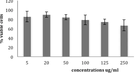
The MTT assay results show that MEE increases WiDr cell’s sensitivity towards 5-FU. MTT assay of MEE as a single agent shows reduced cell viability due to treatment (Figure 1). From this result, concentrations of 5, 50, and 250 µg/ml MEE was chosen as the combination concentrations which represents the whole range of data. Co-chemotherapy treatment using 1000 µM 5-FU with 5, 50, and 250 µg/ml MEE shows further cell viability reduction (Figure 2). Continued co-chemotherapy treatment for 48 hours gave the best results (Figure 2). Percent cell viability of 50% (IC50 value) was obtained
for concentration of 1000 µM 5-FU with 50 µg/ml MEE.
126
Figure 2. Cytotoxicity evaluation of combination treatment post 24 hours and 48 hours treatment. Assay was conducted by incubating WiDr cell in 96-well plate for 24 hours to normalize. Combination treatment series of 5, 50, 250 μg/ml MEE with 1000 μM was administered followed by further incubation for 24 hours or 48 hours as described in the methodology. Cell viability data was obtained thourgh convertion of absorbanse from formazan cyrstals formation due MTT treatment, as described in the methodology. As comparison, single 2500uM 5-FU and EEM data is also presented. Viabilty profile is presented from the mean ± standard error (SE) from 3 experiments. The combination treatment of MEE – 5-FU for 24 hours on WiDr resulted in greater cell viability reduction compared to single treatments. Further cell viability reduction was observed post 48 hours treatment.
These cell deaths were most likely due to apoptosis. To ascertain this postulation, we continued further with our investigation and conducted apoptosis detection assay using the double staining method.
Detection of apoptotic cells using double staining method
127
Figure 3. (A) control (B) – (D) results of double staining following 48 hours post co-chemotherapy treatment concentration of 1000 µM 5-FU with 50 µg/ml MEE. Cells were seeded on cover slips in 24-well plates and treated with combination of 1000 µM 5-FU with 50 µg/ml MEE. After 48 hours of incubation each well was washed with PBS, cover slips removed, then treated with ethidium bromide – acrydin orange fluorescent dye. Living cells emit green fluorescence while apoptotic cells emit orange fluorescence (arrowed).
DISCUSSION
Our hypothesis, that MEE has a potential as a co-chemotherapy agent with 5-FU in colon cancer treatment, was confirmed by the research findings. MTT assay of single MEE treatment show reduction of cell viability. Further and more significant cell viability reduction was observed on the combination treatment cell viability profile.
The mechanism of action of 5-FU – MEE combination is due to enhancement of cancer cell cytotoxicity. As reported by De Angelis et. al. (2006) 5-FU damages the DNA double strand and induce apoptosis of colon cancer cells, which is mediated by the p53 protein. Benzyl isothiocyanate (BITC) and phenethyl isothiocyanate (PEITC), the principle anti-tumoregenesis compounds in Moringa oleifera L., have been previously reported to induce apoptosis. BITC up-regulate the p21 protein, resulting in cell apoptosis (Zhang, R., et. al., 2009). PEITC causes cell apoptosis by down-regulating Bcl-2/XIAP and through the
mitochondrial pathway (Lee, J.W., and Cho, M.K., 2008). Our research findings are in accordance to these previously reported studies.
128 of 1000 uM 5-FU with 50 ug/ml resulted in the
appearance of apoptotic bodies.
To explore the underlying cytotoxicity and apoptosis induction mechanism, further research is needed. Flow cytometric analysis is needed to determine the effect on cell cycle progression. Western Blotting should also be conducted to ascertain which proteins are involved.
CONCLUSION
The anti-tumoregenesis activity of 5-FU is enhanced through combination with MEE through increased cytotoxicity and apoptosis induction. MEE has potential to become a co-chemotherapy agent with 5-FU.
REFERENCES
Bharali, R., Tabassum, J., and Azad, M.R.H., 2003, Chemomodulatory effect of Moringa Oleifera L. on hepatic carcinogen metabolising enzyme, antioxidant parameters, and skin papillomagenesis in mice, Asia Pacific Journal of Cancer Prevention, 4: 131 – 139.
Brenner, D.E., 2002, New paradigms in oncological therapeutics: redefining combination chemotherapy, Annals of Oncology, 13: 1697 – 1698.
De Angelis, P.M., Svendsurd, D.H., Kravik, K.L., and Stokke, T., 2006, Cellular response to fluorouracil (5-FU) in FU-resistant colon cancer cell lines during treatment and recovery, Mol. Cancer,
5: 20.
Hetch, S.S., 2009, Chemoprevention of cancer by isothicyanates, modifiers of carcinogen metabolism, Journal of Nutrition, 129: 768s – 774s.
Kodach, L.L., Bos, C.L., Duran, N., Peppelenbosch, M.P., Ferreira, C.V., and Hardwick, J.C.H., 2006, Violacein synergistically increases 5-fluorouracil
cytotoxicity, induces apoptosis, and inhibits Akt-mediated signal transduction in human colorectal cancer cells, Carcinogenesis, 27(3): 508-516.
Lee, J.W., and Cho, M.K., 2008, Phenethyl Isothiocyanates induced apoptosis via down regulation of Bcl-2/XIAP and triggering the mitochondrial pathway in MCF-7 cells, Arch. Pharm. Res.,
31(12): 1604-1612.
Srivastava, S.K., and Singh, S.V., 2004, Cell cycle arrest, apoptosis induction and inhibition of nuclear factor kappa B activation in antiproliverative activity of benzyl isothiocyanate against human pancreatic cancer cells, Carcinogenesis,
25(9): 1701-1709.