doi: 10.11594/jtls.07.03.15
How to cite:
Astari LF, Cahyono HA, Widjajanto E (2017Correlation of In-terleukin-10, Superoxide Dismutase (SOD) and Malondialdehyde (MDA) Levels with HbA1c in Pediatric Type 1 Diabetes Mellitus. J. Trop. Life. Science 7 (3): 286 – 292.
*Corresponding author: Lina Fitria Astari
Department of Biomedical Sciences, Faculty of Medicine, Brawijaya University
Jalan Veteran, Malang, Indonesia 65145 E mail: [email protected]
THE JOURNAL OF TROPICAL LIFE SCIENCE OPEN ACCESS Freely available online
VOL. 7, NO. 3, pp. 286 – 292, September 2017 Submitted January 2017; Revised February 2017; Accepted February 2017
Correlation of Interleukin-10, Superoxide Dismutase (SOD), and Malondialdehyde (MDA) Levels
with HbA1c in Pediatric Type 1 Diabetes Mellitus
Lina Fitria Astari 1,2 *, Haryudi Aji Cahyono 2, Edi Widjajanto2
1 Department of Biomedical Sciences, Faculty of Medicine, Brawijaya University, Malang, Indonesia 2 Department of Pediatric, Saiful Anwar Public Hospital, Malang, Indonesia
ABSTRACT
Type 1 diabetes mellitus (T1DM) is an autoimmune disease characterized by pancreatic β-cell destruction and
considered to be correlated with oxidative stress. This study aimed to investigate the association of oxidative stress [superoxide dismutase (SOD) and malondialdehyde (MDA) levels], inflammation [interleukin 10 (IL-10)], and gly-cemic control (HbA1c) in pediatric T1DM patients. This study included 25 T1DM subjects and 25 healthy control subjects and was designed as a cross- sectional study. SOD, MDA, and IL-10 levels were measured by ELISA. We obtained that that IL-10 and SOD levels were significantly decreased in the T1DM group, but MDA and HbA1c levels were significantly elevated in the T1DM group. IL-10 levels were positively correlated with SOD levels and negatively correlated with MDA and HbA1c. SOD levels were negatively correlated with HbA1c levels. MDA was positively correlated with HbA1c levels. IL-10 and SOD levels were significantly decreased, but MDA and HbA1c levels were significantly elevated in the T1DM group.
Keywords: Type 1 diabetes mellitus, superoxide dismutase, malondialdehyde, interleukin 10, HbA1c
INTRODUCTION
Type 1 diabetes mellitus (T1DM) is an autoimmune
disease caused by pancreatic β-cell destruction as the
re-sult of several interacting factors, including genetic vul-nerability and environmental exposure [1]. Ninety per-cent of T1DM cases occur in children and adolesper-cents. Its incidence varies worldwide, the highest being in the Finnish population (40 per 100 000 people), and the lowest in the Chinese population (0.1 per 100 000 peo-ple per year) [2]. Data gathered from the Coordination Framework Unit of Pediatric Endocrinology, Indonesian Pediatric Association indicated a prevalence of T1DM was 731 cases in 2012 [3]. Other data from Saiful Anwar General Hospital, Malang showed that there were 35 T1DM patients ranging in age from 1 to 18 years (2005-2013) [4].
Destruction of pancreatic β cells is mediated by
T-cell hyperreactivity, which in turn induces the release of autoantibodies. Impaired immunoregulation causes
T-cell autoreactivity, then induces islet T-cell inflammation,
and finally causes pancreatic β-cell destruction [3, 5].
A previous study showed that oxidative stress plays an important role in diminished insulin action and se-cretion. Oxidative stress in diabetic patients might be caused by decreased antioxidant enzyme activity and el-evated reactive oxygen species (ROS) levels, which in turn cause further lipid peroxidation and glycation [6]. A previous study showed that decreased activity of sev-eral enzymes, such as superoxide dismutase (SOD), roxidase (Px), ceruloplasmin (Cp), and glutathione pe-roxidase (GSH-Px), as well as elevated glutathione di-sulfide (GSSG), were present in erythrocytes and tissues from diabetic patients [7].
Interleukin 10 (IL-10) is an anti-inflammatory cyto-kine with the ability to downregulate inflammatory cy-tokine production and thus inhibit T-cell activation. IL-10 is a potent inhibitor of Th1 cytokines, such as IL-2
elevated and anti-inflammatory cytokines are decreased as compared with the nondiabetic individuals [8]. Gly-cemic control is related to both microvascular and macrovascular complications. Good glycemic control could increase the quality of life of T1DM patients. High HbA1c levels reflect poor glycemic control [9].
To date, a study on the association of oxidative stress and inflammation status with glycemic control in T1DM patients in Indonesia has not been conducted. This study aimed to investigate the association of IL-10, SOD, and malondialdehyde (MDA) levels with HbA1c levels in pediatric T1DM patients.
MATERIALS AND METHODS
Study design
A cross-sectional (observational, analytic) study was conducted to compare the levels of IL-10, SOD, and MDA with those of HbA1c in pediatric T1DM patients. All procedures were approved by the Research Ethics Committee of Saiful Anwar General Hospital, Malang.
Subjects
A total of 50 subjects were included in this research and divided equally into two groups (control and T1DM group). The inclusion criteria for subjects were as fol-lows: diagnosed as T1DM, aged between 1 and 18 years old, and allowed by his/her parents to participate (in-formed consent). The exclusion criteria for subjects were the following: T1DM patients who had other diseases,
such as other autoimmune diseases, liver, and renal im-pairment, and anemia and consumption of antioxidants
within 3 weeks before the study. To establish good matching of subjects and controls, the inclusion criteria for controls were age between 1 and 18 years and al-lowed by his/her parents to participate (informed con-sent). The exclusion criteria for controls were T1DM pa-tients; diagnosed with the autoimmune disease (such as SLE), severe infection, liver and/or renal impairment, or anemia; and consumption of antioxidants within 3 weeks before the study. All subjects (control and T1DM group) were taken from the Pediatric Ward, Saiful Anwar General Hospital, Malang.
Measurement of SOD
SOD was measured by a competitive ELISA method as previously described. Briefly, blood samples to which EDTA was added were centrifuged at 1,000 rpm for 15
minutes. A 50 µL volume of sample or standard was
added to each well of a microtiter plate and an equal volume of biotinylated antibody was added and
incu-bated for 45 min at 37°C. After washing, 100 µL HRP
conjugate was added to each well and then incubated for
30 min at 37°C. After washing, 90 µL
tetramethylbenzi-dine (TMB) substrate was added to each well and then
incubated for 15 minutes at 37°C. Finally, 50 µL stop
solution was added and 30 minutes later, the absorbance was read in a microplate reader at 450 nm. SOD meas-urements were conducted at the Physiology Laboratory, Medical Faculty of Brawijaya University, Malang.
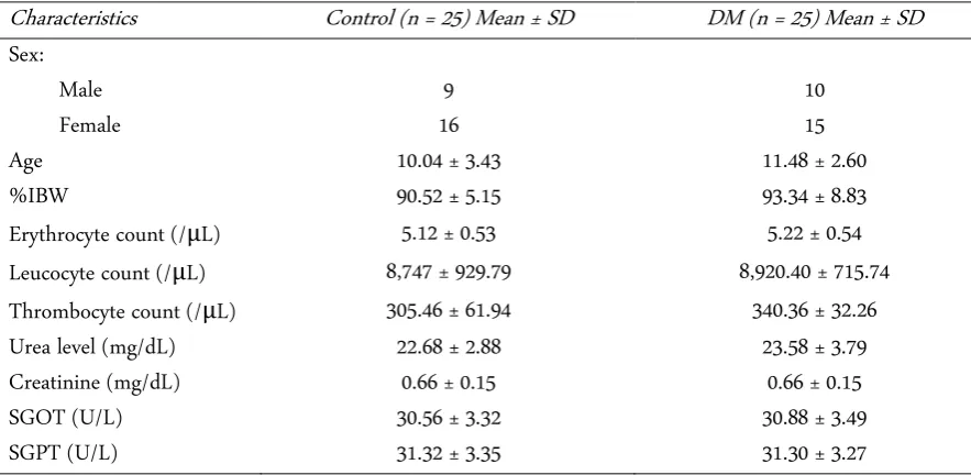
Table 1. Subject characteristics
Characteristics Control (n = 25) Mean ± SD DM (n = 25) Mean ± SD
Sex:
Male 9 10
Female 16 15
Age 10.04 ± 3.43 11.48 ± 2.60
%IBW 90.52 ± 5.15 93.34 ± 8.83
Erythrocyte count (/μL) 5.12 ± 0.53 5.22 ± 0.54
Leucocyte count (/μL) 8,747 ± 929.79 8,920.40 ± 715.74
Thrombocyte count (/μL) 305.46 ± 61.94 340.36 ± 32.26
Urea level (mg/dL) 22.68 ± 2.88 23.58 ± 3.79
Creatinine (mg/dL) 0.66 ± 0.15 0.66 ± 0.15
SGOT (U/L) 30.56 ± 3.32 30.88 ± 3.49
SGPT (U/L) 31.32 ± 3.35 31.30 ± 3.27
Lina F Astari, Haryudi A Cahyono, Edi Widjajanto, 2017
Measurement of MDA
The method of malondialdehyde measurement was based on the reaction of chromogenic reagents N-me-thyl-2-phenylindole (NMPI) with MDA at 45°C as pre-viously described.10 The procedure began by adding 10 mL probucol to each well. After 200 µL sample and 600 µL R1 reagent (NMPI in acetonitrile) were added, the sample was mixed by using a vortex. A 150- µL volume of hydrochloric acid was added to each well, mixed again by using a vortex and incubated for 60 min at 45°C. After incubation, the sample was centrifuged at 10,000 rpm for 10 minutes. The supernatant was removed and the absorbance read at 586 nm. MDA measurements were conducted at the Physiology Laboratory, Medical Faculty of Brawijaya University, Malang.
Measurement of IL-10
IL-10 levels were measured by ELISA as described by the manufacturer.10 A 200-µl volume of assay diluent was added to each well and incubated for 1 h. After that, 100 µl standards was added to control and sample wells, which were covered with an adhesive strip and then in-cubated for 2 h at room temperature. After four washes with wash buffer (400 µL), 100 µL antibody was added to each well and incubated for 1 h. After incubation, 100 µL avidin-horseradish peroxidase (HRP)-conjugated IL-10 was added to each well. The microplate was then cov-ered with an adhesive strip, incubated for 30 min at room temperature, and rewashed. A 100-µL volume of tetramethylbenzidine (TMB) substrate solution was add-ed to each well, incubatadd-ed for 30 min at room tempera-ture, and 100 µL stop solution was added. Optical den-sity was measured at 450 nm by using a microplate read-er. Absorbance values were used to plot a standard curve and to calculate the IL-10 level in each sample. IL-10 measurements were conducted at the Physiology Labor-atory, Medical Faculty of Brawijaya University, Malang.
Measurement of HbA1c
Hemoglobin A1c (HbA1c) levels were measured in whole blood samples to which EDTA was added. A 5-µL blood sample was mixed with 1.5 mL diluent solu-tions before analysis. The level of HbA1c in the sample was measured by using a Bio-Rad D-10TM.10 HbA1c measurements were conducted at the Clinical Pathology Laboratory, Saiful Anwar General Hospital, Malang.
RESULTS AND DISCUSSION
Subject characteristics
In this research, subjects were T1DM patients who
routinely attended the Endocrinology Department of Saiful Anwar General Hospital for outpatient care dur-ing the research period. Table 1 shows subject charac-teristics of the two groups.
SOD and MDA levels
Results showed that both SOD and MDA levels were significantly different between T1DM and control groups (independent samples t-test, p < 0.05). SOD lev-els in the T1DM group were significantly lower as com-pared with control group. Conversely, MDA levels in the T1DM group were significantly higher as compared with the control group.
IL-10 levels
Results showed that the IL-10 level was significantly different between the T1DM and control groups (inde-pendent samples t-test, p < 0.05). IL-10 levels in the T1DM group were significantly higher as compared with the control group.
HbA1c levels
Results showed that HbA1c levels were significantly higher as compared with the control group (independ-ent samples t-test, p < 0.05).
Correlation and path analysis of SOD, MDA, and IL-10 with HbA1c levels
Results showed that all variables were significantly correlated with each other. IL-10 was positively corre-lated with SOD levels (p < 0.05, r = 0.853) and negatively correlated with MDA levels (p < 0.05, r = -0.866). IL-10 was also negatively correlated with HbA1c levels (p < 0.05, r = -0.813). SOD levels were negatively correlated with HbA1c levels (p < 0.05, r = -0.762). MDA levels were positively correlated with HbA1c levels (p < 0.05, r = 0.973) (Table 2).
Table 2. Levels of SOD, MDA, IL-10, and HbA1c between T1DM and control groups
Variable
Control (n = 25) Mean ±
SD
T1DM (n = 25) Mean ± SD
SOD (µmol/L) 384.08 ± 49.60 158.06 ± 56.87*
MDA (µmol/L) 0.40 ± 0.12 1.69 ± 0.87*
IL-10 (pg/mL) 53.85 ± 10.56 17.24 ± 4.14*
HbA1c (µmol/L) 4.96 ± 0.22 10.64 ± 3.17*
Path analysis using multiple regression showed that IL-10, SOD, and MDA levels simultaneously affected HbA1c levels in T1DM patients by as much as 99.4%. Briefly, IL-10 directly increased SOD levels (R2 = 0.853) and directly decreased MDA levels (R2 = -0.866). IL-10 also indirectly decreased HbA1c levels via MDA (R2 = -0.980).
In this study, a total of 25 subjects were included in each of the T1DM and control groups. There were no significant differences in subject characteristics between the two groups, indicating that the T1DM and control groups were appropriately matched. IL-10 levels were significantly lower in the T1DM group as compared with the control group. This result was discordant with a previous study, which reported that IL-10 was pro-duced at higher levels in high-risk diabetes mellitus (DM) patients, suggesting an autocrine role of this cy-tokine in islet cell destruction [5] IL-10 regulates inflam-matory processes through suppression of proinflamma-tory cytokines, chemokines, adhesion molecules, APC (antigen presenting cells), and costimulatory molecules in monocytes/macrophages, neutrophils, and T cells [12]. IL-10 has been reported to act in the phosphatidyl-inositol 3-kinase (PI3K) and Akt/PKB (Protein Kinase B) pathways, whereas PI3K acts in gluconeogenesis sup-pression and glycogen synthesis stimulation [13] The exogenous IL-10 administration could inhibit the pro-gression of T1DM in NOD rats [14]. Another study showed that IL-10, which is produced by pancreatic Th2 cells was not effective in protecting NOD rats from the onset of T1DM [15].
In this study, SOD levels were significantly higher in the control group as compared with the T1DM group. This result was in accordance with a previous study sug-gesting that SOD activity in DM was inhibited [16]. Fur-thermore, another study showed that serum superoxide levels were higher in 47 T1DM patients as compared with the control group, suggesting that oxidative stress was occurring in T1DM patients [17]. Another previous study reported that SOD, CAT, and Cp activity was de-creased in type 1 and 2 DM as compared with the con-trol. A lower level of SOD predicts vascular dysfunction in DM patients [18]. A previous study reported that SOD levels and activity were higher in pediatric T1DM patients and associated with flow-mediated dilatation. High SOD levels in the circulation could protect chil-dren and adolescents with T1DM from endothelial dys-function [19]. The highest activity of SOD was found in pediatric DM patients at the clinical onset of disease [20].
Superoxide anion reacts with nitric oxide to form reactive peroxynitrite [21]. Abundant production of su- peroxide anion-induced by hyperglycemia could stimu-late protein kinase C, the hexosamine and polyol path-ways, and the formation of advanced glycation end prod-ucts (AGE). AGE was important in pathogenesis of DM complications [22, 23].
In this study, MDA levels were significantly higher in the T1DM group as compared with the control group. MDA as a product of lipid peroxidation was increased as a result of oxidative stress, and its level was elevated in DM patients [16, 20, 24] Furthermore, oxidative stress contributed to the formation of oxidized LDL and worsened vascular complications in DM patients [25]. Lipid peroxidation affects membrane function and elas-ticity, which in turn converts enzymatic and receptor ac-tivity and decreases membrane fluidity [26, 27].
In this study, glycemic control of T1DM was re-flected in HbA1c levels. As many as 20 of 25 subjects (T1DM group) had poor glycemic control (HbA1c > 7.5%). HbA1c was reflected in blood glucose concentra-tion within 6 – 12 weeks [28, 29]. This result was in accordance with a previous study [16, 17, 30]. Higher HbA1c levels could be caused by a limited supply of in-sulin and poor blood glucose monitoring [31].
A correlation study of IL-10, SOD, and MDA was carried out. In this study, IL-10 directly increased the SOD level. IL-10 played an important role in endothelial protection after acute inflammation induced by higher superoxide levels. Restoration of vasorelaxation after Polyethylene glycol-superoxide dismutase (PEG-SOD) or allopurinol administration confirmed that endothelial protection was likely mediated by decreased superoxide via xanthine oxidase [32]. A similar study showed that endothelial relaxation in inflammation, atherosclerosis and the diabetic condition was recovered by SOD ad-ministration [33]. Furthermore, IL-10 could inhibit pro-inflammatory cytokine production, which in turn inhib-ited ROS production [34, 35].
Lina F Astari, Haryudi A Cahyono, Edi Widjajanto, 2017
Results also showed that IL-10 decreased HbA1c levels in T1DM patients. A previous study reported that
IFN-γand IL-10 production in diabetic peripheral blood
mononuclear cells were associated with metabolic con-trol. This result indicated that good metabolic control in diabetic patients would improve activation and mainte-nance of the immune response and decrease vulnerabil-ity to infection [37].
Superoxide dismutase levels were negatively corre-lated with HbA1c levels in T1DM patients in this study. This result was in accordance with a previous study, which reported that SOD, both in serum and saliva, was higher in diabetic patients and negatively correlated with glycemic control [38]. A similar study showed that total antioxidant capacity in diabetic patients was signifi-cantly lower as compared with the control. Furthermore, in diabetic patients, there was a significant correlation between total antioxidant capacity and HbA1c levels, fasting blood glucose levels, and duration of DM. How-ever, there was no correlation between SOD and GPx with those parameters as previously stated. The conclu-sion of this study was that total antioxidant capacity could be used as an indicator of glycemic control and progression of complications [39]. Another study con-sidered that the main factor in decreased salivary SOD activity was elevated glycation of this enzyme or a di-minished effect of free radicals by a glycated protein on SOD activity [40].
Malondialdehyde and HbA1c levels were signifi-cantly correlated in this study. A previous study showed similar results [16]. Another study reported significant elevation of total cholesterol, LDL, apolipoprotein A, apolipoprotein B, and MDA levels in T1DM patients. Furthermore, serum MDA levels and the MDA/LDL in-dex were elevated and significantly correlated with met-abolic control in T1DM [41]. A previous study showed that elevated salivary and serum antioxidant levels de-pended on HbA1c levels and the severity of diabetes. Moreover, MDA levels were also correlated with fasting plasma glucose [38]. In accordance with this study, a previous study also showed decreased activity of several antioxidant enzymes such as SOD, GPx, and CAT in T1DM patients, but MDA was increased significantly in the T1DM group relative to the control. Furthermore, MDA was positively correlated with HbA1c, but SOD was negatively correlated with HbA1c [42]. Another study found no correlation between MDA and glycemic control and found that elevation of MDA levels in nondiabetic patients was associated with age, periodon-tal status, and smoking [43, 44].
CONCLUSION
This study can be concluded that IL-10 and SOD levels were significantly decreased, but MDA and HbA1c levels were significantly elevated in the T1DM group. IL-10 levels were positively correlated with SOD levels and negatively correlated with MDA and HbA1c. SOD levels were negatively correlated with HbA1c lev-els. MDA was positively correlated with HbA1c levlev-els.
ACKNOWLEDGMENT
The authors thank to Saiful Anwar Hospital to fa-cilitated this research.
REFERENCES
1. Zóka A, Műzes G, Somogyi A et al. (2013) Altered immune regulation in type 1 diabetes. Clinical and Developmental Immunology 10 (7): 1-17. doi: 10.1155/2013/254874. 2. Craig ME, Hattersley A, Donaghue K (2009) Definition,
ep-idemiology and classification of diabetes in children and ad-olescents. Pediatric Diabetes 7 (10): 3 – 12. doi: 10.1111/j.1399-5448.2009.00568.x.
3. Pulungan AB, Mansyoer R, Batubara JRL, Tridjaja B, (2012) Gambaran diabetes melitus tipe 1. Sari Pediatri 15 (6): 57 – 62.
4. Cahyono HA, Wulandari D, Ratnasari V (2013) Short term complication of type 1 diabetes melitus. Pediatrica Indone-siana 53 (5): 92.
5. Rabinovitch A (2004) Roles of cell-mediated immunity and cytokines in the pathogenesis of type 1 diabetes mellitus. di-abetes mellitus: A fundamental and clinical text 3rd edition. Philadelphia, Lippincott Williams and Wilkins.
6. Maritim AC, Sanders RA, Watkins JB (2003) Diabetes, oxi-dative stress, and antioxidants: a review. Journal of Biochem-ical and Molecular Toxicology 17 (2): 24 – 38. doi: 10.1002/jbt.10058.
7. Bouillon R, Geert C, Lieve V et al. (2008) Vitamin D and human health: Lessons from vitamin D receptor null mice. Endocrine Reviews 29 (6): 726 – 776. doi: 10.1210/er.2008-0004.
8. Costacou T, Zgibor JC, Evans RW et al. (2006) Antioxidants and coronary artery disease among individuals with type 1 diabetes: Findings from the Pittsburgh Epidemiology of Di-abetes Complications Study. Journal of DiDi-abetes and its Complications 20 (1): 387 – 394. doi: 10.1016/j.jdia-comp.2005.10.007.
10. R and D (2011) Human IL-10 Immunoassay. USA and Can-ada. R and D Systems, Inc. http://www.rndsystems.com/ pdf/ D1000B.pdf. Accessed: February 2014
11. Sacks BD, Arnold M, Bakris GL et al. (2002) Guidelines and recommendations for laboratory analysis in the diagnosis and management of diabetes mellitus. Diabetes Care 34 (6): e61 – e99. doi: 10.2337/dc11-9998.
12. Asadullah K, Sterry W, Volk HD (2003) Interleukin-10 ther-apy – Review of a new approach. Pharmacological Reviews 55 (5): 241 – 269. doi: 10.1124/pr.55.2.4.
13. Zeyda M, Stulnig TM (2009) Obesity, inflammation, and in-sulin resistance – A mini-review. Gerontology 55 (4): 379 – 386. doi: 10.1159/000212758.
14. Atkinson MA, Leiter EH (1999) The NOD mouse model of type 1 diabetes: As good as it gets? Nature Medicine 5: 601
– 604. doi: 10.1038/9442.
15. Pennline, KJ, Roque-Gaffney E, Monahan M (1994) Recom-binant human IL-10 prevents the onset of diabetes in the nonobese diabetic mouse. Clinical Immunology and Im-munopathology 71 (2): 169 – 175. doi: 10.1006/clin.1994.1068.
16. Goodarzi MT, Varmaziar L, Navidi AA, Parivar K (2008) Study of oxidative stress in type 2 diabetic patients and its relationship with glycated hemoglobin. Saudi Medical Jour-nal 29 (4): 503 – 506.
17. Hsu WT, Tsai LY, Lin SK et al. (2006) Effects of diabetes duration and glycemic control on free radicals in children with type 1 diabetes mellitus. Annals of Clinical and Labor-atory Science 36 (2): 174 – 178.
18. Abou-Seif MA, Youssef AA (2004) Evaluation of some bio-chemical changes in diabetic patients. Clinica Chimica Acta. 346: 161 – 170. 10.1016/j.cccn.2004.03.030.
19. Suys B, de Beeck LO, Rooman R et al. (2007) Impact of ox-idative stress on the endothelial dysfunction of children and adolescents with type 1 diabetes mellitus: Protection by su-peroxide dismutase?. Pediatric Research 62: 456 – 461. doi: 10.1203/PDR.0b013e318142581a.
20. Dominguez C, Ruiz E, Gussinye M, Carascosa A (1998) Ox-idative stress at onset and in early stages of type 1 diabetes in children and adolescent. Diabetes Care 21 (10): 1736 – 1742. doi: 10.2337/diacare.21.10.1736.
21. Hogg N, Kalyanaraman B (1998) The use of NO gas in bio-logical systems. In: Titheradge MA (eds) Nitric Oxide Pro-tocols. New York, Humana Press. pp 231 – 236. doi: 10.1385/1-59259-749-1:231
22. Nishikawa T, Edelstein D, Du X et al. (2007) Normalizing mitochondrial superoxide production blocks three pathways
of hyperglycemic damage. Nature 404: 787 – 790. doi:10.1038/35008121.
23. Brownlee M (2001) Biochemistry and molecular cell biology of diabetic complications. Nature 414: 813 – 820. doi: 10.1038/414813a.
24. Firoozrai M, Nourbakhsh M, Razzaghy-Azar M (2007) Erythrocyte susceptibility to oxidative stress and antioxidant status in patients with type 1 diabetes.Diabetes Research and Clinical Practice 77 (3): 427 – 432. doi: 10.1016/j.dia-bres.2007.02.001.
25. Siu AW, To CH (2002) Nitric oxide and hydroxyl radical-induced retinal lipid peroxidation in vitro. Clinical and Ex-perimental Optometry 85 (6): 378 – 382. doi: 10.1111/j.1444-0938.2002.tb02389.x.
26. Ferretti G, Bacchetti T, Busni D et al. (2004) Protective effect of paraoxonase activity in high-density lipoproteins against erythrocyte membranes peroxidation: A comparison be-tween healthy subjects and type 1 diabetic patients. The Jour-nal of Clinical Endocrinology and Metabolism 89 (6): 2957
– 2962. doi: 10.1210/jc.2003-031897.
27. Acworth IN, McCabe DR, Maher TJ (1997) The analysis of free radicals, their reaction products, and antioxidants. In: Baskin SI, Salem H (Eds.) Oxidants, Antioxidants and Free Radicals. Washington D.C., Taylor and Francis.
28. Gugliucci A (2000) Glycation as the glucose link to diabetic complications. The Journal of the American Osteopathic As-sociation 100 (10): 621 – 634.
29. Cohen RM, Holmes YR, Chenier TC, Joiner CH (2002) Dis-cordance between HbA1c and fructosamine: Evidence for a glycosylation gap and its relation to diabetic nephropathy. Diabetes Care 26 (1): 163 – 167. doi: 10.2337/dia-care.26.1.163.
30. Sawah SIA (2011) A cross-sectional study of vitamin D, gly-cemic control, and inflammatory cytokines in children and adolescents with type 1 diabetes mellitus. Doctoral Thesis. University of Pennsylvania.
31. Rubio-Cabezas O, Hattersley AT, Njølstad PR et al. (2011) The Diagnosis and Management of Monogenic diabetes. In: Sperling MA (Eds) Diabetes in Childhood and adolescence. ISPAD Clinical Practice Consensus Guidelines 2014. pp 47
– 64. http://www.ispad.org/?page=ISPADClinicalPract. Ac-cessed: February 2014.
Lina F Astari, Haryudi A Cahyono, Edi Widjajanto, 2017
33. Mayhan WG (1997) Superoxide dismutase partially restores impaired dilatation of the basilar artery during diabetes mellitus. Brain Research 760 (1 – 2): 204 – 209.
34. Moore KW, O’Garra A, de Waal Malefyt R et al. (1993) In-terleukin-10. Annual Review of Immunology 11 (1): 165 – 190. doi: 10.1146/annurev.iy.11.040193.001121.
35. Kuga S, Otsuka T, Niiro H et al. (1996) Suppression of su-peroxide anion production by interleukin-10 is accompanied by a downregulation of the genes for subunit proteins of NADPH oxidase. Experimental Hematology 24 (2): 151 – 157.
36. Guizhen Z, Meihua L, Yang S et al. (2007) Effects of micro-nutrients on oxidative stress and islet function in type 1 dia-betes mellitus rats: Relationships to Th2 type cytokines, in-terleukin-4, and interleukin-10. Chemical Research in Chi-nese Universities 23 (6): 701 – 704.
37. Foss-Freitas MC, Foss NT, Donadi EA, Foss MC (2007) Ef-fect of metabolic control on interferon-γ and interleukin-10 production by peripheral blood mononuclear cells from type 1 and type 2 diabetic patients. Brazilian Journal of Medical and Biological Research 40 (5): 671 – 677. doi: 10.1590/S0100-879X2006005000107.
38. Reznick AZ, Shehadeh N, Shafir Y, Nagler RM (2006) Free radicals related effects and antioxidants in saliva and serum of adolescents with type 1 diabetes mellitus. Archives of Oral
Biology 51 (8): 640 – 648. doi: 10.1016/j.archoral-bio.2006.02.004.
39. Rahbani-Nobar ME, Rahimi-Pour, Rahbani-Nobar M et al. (2013) Total antioxidant capacity, superoxide dismutase and glutathione peroxidase in diabetic patients. Medical Journal of Islamic Academy of Sciences 12 (4): 109 – 114.
40. Belce A, Uslu E, Kucur M et al. (2000) Evaluation of salivary sialic acid level and Cu-Zn superoxide dismutase activity in type 1 diabetes mellitus. The Tohoku Journal of Experi-mental Medicine 192 (3): 219 – 225. doi: 10.1620/tjem.192.219.
41. Erciyas F, Taneli F, Arslan B, Uslu Y (2004) Glycemic con-trol, oxidative stress, and lipid profile in children with type 1 diabetes mellitus. Archives of Medical Research 35: 134 – 140. doi: 10.1016/j.arcmed.2003.10.002.
42. Indran M, Rokiah, Chan SP, Kuppusamy UR (2004) Alter-ation of lipid peroxidAlter-ation and antioxidant enzymes in young Malaysian IDDM patients. The Medical Journal of Malaysia 59 (2): 166 – 170.
43. Vessby J, Basu S, Mohsen R et al. (2002) Oxidative stress and antioxidant status in type 1 diabetes mellitus. Journal of Internal Medicine 251 (1): 69 – 76.
44. Celec P, Cervenka T, Hodosy J et al. (2004) Salivary thio-barbituric acid reacting substances and their relation to the gingival inflammation. Timisoara Medical Journal 54 (1): 81