Marine Drugs
1 Department of Biology, Biotechnical Faculty, University of Ljubljana, Večna pot 111, 1000
Ljubljana, Slovenia; E-Mails: [email protected] (T.T.); [email protected] (J.A.A.); [email protected] (G.S.); [email protected] (R.K.);
[email protected] (S.Č.)
2 Institute of Biophysics, Faculty of Medicine, University of Ljubljana, Lipičeva 2, 1000
Ljubljana, Slovenia; E-Mail: [email protected]
3
Senckenberg Research Institute and Natural History Museum, Senckenberganlage 25, D-60325 Frankfurt, Germany; E-Mail: [email protected]
4
Institute of Legal Medicine, University of Frankfurt, Kennedyallee 104, 60596 Frankfurt, Germany; E-Mails: [email protected] (S.K.); [email protected] (D.M.)
* Author to whom correspondence should be addressed; E-Mail: [email protected]; Tel.: +386-1-320-3419; Fax: +386-1-257-3390.
Received: 21 February 2013; in revised form: 5 March 2013 / Accepted: 15 March 2013 / Published: 2 April 2013
Keywords: Antarctic marine sponges; hemolysis; antibacterial activity; acetylcholinesterase inhibition; cytotoxicity
1. Introduction
The marine environment covers around 70% of the Earth surface and has extremely high biodiversity. It is also a rich source of natural compounds that have previously unrecognized chemical structures and biological activities. Since 1974, when the first sponge-derived natural products become part of the pharmacopeia (e.g., cytarabine [Ara-C] and vidarabine [Ara-A]), marine natural products have gained much research interest [1,2]. So far, over 20,000 natural products have been isolated and identified from various marine organisms [3], and about a dozen natural marine-derived compounds and their analogs or derivatives are currently in different phases of clinical trials [1,2].
However, research and isolation of these compounds has been mainly directed towards organisms from temperate and tropical seas. Although the polar marine regions comprise a large portion of the total ocean area of the world, with Antarctica alone representing 10%, the difficulties associated with access to these regions has meant that only 3% of the marine natural products described today are derived from polar environments [4,5]. The harsh environmental conditions in the polar regions have for a long time been considered to have negative effects on biodiversity, interspecies competition and the incidence of chemical defense in polar marine organisms [4]. However, it was shown recently that the biodiversity in these areas can be compared to that in temperate and tropical regions, which applies especially to the marine sponges [4,6].
Sponges are sessile marine feeders and are the predominant species of the Antarctic benthos [7]. They have developed many adaptations that have allowed them to survive in cold waters, like the production of ―antifreeze‖ peptides [8] and their ability to exploit additional nutrient resources [6]. Moreover, Antarctic sponges represent the main prey of various predators, like sea stars and nudibranchs, and they provide a habitat for associated fauna, which range from microorganisms, like bacteria and diatoms, to larger invertebrates, like crustaceans, bivalves and polychaetes [6,9,10]. It is therefore not surprising that marine sponges from polar environments have developed a collection of chemical defense mechanisms that are used as repellents and in territorial competition. For instance, a large number of organohalogens have been detected in Antarctic sponges, which might be released into the seawater by the producers and which eventually enter the food web, since some of these compounds were also discovered in marine mammals [11].
testing their inhibitory potential on ecologically relevant bacteria isolated from marine waters and from Arctic ice.
2. Results and Discussion
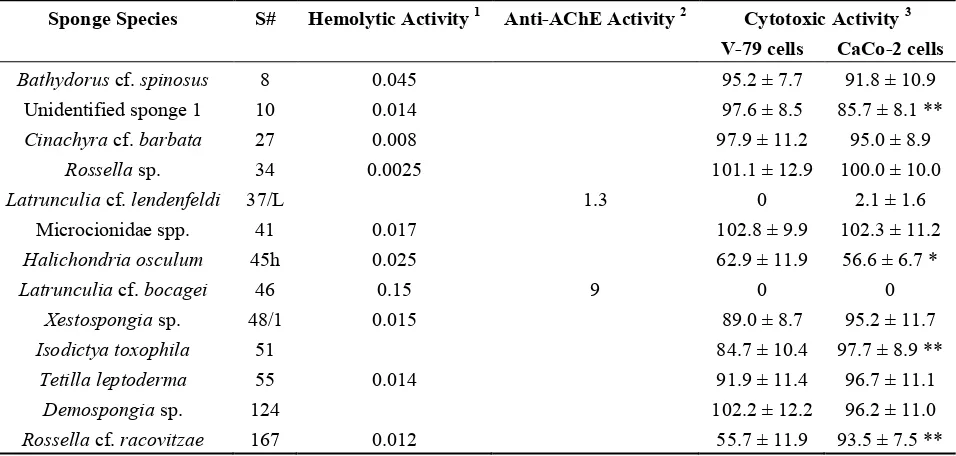
In contrast to our previous study on aqueous and organic extracts from tropical marine sponges, where we described at least one potent biological activity in each of the tested extract [12], ethanolic extracts from the Antarctic sponge species tested in the present study did not show such a wide spectrum of bioactivities. The sponge genus that showed the broadest spectrum and highest activities here was Latrunculia (i.e., extracts #37/L and #46). This sponge genus is known to produce numerous bioactive natural products, such as, for example, the cytotoxic discorhabdins [13,14] and the 2-thiazolidinone macrolides known as the latrunculins, which can disrupt microfilament organization [15].
Hemolytic activity was associated with only ten of the sponge samples and was most prominent in the extract of the sponge Latrunculia cf. bocagei (#46) (Table 1). However, it is interesting to note that the sponge extract from Latrunculia cf. lendenfeldi (#37/L) did not show any hemolytic activity. Although lower than the activity seen for extract #46, high hemolytic activity was detected in the extracts from Bathydorus cf. spinosus (#8), Halichondria osculum (#45h), Xestospongia sp. (#48/1), Rossella cf. racovitzae (#167), Tetilla leptoderma (#55), and from a sponge of the family of Microcionidae (#41). No previous data on hemolytic activities of the marine sponge extracts used in this study have been reported to date. In comparison, in our previous study on the biological activities of extracts from tropical marine sponges [12], hemolytic activity was present in about half of the organic extracts tested.
Table 1. Hemolytic, anti-acetylcholinesterase and cytotoxic activities of the most active sponge extracts. Empty spaces in columns denote that the tested sponge extract exhibited no hemolytic or anti-acetylcholinesterase (AChE) activity.
Sponge Species S# Hemolytic Activity 1 Anti-AChE Activity 2 Cytotoxic Activity 3 V-79 cells CaCo-2 cells
) at 400 μg dried extract/mL in the assay; 2 expressed as concentration of the dried extract (ng/mL) that resulted in 50% inhibition of the enzyme activity; 3 viability of V-79 and CaCo-2 cell lines treated with 100 μg/mL of the dried extract, expressed as % of control; significant difference in cytotoxic activity between V-79 and CaCo-2 cell lines (* p < 0.05; ** p < 0.01).
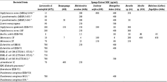
The discovery of new antibiotics is one of the most important goals in biomedical research, as the appearance of multiresistant bacterial strains has made certain human and animal infections virtually untreatable. Sponges are known to contain a high number of compounds that act against terrestrial pathogenic bacteria, while considerably lower activities have been observed against marine bacteria [18]. Furthermore, in comparison with sponges found in temperate and tropical seas, Antarctic sponges have been reported to have a smaller number of antimicrobial secondary metabolites [19] that show generally weaker activities [20]. Previous screenings of crude extracts from 93 Arctic sponges against bacteria and fungi associated with opportunistic infections showed that about 10% of the sponges yielded significant antimicrobial activities, with IC50 values from 0.2 to 5 μg/mL [5]. In the present study, only eight
Table 2. Antibacterial activities (MICs) of the sponge extracts against the laboratory, commensal and clinically relevant bacterial strains. Empty spaces in columns denote that the tested sponge extract exhibited no antibacterial activity.
Bacterial Strain Sponge Extract MIC (μg/mL)
Pseudomonas aeruginosa 06131 A 700 400
Pseudomonas aeruginosa 8591 A
A
Our data are generally in line with those obtained by McClintock and Gauthier [20], who screened non-polar extracts of 17 Antarctic sponges for inhibitory activities against bacteria and fungi. They showed particularly strong inhibitory activities associated with extracts from the sponge species belonging to the genera Latrunculia and Haliclona. The antibacterial activity of the extract from L. cf. lendenfeldi might be related to the presence of Latrunculia-associated natural products that have already been reported to have inhibitory potential against various Gram-positive and Gram-negative bacteria, which are known as the discorhabdins [21–23] and trunculins [24]. Furthermore, sponges of the genus Haliclona, which also include polar species, are known to contain antibacterial 3-alkylpyridinium alkaloids [25,26]. Similarly, sesquiterpenoids halichonadins [27] and a galactoside-specific lectin [28] from Halichondria sponges have been described to have antibacterial and antifungal effects, while extracts from sponges of the genus Hemigellius have not yet been reported to show any antibiotic properties. However, the present study shows antibacterial activities of Hemigellius bidens extract against all of the tested Gram-positive bacterial strains and also against a clinical multiresistant Pseudomonas aeruginosa isolate. Furthermore, the same extract inhibited the growth of E. coli laboratory strains and, to a lesser extent, of an extended-spectrum β-lactamase (ESBL)-producing clinical isolate, although it was ineffective against the pandemic, virulent and multiresistant E. coli ST131 isolate. These data should be borne in mind when screening tests are performed using strains that are solely from nonclinical environments. Weak antimicrobial activities of extracts from sponges belonging to the Antarctic genera of Isodictya have also been reported [20,29]. Moreover, a P. aeruginosa strain that is associated with this sponge was shown to have alkaloids that can inhibit the growth of Gram-positive bacteria [30]. Antimicrobial substances that have been derived from sponge-associated bacteria have also been reported for several other sponge species [18], and these might be responsible in part for the observed antimicrobial activities. Screening of methanolic extracts of Myxilla arenaria from the Indian Ocean did not reveal any activity against pathogenic bacteria and fungi [31]. To the best of our knowledge, there are no other reports on antibacterial activities of this sponge genus.
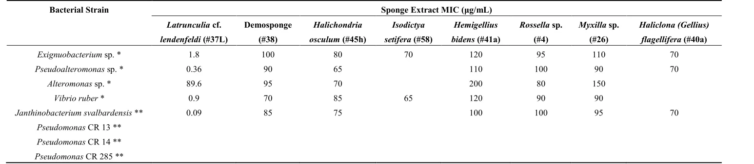
bacteria are probably not a common threat to sponges [29]; indeed, these symbiotic bacteria might instead be used as an alternative source of nutrients when these are scarce in the sponge environment [6].
Table 3. Antibacterial activities (minimal inhibitory concentrations (MICs)) of the sponge extracts against the environmental bacterial strains. Empty spaces in columns denote that the tested sponge extract exhibited no antibacterial activity.
Bacterial Strain Sponge Extract MIC (μg/mL)
Latrunculia cf.
lendenfeldi (#37L)
Demosponge
(#38)
Halichondria
osculum (#45h)
Isodictya
setifera (#58)
Hemigellius
bidens (#41a)
Rossella sp.
(#4)
Myxilla sp.
(#26)
Haliclona (Gellius)
flagellifera (#40a)
Exignuobacterium sp. * 1.8 100 80 70 120 95 110 70
Pseudoalteromonas sp. * 0.36 90 65 110 100 90 70
Alteromonas sp. * 89.6 95 70 200 80 150
Vibrio ruber * 0.9 70 85 65 120 90 90
Janthinobacterium svalbardensis ** 0.09 85 75 100 100 95 70
Pseudomonas CR 13 **
Pseudomonas CR 14 **
Pseudomonas CR 285 **
3.Experimental Section
3.1. Sponge Collection
Thirty-three sponge specimens that represented 28 species were collected in the Antarctic waters (60° to 70° S; 8° to 61° W) by bottom trawling and dredging at depths between 200 m and 900 m, during two cruises of the German Research Vessel ―Polarstern‖ in 2006/07 and 2008. The specimens were identified to at least the family level (Supplementary Table S1), and they were immediately frozen and kept at −20 °C. The sponge samples were lyophilized prior to the extraction.
3.2. Preparation of Extracts
The weights of the lyophilized sponge samples were in the range of 0.52 g to 3.78 g. The lyophilized material was macerated and placed into labelled glass tubes, and 10 mL 96% ethanol (Merck, Germany) was added to each tube. The tubes were sealed with metal stoppers and parafilm and were shaken overnight (600 rpm) at 37 °C. The extracts were then filtered, and the solvent was evaporated to the final volume of 1 mL. The dry weight of each sample was determined by drying an aliquot of a sample in a preweighed round-bottomed flask, with evaporation of the solvent under vacuum at 45 °C. The dry weight was expressed in mg/mL of the 1 mL extract volumes prepared. Stock concentrations were in the range of 3.1 mg/mL to 10.3 mg/mL. The sponge species and the dry weights of the extracts are given in Supplementary Table S1.
3.3. Hemolytic Activity Assay
Fresh bovine erythrocytes were washed three times in physiological saline prior to use and then resuspended in erythrocyte buffer (130 mM NaCl, 20 mM TRIS-HCl, pH 7.4). The erythrocyte suspension had an initial absorption at 650 nm of 1.0 ± 0.01 AU. The hemolytic activity was assayed using a microplate VIS absorption reader (Dynex, USA), as described previously [12]. With 100 μL of erythrocyte buffer in each microplate well, the ethanolic sponge extracts were added to each well at different final dry-extract concentrations, followed by 100 μL erythrocyte suspension. The volume of ethanol in the final reaction mixture did not exceed 20%, a concentration that was tested and shown not to be lytic. The time course of hemolysis was immediately started and monitored for 30 min. The hemolytic activity was expressed as the half-time of hemolysis (t50); e.g., the time in which the apparent
absorbance at 650 nm dropped from 0.5 to 0.25. All of these measurements were carried out in triplicates at 25 °C. The samples showing the highest hemolytic activities were further diluted with ethanol (1:10 and 1:100) for confirmation of the hemolytic activities in further assays.
3.4. Antibacterial Activity Assay
antibacterial activities were also assayed against the following characterized strains of different origins: (1) laboratory strains: E. coli HB101, Bacillus subtilis EXB-V68, Enterobacter EXB-V11, P. aeruginosa EXB-V28 and Staphylococcus epidermidis EXB-V55; (2) commensal isolates from dog skin: S. aureus 10F, Macrococcus 1F, Micrococcus 2F and Acinetobacter 1C; (3) a food isolate: Listeria monocytogenes; and (4) clinical isolates: methicillin-resistant S. aureus (MRSA) S-943, methicillin-resistant Staphylococcus pseudintermedius (MRSP) S-053 and S-043 ESBL-producing E. coli 206 (CTX-M-1 group; ST131), ESBL-E. coli 192 (CTX-M-9; ST131), ESBL-E. coli 30 (CTX-M-2), carbapenemase producing (KPC) Klebsiella pneumoniae, P. aeruginosa 06131 and P. aeruginosa 8591. The strains were obtained from the EX (extremophilic microorganisms) and GM (genetic laboratory microbes) culture collections of the Chair of Molecular Genetics and Microbiology of the Biotechnical Faculty and of the Institute of Microbiology and Parasitology, Veterinary Faculty, University of Ljubljana, Slovenia. The precultured bacteria (laboratory, commensal and clinically relevant strains and strains isolated from Arctic ice) were grown in LB broth (Sigma, USA) and were used for the inoculation of Luria broth agar plates, to a final cell concentration of 5 × 10⁸/mL. Four holes of 1 cm in diameter were made in the agar of each agar plate, which were then filled with 100 μL of an
ethanolic extract. Ethanol was tested for its antimicrobial activity (100 μL) as a control. The inhibition
zone for each sample was determined after overnight incubation of the plates at 37 °C. The plates containing the bacteria isolated from Arctic ice were incubated at 22 °C. The extracts showing the highest inhibition of bacterial growth were further diluted with ethanol and used to determine the minimal inhibitory concentrations (MICs), which were defined as the lowest concentrations in μg/mL that inhibited the growth of tested microorganism 1 mm from the rim of the hole. All of the laboratory, commensal and clinical bacterial strains were also assayed with standard antibiotics (tetracycline, kanamycin, rifampicin, ampicillin and chloramphenicol; Supplementary Table S2). The marine bacteria were precultured in liquid medium prepared by dissolving 5 g peptone and 1 g yeast extract in 1 L of aqueous solution of MgCl2·6H2O (10 mM) and NaCl (300 mM). Agar plates containing this liquid
medium were inoculated with the bacteria to a concentration of 5 × 10⁸/mL, and the antibacterial activities were tested as described above after an overnight incubation of the plates at 37 °C.
3.5. Acetylcholinesterase Inhibition Assay
3.6. Cytotoxic Activity
In contrast to the previous biological tests, the cytotoxic activity was assayed only on selected sponge extracts, as those from: Bathydorus cf. spinosus (#8), non-identified sponge 1(#10), Cinachyra cf. barbata (#27), Rossella sp. (#34), Latrunculia cf. lendenfeldi (#37/L), Microcionidae spp. (#41), Halichondria osculum (#45h), Latrunculia cf. bocagei (#46), Xestospongia (#48/1), Isodictya toxophila (#51), Tetilla leptoderma (#55), Demospongiae (#124) and Rossella cf. racovitzae (#167). The cell lines used were: V-79-379 A (V-79) cells (diploid lung fibroblasts from Chinese hamster) and CaCo-2 cells (human colon adenocarcinoma). The V-79 cells were grown in advanced Eagle’s minimal essential medium (Gibco, Invitrogen, UK) and the CaCo-2 cells in advanced RPMI 1640 (Gibco), both at 37 °C in a CO2 incubator (5% CO2, 95% air, 95% relative humidity). Both of these culture media were
supplemented with 2 mmol/L L-glutamine (Gibco), 100 U/mL penicillin (Gibco), 100 μg/mL streptomycin (Gibco) and 5% fetal bovine serum (Gibco). For the in vitro cytotoxicity assay, the cells were plated in 96-well microtiter plates (100 μL, Costar, USA) at a concentration of 5000 cells/well (V-79 cells) and 10,000 cells/well (CaCo-2 cells). After a 3-h incubation, the ethanol-dissolved extracts prepared in the respective media without serum were added, to a final concentration of 0.1 mg/mL, and the incubations were carried out for 1 h (under cell growth conditions). Ethanol (20 μL) was used as a control. The cells were then washed once with medium, and fresh medium with fetal bovine serum was added for a further 48 h (under cell growth conditions). The cytotoxicity was determined using the MTS (=3-(4,5-dimethylthiazol-2-yl)-5-(3-carboxymethoxyphenyl)-2-(4-sulfophenyl)-2H-tetrazolium) test. To each well, 20 μL of MTS (CellTiter 96 AQueous Reagent, Promega, USA) was added to the cell culture. After 1 h, the absorbance at 490 nm was measured using a Bio-Tek microplate reader (Bio-Tek Instruments Inc., USA). The absorption corresponded to the amount of the soluble formazan product that was formed, which is directly proportional to the number of viable cells. The viability was calculated as the ratio between absorbance at 490 nm of the treated and control cells, expressed as a percentage. The data are presented by means ± SD of 3 independent experiments. The differences were analyzed using Student’s t-tests on two populations, with p < 0.05 and p < 0.01 considered significant.
4. Conclusions
The data from the present study are in line with previous reports on biological activities of polar marine sponges. Although the biological activities of these sponges are in generally associated with a lower number of species as compared to sponges from temperate and tropical regions, they are still widely unexplored and might provide valuable resources for new pharmaceutical lead compounds. One such example is seen with the extracts of the sponges of the genus Latrunculia, which were shown to contain an extremely potent inhibitor of AChE.
Acknowledgments
and ANT-XXIV/2, respectively, and the captain and crew of the RV Polarstern for their constant help and support. The German Research Society (DFG) is acknowledged for financial support to projects on Antarctic sponges (JA-1063/14-1,2; JA-1063/17).
Conflict of Interest
The authors declare that they have no conflicts of interest.
References
1. Molinski, T.F.; Dalisay, D.S.; Lievens, S.L.; Saludes, J.P. Drug development from marine natural products. Nat. Rev. Drug Discov. 2009, 8, 69–85.
2. Mayer, A.M.; Glaser, K.B.; Cuevas, C.; Jacobs, R.S.; Kem, W.; Little, R.D.; McIntosh, J.M.; Newman, D.J.; Potts, B.C.; Shuster, D.E. The odyssey of marine pharmaceuticals: A current pipeline perspective. Trends Pharmacol. Sci. 2010, 31, 255–265.
3. Hu, G.P.; Yuan, J.; Sun, L.; She, Z.G.; Wu, J.H.; Lan, X.J.; Zhu, X.; Lin, Y.C.; Chen, S.P. Statistical research on marine natural products based on data obtained between 1985 and 2008. Mar. Drugs
2011, 9, 514–525.
4. Lebar, M.D.; Heimbegner, J.L.; Baker, B.J. Cold-water marine natural products. Nat. Prod. Rep.
2007, 24, 774–797.
5. Abbas, S.; Kelly, M.; Bowling, J.; Sims, J.; Waters, A.; Hamann, M. Advancement into the Arctic region for bioactive sponge secondary metabolites. Mar. Drugs 2011, 9, 2423–2437.
6. McClintock, J.B.; Amsler, C.D.; Baker, B.J.; van Soest, R.W.M. Ecology of Antarctic marine sponges: An overview. Integr. Comp. Biol. 2005, 45, 359–368.
7. Janussen, D.; Tendal, O.S. Diversity and distribution of porifera in the bathyal and abyssal Weddell Sea and adjacent areas. Deep-Sea Res. II 2007, 54, 1864–1875.
8. Wilkins, S.P.; Blum, A.J.; Burkepile, D.E.; Rutland, T.J.; Wierzbicki, A.; Kelly, M.; Hamann, M.T. Isolation of an antifreeze peptide from the Antarctic sponge Homaxinella balfourensis. Cell. Mol. Life Sci. 2002, 59, 2210–2215.
9. Kunzmann, K. Associated Fauna of Selected Sponges (Hexactinellida and Demospongiae) from the Weddell Sea, Antarctica; Alfred Wegener Institute for Polar and Marine Research: Bremerhaven, Germany, 1996; Volume 210, pp. 1–93.
10. Xin, Y.; Kanagasabhapathy, M.; Janussen, D.; Xue, S.; Zhang, W. Phylogenetic diversity of Gram-positive bacteria cultured from Antarctic deep-sea sponges. Polar Biol. 2011, 34, 1501–1512.
11. Vetter, W.; Janussen, D. Halogenated natural products in five species of Antarctic sponges: Compounds with POP-like properties. Environ. Sci. Technol. 2005, 39, 3889–3895.
12. Sepčić, K.; Kauferstein, S.; Mebs, D.; Turk, T. Biological activities of aqueous and organic extracts from tropical marine sponges. Mar. Drugs 2010, 8, 1550–1566.
14. Na, M.; Ding, Y.; Wang, B.; Tekwani, B.L.; Schinazi, R.F.; Franzblau, S.; Kelly, M.; Stone, R.; Li, X.C.; Ferreira, D.; et al. Anti-infective discorhabdins from a deep-water Alaskan sponge of the genus Latrunculia. J. Nat. Prod. 2010, 73, 383–387.
15. Yarmola, E.G.; Somasundaram, T.; Boring, T.A.; Spector, I.; Bubb, M.R. Actin-latrunculin A structure and function: Differential modulation of actin-binding protein function by latrunculin A. J. Biol. Chem. 2000, 275, 28120–28127.
16. Kaur, J.; Zhang, M.Q. Molecular modelling and QSAR of reversible acetylcholinesterase inhibitors. Curr. Med. Chem. 2000, 7, 273–294.
17. Nèeman, I.; Fishelson, L.; Kashman, Y. Isolation of a new toxin from the sponge Latrunculia magnifica in the Gulf of Aquaba (Red Sea). Mar. Biol. 1975, 30, 293–296.
18. Laport, M.S.; Santos, O.C.; Muricy, G. Marine sponges: Potential sources of new antimicrobial drugs. Curr. Pharm. Biotechnol. 2009, 10, 86–105.
19. Lippert, H.; Brinkmeyer, R.; Mülhaupt, T.; Iken, K. Antimicrobial activity in sub-Arctic marine invertebrates. Polar Biol. 2003, 26, 591–600.
20. McClintock, J.B.; Gauthier, J.J. Antimicrobial activities of Antarctic sponges. Antarc. Sci. 1992, 4, 179–183.
21. Perry, N.B.; Blunt, J.W.; Munro, M. Cytotoxic pigments from New Zealand sponges of the genus Latrunculia: Discorhabdins a, b and c. Tetrahedron 1988, 44, 1727–1734.
22. Copp, B.R.; Fulton, K.F.; Perry, N.B.; Blunt, J.W.; Munro, M.H.G. Natural and synthetic derivatives of discorhabdin C, a cytotoxic pigment from the New Zealand sponge Latrunculia cf. bocagei. J. Org. Chem. 1994, 59, 8233–8238.
23. Ford, J.; Capon, R. Discorhabdin R: A new antibacterial pyrroloiminoquinone from two latrunculiid marine sponges, Latrunculia sp. and Negombata sp. J. Nat. Prod. 2000, 63, 1527–1528. 24. Capon, R.J.; MacLeod, J.K.; Willis, A.C. Trunculins A and B, norsesterterpene cyclic peroxides
from a marine sponge, Latrunculia brevis. J. Org. Chem. 1987, 52, 339–342.
25. Turk, T.; Sepčić, K.; Mancini, I.; Guella, G. 3-Akylpyridinium and 3-alkylpyridine compounds from marine sponges, their synthesis, biological activities and potential use. Stud. Nat. Prod. Chem.
2008, 35, 355–397.
26. Timm, C.; Mordhorst, T.; Kock, M. Synthesis of 3-alkyl pyridinium alkaloids from the arctic sponge Haliclona viscosa. Mar. Drugs 2010, 8, 483–497.
27. Ishiyama, H.; Hashimoto, A.; Fromont, J.; Hoshino, Y.; Mikami, Y.; Kobayashi, J. Halichonadins A–D, new sesquiterpenoids from a sponge Halichondria sp. Tetrahedron 2005, 61, 1101–1105. 28. Kawsar, S.M.A.; Mamun, S.M.A.; Rahman, M.S.; Yasumitsu, H.; Ozeki, Y. In-vitro antibacterial
and antifungal effects of a 30 kDa D-galactoside-specific lectin from the Demosponge, Halichondria okadai. Int. J. Biol. Life Sci. 2010, 6, 31–37.
29. Peters, K.J.; Amsler, C.D.; McClintock, J.B.; Baker, B.J. Potential chemical defenses of Antarctic sponges against sympatric microorganisms. Polar Biol. 2010, 33, 649–658.
30. Jayatilake, G.S.; Thornton, M.P.; Leonard, A.C.; Grimwade, J.E.; Baker, B.J. Metabolites from an Antarctic sponge-associated bacterium, Pseudomonas aeruginosa. J. Nat. Prod. 1996, 59, 293–296.
32. Li, H.; Matsunaga, S.; Fusetani, N. Halicylindramides A–C, antifungal and cytotoxic depsipeptides from the marine sponge Halichondria cylindrata. J. Med. Chem. 1995, 38, 338–343.
33. Zhang, H.J.; Sun, J.B.; Lin, H.W.; Wang, Z.L.; Tang, H.; Cheng, P.; Chen, W.S.; Yi, Y.H. A new cytotoxic cholesterol sulfate from marine sponge Halichondria rugosa. Nat. Prod. Res. 2007, 21, 953–958.
34. Hirata, Y.; Uemura, D. Halichondrins—Antitumor polyether macrolides from a marine sponge. Pure Appl. Chem. 1986, 58, 701–710.
35. Pettit, G.R.; Herald, C.L.; Boyd, M.R.; Leet, J.E.; Dufresne, C.; Doubek, D.L.; Schmidt, J.M.; Cerny, R.L.; Hooper, J.N.; Rützler, K.C. Antineoplastic agents. 219. Isolation and structure of the cell growth inhibitory constituents from the western Pacific marine sponge Axinella sp. J. Med. Chem. 1991, 34, 3339–3340.
36. Ellman, G.L.; Courtney, D.; Andres, V.; Featherstone, R.M. A new and rapid colorimetric determination of acetylcholinesterase activity. Biochem. Pharmacol. 1961, 7, 88–95.