~ 113 ~
Journal of Pharmacognosy and Phytochemistry 2013; 2 (4): 113-115
ISSN 2278-4136 ISSN 2349-8234 JPP 2013; 2 (4): 113-115 © 2013 AkiNik Publications Received: 12-10-2013 Accepted: 28-10-2013
Venkata Sai Prakash Chaturvedula Natural Ingredient Development, Blue California, 30111 Tomas, Rancho Santa Margarita,
CA 92688, USA.
Ruo Huang
Natural Ingredient Development, Blue California, 30111 Tomas, Rancho Santa Margarita,
CA 92688, USA.
Correspondence:
Venkata Sai Prakash Chaturvedula Natural Ingredient Development, Blue California, 30111 Tomas, Rancho Santa Margarita, CA 92688, USA
Email: [email protected]
Tel: +1-949-257-7351
Isolation and NMR Spectral Studies of
Dihydromyricetin
Venkata Sai Prakash Chaturvedula,Ruo Huang
ABSTRACT
From the alcoholic extract of the leaves of Hovenia dulcis, a pentahydroxyl flavanonol has been isolated as a major compound, whose structure has been characterized as dihydromyricetin (DHM) on the basis of extensive 1D and 2D Nuclear Magnetic Resonance (NMR) as well as High Resolution Mass Spectral (HRMS) data. Also, a comparative study of the 1H and 13C NMR spectral data of
dihydromyricetin has been studied in three different solvent systems namely C5D5N (d5-pyridine) or
CD3OD (d4-methanol) or C2D6SO (d6-DMSO).
Keywords: Hovenia dulcis, Flavanonol, Dihydromyricetin, 1D and 2D NMR spectral data, Structure characterization, NMR study
1. Introduction
Hovenia dulcis Thunb, also known as Japanese raisin tree, is commonly found in East Asia
over the eastern China and Korea to the Himalayas. H. dulcis belongs to the family of
Rhamnaceae has a long history as a food supplement and traditional medicine in Japan, China and Korea, but is little known and used in Western countries so far [1]. Extracts from H. dulcis
accelerate possess various medicinal properties including hepatoprotective, antioxidative, antimicrobial and antidiabetic as well as detoxification of ethanol. Although the underlying
molecular mechanisms of H. dulcis are not fully understood, free radical scavenging and
enhancement of ethanol catabolism have been reported in the literature. H. dulcis is a glabrous
tree with lenticular branches, and grows up to 10 m, which cultivated in plantations in China, invasive in South American rainforests and Tanzania. The tree has been introduced as an ornamental tree to several countries, and the fruit is also edible. The taste of the fleshy peduncles is like a combination of raisin, clove, cinnamon and sugar. The peduncles contain
high levels of sugar, while leaves of H. dulcis contain several dammarane-type of terpene
sweetness inhibitors. Earlier phytochemical analysis of H. ducis resulted in the isolation of
several terpenes, phenolic and fatty compounds [2-4].
In our continuing research to discover bioactive natural compounds, we have been on various plant species obtained across the globe. Recently, we have reported several minor diterpene
glycosides from the commercial extracts of Stevia rebaudiana Bertoni and their structures have
been characterized based on the 1D and 2D NMR spectral data as well as chemical studies [5-6].
In this article, we are describing the isolation of a pentahydroxyl flavanonol namely
dihydromyricetin (1) from the leaves of H. dulcis. Further, we are reporting the 1H and 13C
NMR spectral data studies of 1 in the deuterated solvents namely d5-pyridine, d4-methanol, and d6-DMSO.
2. Materials and Methods 2.1. General Instrumentation
The 1D and 2D NMR spectral data were acquired on Bruker Avance DRX 500 MHz or Varian INOVA 600 MHz instrument instruments using standard pulse sequences. The NMR spectra
were performed in C5D5N (d5-pyridine) or CD3OD (d4-methanol) or C2D6SO (d6-DMSO);
chemical shifts are given in (ppm), and coupling constants are reported in Hz. MS and
~ 114 ~ Journal of Pharmacognosy and Phytochemistry
Samples were diluted with methanol and introduced via infusion using the onboard syringe pump.
2.2. Isolation and purification of dihydromyricetin (1)
The leaves of H. dulcis were extracted with ethanol and
concentrated under vacuum at low temperature. The residue obtained has been centrifuged and then finally crystallized to yield a pure compound which has been identified as dihydromyricetin (1).
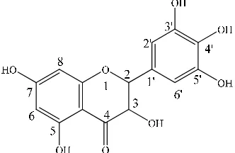
2.3. Identification and spectroscopic data of dihydromyricetin (5, 7, 3′, 4′, 5′-pentahydroxyl flavanonol, 1)
Off-White powder; 1H-NMR (600 MHz,
d5-pyridine/d4-methanol/d6-DMSO, δ ppm) and 13C-NMR (150 MHz,
d5-pyridine/d4-methanol/d6-DMSO, δ ppm) spectroscopic data see
Table 1; HRMS (M+Na) + m/z 343.0426 (calcd. for C
15H12O8Na:
343.0424).
3. Results and Discussion
Compound 1 was isolated as an off-white powder, and its
molecular formula has been was deduced as C15H12O8 from the
adduct ion corresponding to [M+Na]+ ion observed at m/z
343.0426; this composition was further supported by the 13C NMR
spectral data.. The UV spectrum of 1 showed λ max at 261, 319,
and 348 nm suggested a flavonoid structure 7-9. The 1H NMR
spectra data of 1 has been acquired in all three solvents namely
d5-pyridine, d4-methanol and d6-DMSO. The 1H NMR spectra data of
1 showed two doublet signals between δ 4.48 and 5.47 in
d5-pyridine, and d4-methanol, whereas a doublet and doublet of doublets at δ 4.42 and 4.91 in d6-DMSO corresponding to a proton each suggested the 3-hydroxyflavanone or 2, 3-dihydroflavonol
skeleton in the molecular structure of 1. The presence of 2,
3-dihydroflavonol was further supported by the 13C NMR spectral
data which showed the presence of oxymethine groups resonating between δ 71.7 and 85.8. In addition, the 1H NMR spectra data of 1
also showed the presence of two meta-coupled aromatic protons as doublets between δ 5.86 and 6.50, and an additional two meta-coupled aromatic protons δ 6.40 and 7.24 as singlets corresponds to
a pentahydroxyl flavanonol scaffold. The 1H and 13C NMR values
for all the protons and carbons for the compound 1 were assigned
on the basis of COSY, HMQC and HMBC correlations and are given in Table 1. The key HMBC correlations confirmed the placement of all the five hydroxyl groups at 5, 7, 3′, 4′, 5′ positions as shown in Figure 1. On the basis of above 1D and 2D NMR
spectroscopic data, the structure of 1 was determined
unambiguously as dihydromyricetin (5, 7, 3′, 4′, 5′-pentahydroxyl flavanonol) [10].
Table 1. 1H and 13C NMR spectral data (chemical shifts and coupling constants) for dihydromyricetin (1) a-c.
Position
a assignments made on the basis of COSY, HMQC and HMBC correlations; b Chemical shift values are in δ (ppm); c Coupling constants are in Hz.
4. Conclusion
We are herewith reporting the isolation of the main constituent of the alcoholic extract of the leaves of Hovenia dulcis, whose structure has been characterized as dihydromyricetin (1) on the basis of extensive 1D (1H and 13C) and 2D (COSY, HMQC and
HMBC) NMR as well as High Resolution Mass Spectral (HRMS) data. Also, we are herewith reporting for the first time the 1H and 13C NMR spectral data of dihydromyricetin in three different
~ 115 ~ Journal of Pharmacognosy and Phytochemistry
5. Acknowledgements
We wish to thank Dr. Wu Shaoxiong, and Dr. Bing Wang of Emory University, Atlanta, USA for obtaining some selected spectral data and other chemistry related help.
6. References:
1. Hyun TK, Eom SH, Yu CY, Roitsch T. Hovenia dulcis - An Asian
traditional herb. Planta Medica 2010; 76:943-949.
2. Cho JY, Moon JH, Eun JB, Chung SJ, Park KH. Isolation and
characterization of 3(Z)-dodecenedioic acid as an antibacterial
substance from Hovenia dulcis Thunb. Food Science and
Biotechnology 2004; 13:46-50.
3. Cho JY, Hyun SH, Moon JH, Park KH. Isolation and structural
determination of a novel flavonol triglycoside and seven
compounds from the leaves of oriental raisin tree (Hovenia dulcis)
and their antioxidative activity. Food Science and Biotechnology 2013; 22:115-123.
4. Suttisri R, Lee IS, Kinghorm AD. Plant-derived triterpenoid
sweetness inhibitors. Journal of Ethnopharmacology 1995; 47:9–26.
5. Chaturvedula VSP, Chen S, Yu O, Mao G. NMR spectral analysis
and hydrolysis studies of rebaudioside N, a minor steviol glycoside of
Stevia rebaudiana Bertoni. Food and Nutrition Sciences 2013;
4:1004-1008.
6. Chaturvedula VSP, Chen S, Yu O, Mao G. Isolation, NMR spectral
analysis and hydrolysis studies of a hepta pyranosyl diterpene
glycoside from Stevia rebaudiana Bertoni. Biomolecules 2013;
3:733-740.
7. Nakano K, Takatani M, Tomimatsu T, Nohara T. Four kaempferol
glycosides from the leaves of Cinnamomum sieboldi. Phytochemistry
1983; 22:2831-2833.
8. Chaturvedula VSP, Prakash I. Kaempferol glycosides from Siraitia
grosvenorii,Journal of Chemical and Pharmaceutical Research 2011;
3:799-804.
9. Chaturvedula VSP, Prakash I. Flavonoids from Astragalus
propinquus, Journal of Chemical and Pharmaceutical Research 2012; 4:2666-2670.
10. Zhang YS, Zhang QY, Wang B, Li LY, Zhao YY. Chemical
constituents from Ampelopsis grossedentata. Journal of Chinese