www.elsevier.com / locate / bres
Interactive report
1
The neurobiology of stress: from serendipity to clinical relevance
*
Bruce S. McEwen
Harold and Margaret Milliken Hatch Laboratory of Neuroendocrinology, The Rockefeller University, 1230 York Avenue, Box 165, New York,
NY10021, USA Accepted 25 September 2000
Abstract
The hormones and other physiological agents that mediate the effects of stress on the body have protective and adaptive effects in the short run and yet can accelerate pathophysiology when they are over-produced or mismanaged. Here we consider the protective and damaging effects of these mediators as they relate to the immune system and brain. ‘Stress’ is a principle focus, but this term is rather imprecise. Therefore, the article begins by noting two new terms, allostasis and allostatic load that are intended to supplement and clarify the meanings of ‘stress’ and ‘homeostasis’. For the immune system, acute stress enhances immune function whereas chronic stress suppresses it. These effects can be beneficial for some types of immune responses and deleterious for others. A key mechanism involves the stress–hormone dependent translocation of immune cells in the blood to tissues and organs where an immune defense is needed. For the brain, acute stress enhances the memory of events that are potentially threatening to the organism. Chronic stress, on the other hand, causes adaptive plasticity in the brain, in which local neurotransmitters as well as systemic hormones interact to produce structural as well as functional changes, involving the suppression of ongoing neurogenesis in the dentate gyrus and remodelling of dendrites in the Ammon’s horn. Under extreme conditions only does permanent damage ensue. Adrenal steroids tell only part of the story as far as how the brain adapts, or shows damage, and local tissue modulators — cytokines for the immune response and excitatory amino acid neurotransmitters for the hippocampus. Moreover, comparison of the effects of experimenter-applied stressors and psychosocial stressors show that what animals do to each other is often more potent than what experimenters do to them. And yet, even then, the brain is resilient and capable of adaptive plasticity. Stress-induced structural changes in brain regions such as the hippocampus have clinical ramifications for disorders such as depression, post-traumatic stress disorder and individual differences in the aging process. 2000 Elsevier Science B.V. All rights reserved.
Keywords: Stress; Homeostasis; Allostasis; Immune function; Adaptive plasticity; Hippocampus; Dendrite; Neurotransmitter; Glucocorticoid; Adrenalec-tomy; Adrenal steroid; Learning; Memory; Cognitive function; Psychiatric disorder
1. Introduction hormones are protective in the short run and yet can participate in damage when they are overproduced or not Stress is an aspect of our daily lives and conversations, shut off when no longer needed.
and yet there is considerable ambiguity in the meaning of Animals are continually learning and some experiences this word. The brain is the master controller of the are classified as ‘stressful’ in part because stress hormones interpretation of what is stressful and the behavioral and are produced. Contrary to the late Hans Selye, who physiological responses that are produced. The brain is emphasized physical stressors [133], psychological and also a target of stress, along with the immune system, experiential factors are among the most powerful of metabolic and cardiovascular systems and other systems of stressors: e.g., novelty, withholding of reward, and antici-the body. Stress hormones play a major role in mediating pation of punishment rather than the punishment itself are both adaptive and maladaptive responses, and they do so among the most potent activators of HPA and ANS activity by interacting with specific aspects of the physiology of [89,90].
each tissue. What is often overlooked is that the stress Although stress is often thought about as bad and damaging, recent studies paint a different picture as far as
1 the brain and also the immune system are concerned. The
Published on the World Wide Web on 22 November 2000.
main point is that the brain appears to handle repeated *Tel.:11-212-327-8624; fax:11-212-327-8634.
E-mail address: [email protected] (B.S. McEwen). stress over weeks by showing adaptive plasticity in which
local neurotransmitters as well as systemic hormones Behaviorally, the response to stress may consist of fight-interact to produce structural as well as functional changes. or-flight reactions or potentially health-related behaviors Likewise, the immune system responds to acute stress by such as eating, alcohol consumption, smoking and other showing enhanced responses, and this is mediated by forms of substance abuse. Another type of reaction to a adrenal steroids and catecholamines, as well as by locally- potentially stressful situation is an increased state of produced cytokines and cell adhesion molecules. Thus, vigilance, accompanied, at least in our own species, by systemic levels of adrenal steroids and catecholamines, the enhanced anxiety and worrying, particularly when the classical stress hormones, do not tell the whole story as far threat is ill-defined or imaginary and when there is no clear as how the brain adapts. Moreover, comparison of the alternative behavioral response that would end the threat. effects of experimenter-applied stressors and psychosocial The behavioral responses to stress and these states of stressors show that what animals do to each other is often anxiety are both capable of exacerbating and potentiating more potent than what we, as experimenters, do to them. the production of the physiological mediators of health Yet, even then, there is reason to believe that the brain is outcomes.
resilient and capable of adaptive plasticity. The changes in
the brain and immune system produced by acute and 2.2. Homeostasis repeated stress and the underlying mechanisms have turned
out to have unexpected clinical ramifications. After discus- Homeostasis, in a strict sense, applies to a limited sing these, the article will consider future directions of this number of systems like pH, body temperature and oxygen research and consider important unanswered questions. tension, components of the internal milieu, that are truly essential for life and are, therefore, maintained over a narrow range, as a result of their critical role in survival.
2. Protective and damaging effects of stress These systems are not activated or varied in order to help
mediators: homeostasis and allostasis the individual adapt to its environment. In contrast, systems that show ‘variation to meet perceived / anticipated Before discussing the brain and its adaptive responses to demands’ [145] characterizes the state of the organism in a stress, it is important to consider the definition of some key changing world and reflects the operation of most body terms. Stress is often defined as a threat, real or implied, to systems in meeting environmental challenges, e.g., through homeostasis, and homeostasis refers to the maintenance of fluctuating hormones, heart rate and blood pressure, cyto-a ncyto-arrow rcyto-ange of vitcyto-al physiologiccyto-al pcyto-arcyto-ameters necesscyto-ary kines of the immune system, and other tissue mediators for survival. In common usage, stress usually refers to an like neurotransmitters and hormones. Those mediators are event or succession of events that cause a response, often most certainly not held constant, although their levels may in the form of ‘distress’ but also, in some cases, referring usually operate within a range, and they participate in to a challenge that leads to a feeling of exhilaration, as in processes leading to adaptation as well as contributing to ‘good’ stress. But, the term ‘stress’ is full of ambiguities. It pathophysiology when they are produced insufficiently or is often used to mean the event (stressor) or the response in excess, i.e., outside of the normal range.
(stress response). It is frequently used in the negative sense
of ‘distress’, and sometimes it is used to describe a chronic 2.3. Allostasis state of imbalance in the response to stress. In this article,
‘stress’ will be used to describe an event or events that are Allostasis is a term introduced by Sterling and Eyer interpreted as threatening to an individual and which elicit [145] to characterize how blood pressure and heart rate physiological and behavioral responses. The brain is the responses vary with experiences and time of day and also key organ involved in interpretation and responding to to describe changes in the set point of these parameters in potential stressors. But before considering its role, the hypertension. The change in set point was used by them as following are some additional key terms and the way they the primary example that distinguishes allostasis from
are used in this article. homeostasis. Yet there is a much broader implication of
what they wrote. In their paper, they state: ‘‘Allostasis
2.1. Stress response emphasizes that the internal milieu varies to meet
Therefore, we propose that allostasis is a much better the short run by replenishing energy reserves after a period term for physiological coping mechanisms than is homeo- of activity, like running away from a predator. Glucocor-stasis, which should be reserved for the parameters that are ticoids also act on the brain to increase appetite for food essentially maintained for survival. Therefore, allostasis is and to increase locomotor activity and food seeking the process that keeps the organism alive and functioning, behavior [69], thus regulating behaviors which control i.e., maintaining homeostasis or ‘maintaining stability energy input and expenditure. This is very useful when we through change’ and promoting adaptation and coping, at do manual labor or play active sports, but it is not least in the short run [94,99]. beneficial when we grab a pizza and a beer while watching We note, however, that another view of homeostasis is television or writing a paper. Inactivity and lack of energy that it can also mean the operation of coordinated physio- expenditure creates a situation where chronically-elevated logical processes which maintain most of the steady states glucocorticoids that may result from either poor sleep, of the organism [16]. In this interpretation, homeostasis ongoing stress, or as side effects of rich diet can impede and allostasis might seem to mean almost the same thing. the action of insulin to promote glucose uptake. One of the The problem with this use of ‘homeostasis’ is that it does results of this interaction is that insulin levels increase, not distinguish between those systems essential for life and and, together, insulin and glucocorticoid elevations
pro-those that maintain them. mote the deposition of body fat and this combination of
What are some examples of allostasis? Sterling and Eyer hormones also promotes the formation of atherosclerotic [145] used variations in blood pressure as an example: e.g., plaques in the coronary arteries [6].
in the morning, blood pressure rises when we get out of For the heart, we see a similar paradox. Getting out of bed and blood flow is maintained to the brain when we bed in the morning requires an increase in blood pressure stand up in order to keep us conscious. This type of and a reapportioning of blood flow to the head so we can allostasis helps to maintain oxygen tension in the brain. stand up and not faint [145]. Our blood pressure rises and There are other examples: e.g., catecholamine and falls during the day as physical and emotional demands glucocorticoid elevations during physical activity mobilize change, providing adequate blood flow as needed. Yet and replenish, respectively, energy stores needed for brain repeatedly elevated blood pressure promotes generation of and body function under challenge. These adaptations atherosclerotic plaques, particularly when combined with a maintain essential metabolism and body temperature. supply of cholesterol and lipids and oxygen free radicals Examples of allostasis go beyond the immediate control that damage the coronary artery walls [87]. Beta adrenergic of body temperature and pH to broader aspects of in- receptor blockers are known to inhibit this cascade of dividual survival, e.g., from pathogens or physical danger. events and to slow down the atherosclerosis that is In the immune system, we will see below that acute accelerated in dominant male cynomologus monkeys ex-stress-induced release of catecholamines and glucocor- posed to an unstable dominance hierarchy [88]. Thus, ticoids facilitates the movement of immune cells to parts of catecholamines and the combination of glucocorticoids and the body where they are needed to fight an infection or to insulin can have dangerous effects on the body, besides produce other immune responses [25]. Finally, in the brain, their important short-term adaptive roles [6].
glucocorticoids and catecholamines act in concert to We now shall consider protective and damaging effects promote the formation of memories of events of potentially of mediators of allostasis in the immune system and brain, dangerous situations so that the individual can avoid them two systems that are less well-understood.
in the future [125]. Yet, each of these adaptive processes has a potential cost to the body when allostasis is either called upon too often or is inefficiently managed, and that
cost is referred to as ‘allostatic load’. 3. Stress and immune function
2.4. Allostatic load The immune system is regulated by neural input from
defense system should acutely gear up to protect the organism from infections and accelerate wound healing.
A primary underlying mechanism for these effects is the translocation or ‘trafficking’ of immune cells between the blood and different primary, secondary and tertiary im-mune tissues (see [7,142]). Elevations of stress hormones, both glucocorticoids and catecholamines, direct the move-ment of various cell types of the immune system. Lympho-cytes, monocytes and NK cells are all reduced in number in blood and increased in number in tissues, such as the skin, as a result of acute stress or acute glucocorticoid
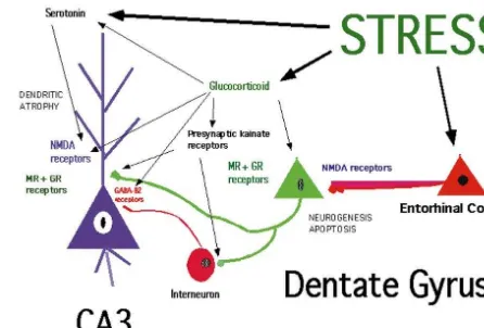
Fig. 2. Physiological doses of corticosterone mimic effects of stress in administration [27,29] (see Fig. 1).
enhancing the DTH response in the ear, whereas high doses of corticos-Once immune cells have marginated and begun to enter
terone and dexamethasone, which does not bind to CBG, suppresses the the tissue, other factors become involved as local medi- DTH response. From [25].
ators of further activation of immune function. Interferon gamma is an important factor in the stress-enhancement of the DTH response, and this has been shown by a lack of a
inverted U dose response curve and suppression of the stress effect in mice lacking the receptor for IFN gamma
DTH response is seen [25] (see Fig. 2). Dexamethasone, [30]. Furthermore, immunoneutralization of IFN gamma in
which does not bind to serum corticosteroid binding normal mice also blocks the stress effect. Thus, although
globulin (CBG), mimics the suppressive effects of high IFN gamma is not required for the baseline DTH response,
dose glucocorticoids [25]. Chronic stress over 3–5 weeks it is evidently important for the manifestations of the
produces a suppression of the DTH response and also effects of acute stress. IFN gamma is known to induce
suppressed the initial sensitization of the response [28] (see expression of antigen-presenting and cell-adhesion
mole-Fig. 3). An important factor in this suppression is the lack cules on endothelial cells and macrophages and cell
of immune cell trafficking, and this may be due, at least in adhesion molecules on leukocytes. It is also significant that
part, to habituation of the corticosterone response to stress glucocorticoids induce IFN gamma receptors on
mono-[28]. cytes. (For discussion and refererences, see [30]).
These findings are relevant to the extensive literature on What about the effects of chronic stress? Acute stress
the effects of stress on immune function in animals and shows a dose dependency to activate the DTH response,
humans [142]. Enhancement of immune function, in the and this is related to the magnitude of glucocorticoid
case of an autoimmune disease, may be deleterious, secretion [28]. Exogenous glucocorticoids mimic this dose
whereas it may be beneficial where there is a pathogen dependency, but at higher glucocorticoid doses, there is an
involved; conversely, suppression of immune function may be beneficial where an autoimmune disorder is concerned, whereas it may be dangerous where a pathogen is involved [25]. Thus, the immune system exemplifies the contrasting aspects of ‘protection’ and ‘damage’ and the effects of stress and stress hormones are highly relevant to human disease. Now we turn to the brain and consider how it handles both acute and repeated stress.
Fig. 1. Immune cells are depleted reversibly from the blood during
4. Stress, adaptive plasticity and the hippocampus
The brain is the key to interpreting and responding to potentially stressful events; it is also a target for the actions of stress hormones, particularly glucocorticoids. In the short run, acute elevation of both glucocorticoids and catecholamines facilitates the formation of memories of events associated with strong emotions [102,125]. Chroni-cally, however, stress hormones, and glucocorticoids, in particular, contribute to impairment of cognitive function and promote damage to brain structures such as the hippocampus [78,98,127].
The response of the brain to both acute and chronic
stress must be regarded in terms of its capacity to show Fig. 4. Schematic diagram of the role of neurotransmitters and glucocor-adaptive plasticity. The adult brain is more plastic than ticoids in regulating neurogenesis and dendritic remodeling in the dentate previously believed. Remodeling of synaptic contacts and gyrus-CA3 system of the hippocampal formation. Granule neurons are replaced in adult life, and neurogenesis, as well as apoptotic neuronal dendrites in the hypothalamus with the onset of lactation
death, are regulated by stress as well as by seizure-like activity. Granule [108,146] and growth and branching of dendrites of
neurons send mossy fibers to both the CA3 pyramidal neurons and to cerebrocortical neurons in an enriched environment and interneurons in the hilus, which, in turn, send inhibitory projections to the after training [54,155] are two examples of such plasticity. CA3 pyramidal neurons. The balance between the excitatory input and the Recent studies on the hippocampal formation of the brain inhibitory tone from the interneurons is presumed to be very important to the excitability of CA3 neurons. Evidence summarized in the text provides further examples of adult brain plasticity which is
indicates that excitatory amino acid release during repeated stress, aided regulated by hormones in adult life and during brain
by circulating glucocorticoids, leads to a reversible remodeling of apical development. The hippocampus is involved in episodic, dendrites over 3–4 weeks in rats and tree shrews. Serotonin also declarative, contextual and spatial learning and memory, as participates, possibly by aiding the excitatory amino acid activity at the well as a component in the control of autonomic and NMDA receptor, and reduced GABA-benzodiazepine-mediated inhibitory activity at synapse from the interneurons on CA3 pyramidal neurons may vegetative functions such as ACTH secretion [34,57,118].
also exacerbate the remodeling. Excitatory input to the dentate granule The hippocampus is also vulnerable to damage by stroke
neurons from the entorhinal cortex acts via NMDA receptors in concert and head trauma and susceptible to damage during aging with circulating adrenal steroids to regulate the rate of neurogenesis and and repeated stress [127]. apoptotic cell death, and both acute and chronic stress appear to be
Hippocampal neurons express receptors for circulating capable of inhibiting neurogenesis in the dentate gyrus. adrenal steroids [101], and work in many laboratories has
shown that the hippocampus has two types of adrenal
steroid receptors, Type I (mineralocorticoid) and Type II chronic elevation of glucocorticoids and the inhibition of (glucocorticoid) which mediate a variety of effects on neurogenesis of granule cells in the dentate gyrus (see Fig. neuronal excitability, neurochemistry and structural plas- 4).
ticity [24]. These effects, which involve hormone-mediated effects on gene expression, include the regulation of
branching and length of dendrites in the pyramidal cells of 5. Remodelling of dendrites in hippocampal neurons
Ammon’s horn and the replacement of nerve cells in the
dentate gyrus. The hippocampus is also sensitive to 5.1. Role of excitatory amino acids gonadal hormones and expresses both intracellular
an-drogen and estrogen receptors [63,154]. Gonadal and Remodelling of dendrites in hippocampal neurons was adrenal hormones participate in functional and structural first described after treatment of adult male rats for 21 days changes in adult life, as well as in developmental events, with exogenous glucocorticoids [157] (reviewed in [96]). which include sexual differentiation and influences of early Subsequently, chronic restraint stress for 21 days in rats
stressful life experiences. produced apical dendrites of CA3 pyramidal neurons to
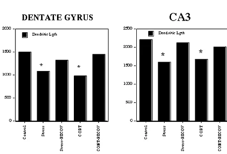
blockade is also effective in preventing stress-induced neurons (see [96]), serotonin released by stress or by dendritic atrophy (see [96] for review). corticosterone may interact pre- or post-synaptically with glutamate released by stress or by corticosterone, and the 5.2. Dendritic remodelling throughout the hippocampus? final common path may involve interactive effects between serotonin and glutamate receptors on the dendrites of CA3 A recent study using a multiple stress paradigm over 4 neurons innervated by mossy fibers from the dentate gyrus. weeks has demonstrated that the remodelling of dendrites There is evidence for interactions between serotonin and found in CA3 also occurs in the dentate gyrus and in CA 1, NMDA receptors, indicating that serotonin potentiates although the effects in the CA3 tend to be the greatest NMDA receptor binding as well as activity of NMDA [141] (see Fig. 5). This important finding provides the receptors and may do so via 5-HT receptors [107,120].2
basis for expecting that remodelling of dendrites could be a
factor in the shrinkage of the hippocampus reported in a 5.4. Involvement of glucocorticoids number of disorders such as recurrent major depression
and aging with mild cognitive impairment (see [95,129] for Glucocorticoid treatment causes dendritic atrophy, and
review). stress-induced atrophy is blocked by treatment with an
adrenal steroid synthesis blocker, cyanoketone (see [96]), 5.3. Importance of other neurotransmitters indicating a role of endogenous glucocorticoids in stress-induced dendritic atrophy. There appear to be several ways Besides glutamate, other participating neurotransmitters in which glucocorticoids affect the excitatory amino acid include GABA and serotonin. As far as GABA, inhibitory system. First, adrenal steroids modulate expression of interneurons have a significant role in controlling hip- NMDA receptors in hippocampus [2,152], with chronic pocampal neuronal excitability [39], and the involvement glucocorticoid exposure leading to increased expression of of the GABA-benzodiazepine receptor system is revealed NMDA receptor binding and both NR2A and NR2B by the ability of a benzodiazepine, adinazolam, to block subunit mRNA levels [153]. Second, there are glucocor-dendritic atrophy [79]. Serotonin is released by stressors, ticoid effects on the expression of mRNA levels for and tianeptine, an atypical tricyclic antidepressant that specific subunits of GABAa receptors in CA3 and the enhances serotonin reuptake and thus reduces extracellular dentate gyrus; both low and high levels of CORT have 5HT levels. Tianeptine prevented both stress- and corticos- different effects on GABAa receptor subunit mRNA levels terone-induced dendritic atrophy of CA3 pyramidal neu- and receptor binding ([114], Orchinik, Weiland and rons [149], whereas fluoxetine and fluvoxamine, inhibitors McEwen, unpublished), suggesting corticosterone may of serotonin reuptake, and desipramine, an inhibitor of alter the excitability of hippocampal neurons through noradrenaline uptake, failed to block atrophy [79]. Further regulation of GABAa receptor expression. However, it evidence for serotonin involvement in dendritic atrophy remains to be seen if the corticosteroid effects on neuronal comes from studies of psychosocial stress in rats, summa- morphology involve changes in the number or
pharmaco-rized below. logical properties of GABAa receptors.
Because both phenytoin and tianeptine block corticos- Third, adrenal steroids regulate the release of glutamate, terone- and stress-induced atrophy of CA3 pyramidal since adrenalectomy markedly reduces the magnitude of the EAA release evoked by restraint stress [72]. Mossy fiber terminals in the stratum lucidum contain presynaptic kainate receptors that positively regulate glutamate release [17]; these presynaptic kainate receptors are decreased in density by ADX and restored to normal by corticosterone replacement [151]. Moreover, repeated stress causes a reorganization of synaptic vesicles within mossy fiber terminals, as reported recently using electron microscopy [85]. Whereas mossy fiber terminals (MFT) from control rats were packed with small, clear synaptic vesicles, terminals from rats receiving 21 days of restraint stress showed a marked rearrangement of vesicles, with more densely packed clusters localized in the vicinity of active synaptic zones. Moreover, compared with controls, re-straint stress increased the area of the mossy fiber terminal occupied by mitochondrial profiles, which implies a great-Fig. 5. One month of a multiple stress paradigm or daily corticosterone
er, localized energy-generating capacity. A single stress treatment causes dendrites in CA3 and dentate gyrus to become shorter in
session did not produce these changes either immediately total length. Note that the effects of both stress and corticosterone
6. Neurogenesis in the dentate gyrus activity [116] and the stimulus for this neurogenesis is likely to be apoptotic cell death because seizures kill 6.1. Effects of adrenalectomy and adrenal steroids granule neurons [3] and local increases in apoptosis simulate local neurogenesis [13]. Granule neuron birth is Neurogenesis in the dentate gyrus of adult rodents was also accelerated by blocking NMDA receptors or lesioning reported [58,59] but never fully appreciated until recently, the excitatory perforant pathway input from the entorhinal and the reactivation of this topic occurred in an unusual cortex [14]. Unlike ADX, these treatments do not increase manner. First, bilateral adrenalectomy (ADX) of an adult granule neuron apoptosis, and a single dose of an NMDA-rat was shown to increase granule neuron death by blocking drug results in a 20% increase in dentate gyrus apoptosis [53,140]. Subsequently, neurogenesis was also neuron number several weeks later [14]. Thus, although found to increase following ADX in adults rats [11], as increased apoptosis leads to increased neurogenesis [49], well as in the developing dentate gyrus [12]. In adult rats, the two processes occur in different regions of the granule very low levels of adrenal steroids, sufficient to occupy cell layer and can be uncoupled from each other. Neverthe-Type I adrenal steroid receptors completely blocks dentate less, the adrenal steroid suppression of neurogenesis is gyrus neuronal loss [156]; but, in newborn rats, Type II through an NMDA-receptor mechanism [50,111].
receptor agonists protect against neuronal apoptosis [51]. Very recently it was reported that serotonin may be a This is consistent with the fact that dentate neuronal loss in positive signal for neurogenesis in the adult dentate gyrus. the developing rat occurs at much higher circulating Treatment with the serotonin-releasing drug, d-fen-steroid levels than in the adult and it represents another fluramine, increased neurogenesis [46]. Likewise, the example of the different ways that the two adrenal steroid 5HT1A agonist, 8-hydroxy-DPAT, stimulated neuro-receptor types are involved in hippocampal function [78]. genesis, whereas blockade of 5HT1A receptors had the In adult rats, newly-born neurons arise in the hilus, very opposite effect and prevented the effect of d-fenfluramine close to the granule cell layer, and then migrate into the treatment [46], as well as preventing increased neuro-granule cell layer, presumably along a vimentin-staining genesis caused by pilocarpine-induced seizures [119]. radial glial network that is also enhanced by ADX [10].
3
Most neuroblasts labeled with [ H] thymidine lack both 6.5. Role in learning and memory? Type I and Type II adrenal steroid receptors [10],
indicat-ing steroidal regulation occurs via messengers from an One reason for turnover of dentate gyrus granule unidentified steroid-sensitive cell that may involve a neurons in adult life is to adjust needs for hippocampal signalling role for TGF alpha and the EGF receptor system function in spatial learning and memory to environmental
[147]. demands [137]. Birds that use space around them to hide
and locate food, and voles as well as deer mice that 6.2. Aging, glucocorticoids and neurogenesis traverse large distances to find mates, have larger hip-pocampal volumes than closely-related species which do It has been reported that neurogenesis declines in the not; moreover, there are indications that hippocampal aging rodent [61] and rhesus monkey [36] dentate gyrus. volume may change during the breeding season [41,137]. Recent studies of aging rats showed that adrenalectomy Indeed, the rate of neurogenesis in the male and female could reverse the decline in dentate gyrus neurogenesis prairie vole varies according to the breeding season [42]. [15], suggesting that they are the result of age-related In contrast, an enriched environment has been found to increases in HPA activity and glucocorticoid levels that increase dentate gyrus volume in mice by increasing have been reported [68,93,127,128]. neuronal survival without altering the rate of neurogenesis [60]. Thus, there are several ways to maintain the balance 6.3. Neurogenesis in different mammalian species between neuronal apoptosis and neurogenesis.
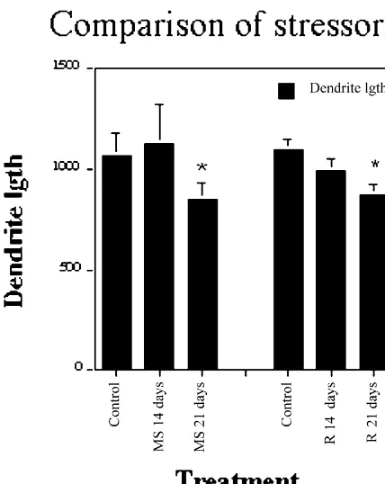
neurons and dentate granule neurons (see above) as to We showed previously that a similar multiple stress reduced dentate gyrus neurogenesis. paradigm, consisting of daily 1-h restraint, 1-h shaking, 309 swim in body-temperature water, did indeed produce
continued elevation of glucocorticoids throughout the
21-7. Experimenter-applied stressors: restraint and day paradigm, in contrast to 21 days of 6 h / day restraint
multiple stress effects stress which did produce habituation of the glucocorticoid stress response [80] (see Fig. 6). Yet both treatments A closer examination of the effects of stressors on caused similar degrees of dendritic remodelling in CA3 structural plasticity in the hippocampus and neurochemical (Fig. 7) and both treatments produced non-significant changes in other brain regions reveals some important atrophy of the thymus and hypertrophy of the adrenal properties of both experimenter-applied stressors and gland and moderate decrease in body weight gain over 21 natural psychosocial stressors which emphasize the adap- days (Table 1). In spite of the minimal changes in the tive nature of these changes in brain structure and the expected parameters of chronic stress, blockade of complex interactions between circulating stress hormones glucocorticoid synthesis with cyanoketone prevented
de-and neurotransmitters. ndritic remodelling [81] and oral administration of
Contrary to expectations, there is no consistent relation- glucocorticoids, which does not involve the stress of ship between stress-induced dendritic remodelling in hip- injection, causes dendritic remodelling [79,84].
pocampus and elevated glucocorticoid secretion or other We noted above that dendritic remodelling is reversible signs of chronic stress such as thymus atrophy, adrenal following the termination of stress or corticosterone treat-hypertrophy and body weight reduction. As noted above, a ment [19,141]. Moreover, we also noted that both Dilantin recent study showed that a chronic multiple stress and tianeptine prevent dendritic remodelling caused by paradigm, which is designed to avoid habituation of repeated restraint stress and glucocorticoid injections. glucocorticoid responses to the same stressor, produced Treatment with tianeptine is able to reverse the remodel-dendritic remodelling not only in the CA3 region of the ling produced by 21 days oral corticosterone and to do so hippocampus but also in the dentate gyrus and CA1 [141]. within 2 weeks even while the corticosterone treatment is Glucocorticoid treatment mimicked the effects of stress, continuing.
and the effects of both treatments were largely reversible A final note on the structural plasticity produced by after termination of the treatment [141]. See Fig. 5. repeated restraint stress is that dentate gyrus neurogenesis
the hippocampus. As noted above, serotonin and NMDA receptors are both implicated as mediators of dendritic remodelling, and GABA-benzodiazepine receptors play an opposing role. Moreover, as noted earlier, in the regulation of neurogenesis, serotonin acting via 5HT1A receptors plays an opposing role to glucocorticoids and NMDA receptors.
The multiplicity of regulatory factors for structural plasticity in the hippocampus is consistent with the inter-pretation of these changes as reflecting adaptive plasticity, rather than as a pathway leading to damage. Behavioral changes accompanying repeated restraint stress fit with this view and provide additional surprises. In spite of the apparent habituation of the HPA response to repeated restraint stress, these animals develop increased fear in an open field apparatus and showed increased fear condition-ing to both tone and contextual cues [19]. Moreover, repeatedly restrained rats show progressively increasing aggression towards their cage mates after they are released from their restrainers (G. Wood and B. McEwen, un-published).
Fig. 7. CA3 apical dendritic length over 3 weeks of either a multiple stress paradigm or a repeated restraint stress paradigm. Note that both
8. Animal to animal stress
types of stress cause shortening of dendrites but only after 3 weeks. Data from [80].
The theme of adaptive plasticity in the face of repeated stress is further documented by studies of more naturalistic types of stressors, and these studies provide additional is not inhibited by a single acute restraint stress but is clues as to the multiplicity of neurochemical pathways impaired after 21 days of daily restraint [117]. The affected by chronic stressors. In the tree shrew, a resident– reduction in neurogenesis is not as large as has been intruder paradigm was used to produce chronic psycho-reported in rats after natural predator odor [44] or in tree social stress in the intruder over 28 days. This procedure shrews after acute or chronic psychosocial stress [48]. We caused the same type of dendritic remodelling of CA3 will return to this topic in the next section. neurons in the tree shrew as was found in the rat; moreover We interpret these effects as further evidence of the the effects of this type of stress were prevented by importance of endogenous neurotransmitters along with concurrent treatment of intruder tree shrews with Dilantin glucocorticoids in the regulation of structural plasticity in [83]. Dilantin treatment did not prevent the stress-induced
elevation in catecholamines and glucocorticoids, and the HPA response was very robust and showed no signs of habituating over time, in contrast to the story with repeated Table 1
restraint stress (see above) [83]. Summary of changes in body weight, thymus and adrenal weights after
a Neurogenesis was inhibited by both acute and chronic
repeated stress
psychosocial stress in the intruder tree shrew [48], and this Body weight 1 Adrenal wt. 2 Thymus wt. 3
effect was substantially larger than that seen in rats after Restraint stress, 21 days
chronic restraint stress (see above) (see Fig. 8). Moreover,
Control 100% 1661 138613
the dentate gyrus is 30% smaller in the chronically-stressed *
1 week 90 n.d. n.d.
tree shrew, although granule neuron number shows a *
2 weeks 88 1761 150617
*
3 weeks 88 1661 122615 smaller reduction (Gould, Fuchs and McEwen, unpub-lished). This finding suggests that there may be other Chronic multiple stress, 21 days
changes such as atrophy of dendritic branching to account
Control 100% 1160.4 176611
for the decrease in dentate gyrus volume. It should also be *
1 week 93 n.d. n.d.
noted that neurogenesis is inhibited in the marmoset after
* *
2 weeks 89 1460.7 140612
*
3 weeks 100 1260.4 126610 acute psychosocial stress [48].
a Another important animal model to study psychosocial
Data from [80].
Table 2
Summary of changes in dominant and subordinate rats under psychosocial stress in the visible burrow
Endocrine parameters:
Dominants and subordinates show: Elevated CORT
Adrenal hypertrophy Thymic involution
Subordinates show: Progressive weight loss Greater thymic involution Decreased CBG Decreased testosterone Decreased insulin and glucose
Non-responsive subordinates show: Largest body weight loss Lowest testosterone Lowest CBG
No response to novel stressor
Neural parameters:
Dominants and subordinates show: CA3 dendritic remodelling
Down regulation of 5HTT in hippocampus
Down regulation of 5HT1a receptors in hippocampus Fig. 8. Acute psychosocial stress inhibits neurogenesis in the dentate
Decreased 5HT1A receptors gyrus of an adult tree shrew. The stress consisted of putting a tree shrew
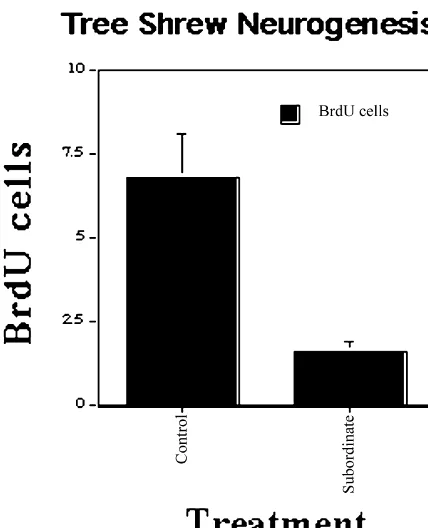
Increased CRF mRNA in hypothalamus into the territory of a dominant tree shrew. Data from [48].
Dominants show:
Somewhat more dendritic remodelling
Somewhat greater down-regulation of 5HTT in hippocampus
Subordinates show: Five males and two females are placed in an apparatus that
Increased CRF mRNA levels in central nucleus of amygdala has an open chamber and tunnels and compartments, and Increased tyrosine hydroxylase mRNA and protein
there is a videocamera to record the animals’ behavior. A in locus coeruleus
dominant male usual emerges within several days and Increased 5HT2A receptor binding in layer IV of parietal cortex Down-regulation of GR and MR mRNA in CA1 of hippocampus controls access to the food and water cups and to the
females. Within the 2-week span of this study, it is not Non-responsive subordinates show: uncommon for one or two of the subordinate males to die, Decreased CRF mRNA in hypothalamus
Somewhat less decrease of 5HT1A receptors in hippocampus usually not so much from wounds as from autonomic
Somewhat less down-regulation of GR and MR mRNA collapse related to defeat. A complex set of neuroendocrine
in CA1 of hippocampus and neurochemical changes has been found for the
domi-nants and subordinates, which are both experiencing stress compared to control animals living in the normal cage environment (see Table 2). The most striking difference is
among the subordinates, in which two types have been shrews described above [38,96,104]. These stress-induced recognized. The non-responders are so identified because changes are relevant to the suspected role of 5HT1A they show an attenuated HPA response to acute restraint receptors in anxiety disorders and depression [71]. For stress and also show a reduced level of CRH mRNA in the example, deletion of the 5HT1A receptor in mice results in
hypothalamus [1]. increased anxiety, due in part to an impairment of the
For rats in the VBS and the intruder tree shrew there is a GABA-benzodiazepine system [122,138].
are relevant to human psychiatric and neurological dis- performance on an eight-arm radial maze when they were
orders. trained starting one day after the end of stress but not when
trained 18 days later [74]. We now know that dendritic atrophy is reversible within 7–10 days after the end of
9. Implications for cognitive function and human stress [19]. The impairment was in the same direction, but
psychiatric disorders not as great as, impairment found in aging rats. Moreover, stress effects were prevented by prior treatment of rats We have seen that the serendipitous discovery of adrenal with phenytoin or with tianeptine under the same con-steroid receptors in hippocampus led to findings about the ditions in which both drugs prevented the stress-induced vulnerability of the hippocampus in relation to stress, atrophy of CA3c pyramidal neurons [74,149,150]. A aging, seizures and head trauma in animal models of these subsequent study showed that 21 days of repeated restraint conditions. The advent of magnetic resonance imaging stress impaired the short-term (4 h) retention of a spatial (MRI) and positron emission tomography (PET) has recognition memory in a hippocampus-dependent Y-maze opened the door for translating information based upon task; again, stress impairment was prevented by tianeptine animal models of neurological and psychiatric disorders to treatment during the stress regimen [18].
directly studying patients with those disorders. In
par-ticular, changes in hippocampus in animal models provide 9.2. Atrophy of the human hippocampus and other brain insights into altered human hippocampal structure and structures
function in depression and aging. First, we will consider
the effects of stress and glucocorticoids on cognitive The human brain shows signs of atrophy as a result of function and then review some of the evidence from brain elevated glucocorticoids and severe, traumatic stress (e.g., imaging for structural changes in the hippocampus, holocaust survivors, see [127]). However, it has been very amygdala and prefrontal cortex. recently only that brain-imaging techniques have allowed for a regional analysis of the atrophy of various brain 9.1. Stress, glucocorticoids and cognition structures to see which ones are most affected. Recent evidence indicates that the human hippocampus is par-Stress and glucocorticoids have specific effects on ticularly sensitive in this respect and tends to show greater cognitive function in humans and in animal models. changes than other brain areas, in particular in Cushing’s Adrenal steroids and stressful experiences produce short- syndrome, recurrent depressive illness, post-traumatic term and reversible deficits in episodic and spatial memory stress disorder (PTSD, schizophrenia and aging prior to in animal models and in humans [78], whereas repeated overt dementia [5,40,55,126,135,136,143,144]).
stress also impairs cognitive function in animal models and For example, declines of hippocampally-related cogni-repeated glucocorticoid elevation or treatment in humans is tive functions, such as spatial and episodic memory, occur accompanied by cognitive dysfunction [98]. There are also in human subjects and are correlated with increases in HPA declines in cognitive function in aging humans that are activity over 4–5 years [76,132]. Recent evidence has correlated with progressive elevations in HPA activity over revealed that the most severely impaired individuals have a
3–4 years [76,132]. significantly smaller hippocampal volume compared to the
Acute effects of stress or glucocorticoid administration least impaired individuals [75]. This result is consistent are evident within a time span ranging from a few hours to with other findings of individual differences in cognitive a day and are generally reversible and quite selective to the function correlated with hippocampal volume reductions in task or particular situation [66,78]. Adrenal steroid effects elderly humans [20,45].
are implicated in both selective attention as well as in In women with recurrent depressive illness, the mag-memory consolidation [78], and such actions are consistent nitude of hippocampal atrophy is related to the duration of with the effects of adrenal steroids on the modulation of the symptoms of the disorder and not the age of the long-term potentiation and primed-burst potentiation (see individuals [135].
and amygdala [134]. Moreover, new evidence suggests that gression of neuronal structural changes, involving atrophy glial cell depletion may contribute to atrophy of brain that might lead to permanent damage, including neuronal regions like the prefrontal cortex and amygdala loss.
[32,113,121,134,135] and the contribution of glial cell Regarding reversibility, treatment with drugs like pheny-changes must now be considered in the hippocampus. toin or tianeptine, both of which block stress-induced It is tempting to attribute the occurrence of hippocampal atrophy, is a potential means of testing both the mecha-atrophy to glucocorticoids. This is because the hippocam- nism and at the same time, demonstrating the reversibility pus is a primary target area for adrenal steroids in brain, of human hippocampal atrophy. There is already some and adrenal steroids have been shown to have effects on indication that hippocampal atrophy in Cushing’s hippocampal neuronal plasticity and on the loss of hip- syndrome is reversible [144]. On the other hand, there may pocampal neurons in conditions like ischemia and aging be irreversible loss of hippocampal neurons, and some of [68,96,127,128]. However, we have seen earlier in this the evidence from MRI concerning hippocampal abnor-article that other factors play a role, including the endogen- malities in recurrent depressive illness is consistent with ous excitatory amino acid neurotransmitters. Moreover, this possibility [136]. In so far as atrophy of the hippocam-changes in dentate gyrus neuron number may be involved pus and accompanying cognitive impairment are signs of along with atrophy of dendritic processes. Nevertheless, reversible neuronal atrophy, they may be treatable with the role of glucocorticoids should not be ignored. agents that block the neuronal atrophy in animal models. Glucocorticoids are elevated in Cushing’s syndrome and On the other hand, where atrophy involves neuronal loss, may also be somewhat elevated in depressive illness, but treatment strategies should focus on the earlier traumatic or this is probably not the case for PTSD, at least at the time recurrent events, and it may be possible to devise strategies they are studied, except as there are elevations in glucocor- to reduce or prevent neuronal damage.
ticoids associated with the diurnal rhythm and stressful experiences that take place on a daily basis.
Sustained stress-level of Cushingoid elevations of adren- 10. Future directions
al steroids are not required for atrophy of hippocampal
neurons, since we have already noted above that in the Research on the hippocampus in animal models and animal models of stress-induced atrophy, ordinary, period- human disorders has established that the brain is a resilient ic adrenocortical stress responses are all that is needed for structure that is capable of adaptive plasticity but is also the process to occur with daily stress. With regard to vulnerable to damage. While illuminating many aspects of human hippocampal atrophy, individual differences in mechanisms underlying plasticity and opening the door for stress responsiveness may play a role in making some therapeutic interventions, recent research on this topic has people more vulnerable to their own stress hormones: e.g., also raised some important questions that form the basis of some individuals who are exposed to repeated psycho- future studies. First, what are the conditions that determine social stress (e.g., public speaking) fail to habituate their whether brain structures will respond with adapative cortisol elevation; these individuals lack self-esteem and plasticity or show permanent damage? Second, how does self-confidence [65]. Therefore, one could imagine that the vulnerability to permanent damage change in the aging individuals with a more reactive stress hormone profile brain? And third, how do gender differences in the will expose themselves to more cortisol and experience response to stressful challenges influence the brain’s more stress-elevated neural activity, than other people who response in terms of resilience or damage?
can more easily habituate to psychosocial challenges.
In this regard, events related to trauma leading to PTSD 10.1. From protection to damage and the course of illness in recurrent depressive illness
may involve very distinct pathways of selective and The concept of allostasis and allostatic load imply that repeated elevations of glucocorticoid hormones in relation normal attempts of the body to adapt to stressful chal-to the individual experiences and reactivities. In the case of lenges and a changing environment exact a cost on the PTSD, we are ignorant of stress responses and neuro- body in terms of wear and tear on tissues and organs that chemical changes accompanying the initial trauma, which impair their normal function, as well as a progressive may have taken place 10–20 years ago, as well as the failure of the systems that mediate allostasis to operate ongoing stress responsiveness and neurochemical activity efficiently. An example of wear and tear is the stress-(e.g., brain glucose metabolism) of traumatized individuals. induced remodelling of hippocampal dendrites and reduc-Likewise, for recurrent depressive illness, we are largely tion of dentate gyrus neurogenesis, described above, in ignorant of the history of the depressed individual as far as which the combined actions of circulating glucocorticoids endocrine function and neurochemical activity, as well as and excitatory amino acids are involved in changes that responses to stressful life experiences. In both disorders, a lead to cognitive impairment and other behavioral altera-long-term pattern of increased neurochemical, autonomic tions.
allostasis, the aging process provides several good exam- and coworkers have had some success in reducing ex-ples as far as the hippocampus is concerned. Aging is citotoxic damage by introducing glucose transporters in accompanied in some individuals by an elevated level of viral vectors [115]. Diabetes is a condition in which tissues glucocorticoids and a failure of the HPA axis to shut off may be unable to obtain sufficient glucose. The strep-efficiently in the aftermath of activation tozotocin (STZ) model of Type I diabetes has provided a [67,93,105,127,128]. According to the glucocorticoid cas- model in which the absence of insulin creates a hy-cade hypothesis of stress and aging [130], the progressive perglycemic state as well as an elevation of glucocor-failure of the hippocampus to exercise its role in shutting ticoids. STZ diabetes results in an acceleration of the off the HPA axis leads to further elevation of glucocor- remodelling of dendrites in the hippocampus, in that ticoids and further damage to the hippocampus during the diabetic animals without additional stress display within 10 aging process. We have seen that evidence for individual days the same morphological changes that non-diabetic differences in human hippocampal aging is consistent with rats show after 21 days of stress or glucocorticoid
treat-this model [77]. ment; moreover, restraint stress causes additional dendritic
Long-term stress accelerates a number of biological remodelling within 7 days [82]. In addition, there are signs markers of aging in rats, including increasing the ex- of astroglial reactivity and the appearance of damaged citability of CA1 pyramidal neurons via a calcium-depen- synapses and neuron cell bodies that suggest the beginning dent mechanism and causing loss of hippocampal pyrami- of permanent damage (Magarinos, unpublished). Studies dal neurons [64]. An important factor may be the enhance- are underway to evaluate the levels of glucose transporter ment by glucocorticoids of calcium currents in hippocam- proteins and mRNA in the hippocampus [124], as well as pus [62], in view of the key role of calcium ions in insulin-like growth factor expression [123].
destructive as well as plastic processes in hippocampal
neurons [91,92]. Another aspect making the aging hip- 10.2. Determinants of individual differences in stress pocampus more vulnerable may be the persistence of reactivity in early development
excitatory amino acid release after the termination of a
stressful experience [73]. Individual differences in rates of brain aging and
We have seen earlier in this article that, besides hippocampal atrophy are evident in longitudinal studies on glucocorticoids, excitatory amino acids are also important human subjects (see above and [76,77,132]). Individual players in the structural remodelling of the hippocampus. differences in stress reactivity are also seen in rats, and this The same is true for hippocampal damage produced by reactivity can be increased by early maternal deprivation ischemia, seizures and head trauma [127]. The release of and decreased by separating pups from their mothers for glutamate as a neurotransmitter is a good example of brief periods of time, resulting in increased maternal care allostasis that leads to transmission of nerve impulses and when the pups are reunited with their mothers [8,9,70]. As learning and memory, and the failure to efficiently termi- discussed elsewhere [105,106], such early experiences can nate this release and / or recapture the glutamate after either increase or decrease the rate of brain aging through a release leads to an example of allostatic load. Such a mechanism in which the activity of the HPA axis appears situation has been encountered in the aging rat brain. to be involved. The early experiences are believed to set Under restraint stress, rats show increased extracellular the level of responsiveness of the HPA axis and autonomic levels of glutamate in hippocampus, as determined by nervous system in such a way that these systems either microdialysis, and adrenalectomy markedly attenuates this over-react in animals subject to early unpredictable stress elevation [72]. Glucocorticoids appear to be involved in or under-react in animals exposed to the neonatal handling potentiating the increased extracellular levels of excitatory procedure.
amino acids under stress [110]. Similar results have been There are situations in both the animal model literature reported using lactography, a method that is based upon and in studies of children exposed to early trauma or the stimulation of glucose metabolism by increased neuro- neglect in which alterations in emotionality and HPA axis nal activity [23,131]. In aging rats, hippocampal release of functioning have been described. In studies on infrahuman excitatory amino acids during restraint stress is markedly primates, maternal deprivation and peer rearing and the potentiated [73], and this constitutes an example of allos- variable foraging demand model have both been linked to tatic load in the brain, representing a failure to shut off the later increases in psychopathology [21,22,56]. In children production and / or removal of a mediator of neuronal exposed to physical and sexual abuse, there are indications
activation. of later life problems such as substance abuse, hostility and
In considering the resilient brain that shows reversible antisocial behavior, suicide and a host of physical illnesses remodelling of dendrites in the hippocampus after repeated reflecting a general systemic disregulation [37].
stress, we must consider the mechanisms that protect the
brain and counteract the potential for permanent damage. 10.3. Sex differences in response to stress and Glucose availability is a key factor, and Sapolsky has glucocorticoids
referrred to an ‘energy crisis’ in which inability to obtain
above and imply that either the sexual differentiation of the volume remains to be determined, but it is the continuing hippocampus or the presence of circulating hormones work in animal models on processes such as neurogenesis, affects how the hippocampus responds to stress. Stress dendritic remodelling, glial cell plasticity and neuron loss effects on dendritic atrophy occur in males but not in that will continue to provide the essential information for females [43]. Moreover, pyramidal neuron loss was evi- interpreting changes in the living human brain seen by dent in subordinate male vervet monkeys, but not in imaging procedures.
females, after prolonged psychosocial stress [148], and evidence of neuronal damage in rats undergoing cold-swim
stress was evident in males, but not in females [109]. Acknowledgements However, after estrogens are absent, the female
hippocam-pus may be vulnerable to functional impairment, judging Research in the author’s laboratory described in this from the report that women who showed an increase in paper is supported by NIH grants MH41256 and overnight urinary cortisol over 2.5 years showed declines MH58911, NSF IBN9815480, and by grants from Servier in cognitive performance on hippocampal-related memory (France) and the Health Foundation (New York). The
tasks [132]. author wishes to thank his many laboratory colleagues for
their many contributions to the work summarized in this review and to his colleagues in the MacArthur Foundation
11. Conclusions Research Network for Socioeconomic Status and Health
for their contributions to the concepts of allostasis and Studies of how acute and repeated stress affect the allostatic load discussed in this article.
hippocampus and immune system reveal a degree of resilience that is not normally appreciated. The protective
and adaptive effects of the mediators of allostasis are References capable of promoting adaptive plasticity. In the case of the
immune system, this takes the form of the movement of [1] D.S. Albeck, C.R. McKittrick, D.C. Blanchard, R.J. Blanchard, J. immune cells to sites where they are needed to respond to Nikulina, B.S. McEwen, R.R. Sakai, Chronic social stress alters a challenge. Where the immune response is to a pathogen, expression of corticotrophin releasing factor and arginine vasopres-sin mRNA expression in rat brain, J. Neurosci. 17 (1997) 4895– the enhancement of the response is beneficial; where an
4903. autoimmune response is involved, the outcome may by
[2] V. Bartanusz, J.M. Aubry, S. Pagliusi, D. Jezova, J. Baffi, J.Z. Kiss, deleterious. Glucocorticoids and catecholamines play an Stress-induced changes in messenger RNA levels of N-methyl-D -important role but act in concert with other tissue-specific aspartate and ampa receptor subunits in selected regions of the rat regulators such as cytokines and cell surface molecules. hippocampus and hypothalamus, Neuroscience 66 (1995) 247–252. [3] J. Bengzon, Z. Kokaia, E. Elmer, A. Nanobashvili, M. Kokaia, O. In the case of the brain, the enhancement of fear-related
Lindvall, Apoptosis and proliferation of dentate gyrus neurons after learning is generally beneficial to an individual’s ability to
single and intermittent limbic seizures, Proc. Natl. Acad. Sci. USA remember danger and act accordingly. Even the stress- 94 (1997) 10432–10437.
induced remodelling of hippocampal dendrites and sup- [4] D.C. Blanchard, Behavioral correlates of chronic dominance-pression of neurogenesis may serve a protective function in subordination relationships of male rats in a seminatural situation,
Neurosci. Biobehav. Rev. 14 (1990) 455–462. the short run against outright excitotoxic damage, and it
[5] B. Bogerts, J.A. Lieberman, M. Ashtair, R.M. Bilder, G. De Greef, also appears to go hand-in-hand with the adjustments of
G. Lerner, C. Johns, S. Masiar, Hippocampus-amygdala volumes the animal’s behavior to a long-term change in the social and psychpathology in chronic schizophrenia, Biol. Psychiatry 33 and physical environment. For these changes, circulating (1993) 236–246.
glucocorticoids and catecholamines act together with local [6] D. Brindley, Y. Rolland, Possible connections between stress, diabetes, obesity, hypertension and altered lipoprotein metabolism tissue mediators such as the excitatory amino acids and
that may result in atherosclerosis, Clin. Sci. 77 (1989) 453–461. serotonin, among other neurotransmitters.
[7] K. Bulloch, Regional neural regulation of immunity: anatomy and The findings of plasticity, as well as damage, in hip- function, in: Handbook of Physiology, Coping with the Environ-pocampus after prolonged psychosocial stress in animal ment: Neural and Endocrine Mechanisms, Oxford University Press, models have encouraged investigations of the human New York, 2000.
[8] C. Caldji, D. Francis, S. Sharma, P.M. Plotsky, M.J. Meaney, The hippocampus in stress-related disorders such as depressive
effects of early rearing environment on the development of GABAA
illness, post-traumatic stress disorder and schizophrenia, as
and central benzodiazepine receptor levels and novelty-induced well as in relation to individual differences in human fearfulness in the rat, Neuropsychopharmacology 22 (2000) 219– cognitive aging. These investigations have uncovered the 229.
phenomenon of atrophy, not only of the hippocampus, but [9] C. Caldji, D. Liu, S. Sharma, J. Diorio, D. Francis, M.J. Meaney, P.M. Plotsky, Development of individual differences in behavioral also of structures such as the amygdala and prefrontal
and endocrine responses to stress: role of the postnatal environment, cortex in some of the conditions. Other studies have
in: Handbook of Physiology Coping with the Environment: Neural provided initial suggestions that the human hippocampus and Endocrine Mechanisms, Oxford University Press, New York, may increase in volume as a result of spatial information 2000.
newly born neurons and glia in the dentate gyrus of the adult rat, [31] D.M. Diamond, M. Fleshner, G.M. Rose, Psychological stress Neuroscience 56 (1993) 337–344. impairs spatial working memory: relevance to electrophysiological studies of hippocampal function, Behav. Neurosci. 110 (1996) [11] H.A. Cameron, E. Gould, Adult neurogenesis is regulated by adrenal
661–672. steroids in the dentate gyrus, Neuroscience 61 (1994) 203–209.
[32] W.C. Drevets, D. Ongur, J.L. Price, Neuroimaging abnormalities in [12] H.A. Cameron, E. Gould, The control of neuronal birth and survival,
the subgenual prefrontal cortex: implications for the pathophysiol-in: C.A. Shaw (Ed.), Receptor Dynamics in Neural Development,
ogy of familial mood disorders, Mol. Psychiatry 3 (1998) 220–226. CRC Press, New York, 1996, pp. 141–157.
[33] W.C. Drevets, J.L. Price, J.R. Simpson Jr, R.D. Todd, T. Reich, M. [13] H.A. Cameron, E. Gould, Distinct populations of cells in the adult
Vannier, M.E. Raichle, Subgenual prefrontal cortex abnormalities in dentate gyrus undergo mitosis or apoptosis in response to
adrenalec-mood disorders, Nature 386 (1997) 824–827. tomy, J. Comp. Neurol. 369 (1996) 56–63.
[34] H. Eichenbaum, T. Otto, The hippocampus — what does it do?, [14] H.A. Cameron, B.S. McEwen, E. Gould, Regulation of adult
Behav. Neural. Biol. 57 (1992) 2–36. neurogenesis by excitatory input and NMDA receptor activation in
the dentate gyrus, J. Neurosci. 15 (1995) 4687–4692. [35] P.S. Eriksson, E. Permlieva, T. Bjork-Eriksson, A.-M. Alborn, C. Nordborg, D.A. Peterson, F.H. Gage, Neurogenesis in the adult [15] H.A. Cameron, D.G. McKay, Restoring production of hippocampal
human hippocampus, Nature Medicine 4 (1998) 1313–1317. neurons in old age, Nature Neurosci. 2 (1999) 894–897.
[36] M. Fallah, E. Fuchs, P. Tanapat, A.J. Reeves, E. Gould, Hippocam-[16] W. Cannon, The wisdom of the body, Physiol. Rev. 9 (1929)
pal neurogenesis in old world primates declines with aging, Abstr. 399–431.
Soc. Neurosci. 24 (1998) 796.9, p. 1993. [17] R. Chittajallu, M. Vignes, K.K. Dev, J.M. Barnes, G.L. Collingridge,
[37] V.J. Felitti, R.F. Anda, D. Nordenberg, D.F. Williamson, A.M. Spitz, J.M. Henley, Regulation of glutamate release by presynaptic kainate
V. Edwards, M.P. Koss, J.S. Marks, Relationship of childhood abuse receptors in the hippocampus, Nature 379 (1996) 78–81.
and household dysfunction to many of the leading causes of death in [18] C.D. Conrad, L.A.M. Galea, Y. Kuroda, B.S. McEwen, Chronic
adults. The adverse childhood experiences (ACE) study, Am. J. stress impairs rat spatial memory on the Y-Maze and this effect is
Prev. Med. 14 (1998) 245–258. blocked by tianeptine pre-treatment, Behav. Neurosci. 110 (1996)
[38] G. Flugge, Dynamics of central nervous 5-HT -receptors under
1321–1334. 1A
psychosocial stress, J. Neurosci. 15 (1995) 7132–7140. [19] C.D. Conrad, A.M. Magarinos, J.E. LeDoux, B.S. McEwen,
Re-peated restraint stress facilitates fear conditioning independently of [39] T.F. Freund, G. Buzsaki, Interneurons of the hippocampus, Hip-causing hippocampal CA3 dendritic atrophy, Behav. Neurosci. 113 pocampus 6 (1996) 345–470.
(1999) 902–913. [40] H. Fukuzako, T. Fukuzako, T. Hashiguchi, Y. Hokazono, K. [20] A. Convit, M.J. DeLeon, C. Tarshish, S. De Santi, A. Kluger, H. Takeuchi, K. Hirakawa, K. Ueyama, M. Takigawa, Y. Kajiya, M. Rusinek, A.J. George, Hippocampal volume losses in minimally Nakajo, T. Fujimoto, Reduction in hippocampal formation volume is impaired elderly, Lancet 345 (1995) 266. caused mainly by its shortening in chronic schizophrenia: assess-[21] J.D. Coplan, M.W. Andrews, L.A. Rosenblum, M.J. Owens, S. ment by MRI, Biol. Psychiatry 39 (1996) 938–945.
Friedman, J.M. Gorman, C.B. Nemeroff, Persistent elevations of [41] L.A.M. Galea, M. Kavaliers, K.-P. Ossenkopp, D. Innes, E.L. cerebrospinal fluid concentrations of corticotropin-releasing factor in Hargreaves, Sexually dimorphic spatial learning varies seasonally in adult nonhuman primates exposed to early-life stressors: Implica- two populations of deer mice, Brain Res. 635 (1994) 18–26. tions for the pathophysiology of mood and anxiety disorders, Proc. [42] L.A.M. Galea, B.S. McEwen, Sex and seasonal differences in the Natl. Acad. Sci. USA 93 (1996) 1619–1623. rate of cell proliferation in the dentate gyrus of adult wild meadow [22] J.D. Coplan, R.C. Trost, M.J. Owens, T.B. Cooper, J.M. Gorman, voles, Neuroscience 89 (1999) 955–964.
C.B. Nemeroff, L.A. Rosenblum, Cerebrospinal fluid concentrations [43] L.A.M. Galea, B.S. McEwen, P. Tanapat, T. Deak, R.L. Spencer, of somatostatin and biogenic amines in grown primates reared by F.S. Dhabhar, Sex differences in dendritic atrophy of CA3 pyrami-mothers exposed to manipulated foraging conditions, Arch. Gen. dal neurons in response to chronic restraint stress, Neuroscience 81
Psychiatry 55 (1998) 473–477. (1997) 689–697.
[23] L.A. De Bruin, M.C. Schasfoort, A.B. Stefens, J. Korf, Effects of [44] L.A.M. Galea, P. Tanapat, E. Gould, Exposure to predator odor stress and exercise on rat hippocampus and striatum extracellular suppresses cell proliferation in the dentate gyrus of adult rats via a lactate, Am. J. Physiol. 259 (1994) R773–R779. cholinergic mechanism, Abstr. Soc. Neurosci. 22 (1996)[474.8, p. [24] E.R. DeKloet, E. Vreugdenhil, M.S. Oitzl, M. Joels, Brain cortico- 1196.
steroid receptor balance in health and disease, Endocrine Rev. 19 [45] J. Golomb, A. Kluger, M.J. DeLeon, S.H. Ferris, A. Convit, M.S. (1998) 269–301. Mittelman, J. Cohen, H. Rusinek, S. De Santi, A.E. George, [25] F. Dhabhar, B. McEwen, Enhancing versus suppressive effects of Hippocampal formation size in normal human aging: a correlate of stress hormones on skin immune function, Proc. Natl. Acad. Sci. delayed secondary memory performance, Learn. Mem. 1 (1994)
USA 96 (1999) 1059–1064. 45–54.
[26] F.S. Dhabhar, Stress-induced Enhancement of Antigen-specific Cell- [46] E. Gould, Serotonin and hippocampal neurogenesis, Neuro-mediated Immunity: The Role of Hormones and Leukocyte Traffick- psychopharmacology 21 (1999) 46S–51S.
ing, Rockefeller University, New York, 1996. [47] E. Gould, A. Beylin, P. Tanapat, A. Reeves, T.J. Shors, Learning [27] F.S. Dhabhar, B.S. McEwen, Stress-induced enhancement of an- enhances adult neurogenesis in the hippocampal formation, Nature
tigen-specific cell-mediated immunity, J. Immunol. 156 (1996) Neurosci. 2 (1999) 260–265.
2608–2615. [48] E. Gould, B.S. McEwen, P. Tanapat, L.A.M. Galea, E. Fuchs, [28] F.S. Dhabhar, B.S. McEwen, Acute stress enhances while chronic Neurogenesis in the dentate gyrus of the adult tree shrew is stress suppresses cell-mediated immunity in vivo: a potential role for regulated by psychosocial stress and NMDA receptor activation, J. leukocyte trafficking, Brain Behav. Immun. 11 (1997) 286–306. Neurosci. 17 (1997) 2492–2498.
[29] F.S. Dhabhar, A.H. Miller, B.S. McEwen, R.L. Spencer, Effects of [49] E. Gould, P. Tanapat, Lesion-induced proliferation of neuronal stress on immune cell distribution: dynamics and hormonal mecha- progenitors in the dentate gyrus of the adult rat, Neuroscience 80 nisms, J. Immunol. 154 (1995) 5511–5527. (1997) 427–436.
[30] F.S. Dhabhar, A.R. Satoskar, H. Bluethmann, J.R. David, B.S. [50] E. Gould, P. Tanapat, H.A. Cameron, Adrenal steroids suppress McEwen, Stress-induced enhancement of skin immune function: A granule cell death in the developing dentate gyrus through an role for interferon, Proc. Natl. Acad. Sci. USA 97 (2000) 2846– NMDA receptor-dependent mechanism, Dev. Brain Res. 103 (1997)
![Fig. 3. Acute stress enhances, whereas chronic stress suppresses, theDTH response. From [25,28].](https://thumb-ap.123doks.com/thumbv2/123dok/3138078.1382514/4.612.76.255.494.692/acute-stress-enhances-chronic-stress-suppresses-thedth-response.webp)