OPTIMIZING STRUCTURE-BASED VIRTUAL SCREENING
PROTOCOL TO IDENTIFY PHYTOCHEMICALS AS
CYCLOOXYGENASE-2 INHIBITORS
Enade Perdana Istyastono
Division of Drug Design and Discovery, Faculty of Pharmacy and Center for Environmental Studies Sanata Dharma University, Yogyakarta, Indonesia. Kampus III Universitas Sanata Dharma, Paingan, Maguwoharjo, Depok, Yogyakarta 55284
Submitted: 14-04-2016 Revised: 13-06-2016 Accepted: 28-07-2016
*Corresponding author Enade Perdana Istyastono
Email:
ABSTRACT
By employing Databases of Useful Decoys (DUD) and its enhanced version (DUD-E), several attempts to construct validated Structure-based Virtual Screening (SBVS) protocols to identify cyclooxygenase-2 (COX-2) inhibitors have been performed. Both databases tagged active COX-2 inhibitors for compounds with IC50 values < 1M. In the search for
phytochemicals as natural COX-2 inhibitors, however, most of their IC50 values are in the micromolar range, which will likely be
identified as non-inhibitors for COX-2 by the available SBVS protocols. In this article, validation of an SBVS protocol by adding marginal active COX-2 inhibitors from DUD-E as active compounds is presented. Binary quantitative-structure activity relationship analysis by using recursive partition and regression tree method was performed subsequently to optimize the predictive ability of the protocol. The enrichment factor and the
F-measure values of the optimized protocol could reach 44.78 and 0.47, respectively. The optimized protocol could identify 1 out of 9 phytochemicals as COX-2 inhibitors.
Key words: Structure-based virtual screening (SBVS), phytochemical, cyclooxygenase-2 (COX-2).
INTRODUCTION
Enzyme cyclooxygenase-2 (COX-2) plays important role in several inflammation-related pathophysiological processes (Chakraborti et al., 2010; Penning et al., 1997; Willoughby et al., 2000). Moreover, besides being employed in the therapy for inflammation, a blockbuster COX-2 selective inhibitor celecoxib (Maggon, 2005; Penning et al., 1997; Sadée and Bohn, 2006) was reported could interfere the apoptosis pathways in cancer (Jendrossek, 2013), bind to estrogen receptor alpha (Dai et al., 2012; Istyastono et al., 2015a) and reducing scar formation during
wound healing processes (Wilgus et al., 2004, 2003). Targeting COX-2 in drug discovery and development programs has therefore become of considerable interest, which has also been shown by several attempts to construct validated in silico protocols, including Structure-Based Virtual Screening (SBVS) protocols, and to employ the protocols to identify and design potent inhibitors for COX-2, not only by academia but also in companies related to drug discovery and development (Cappel et al., 2015;
Chakraborti et al., 2010; Cianchi et al., 2005; Kaserer et al., 2015; Krüger and Evers, 2010; Larsson et al., 2005; Pany et al., 2013; Rao et al., 2006; Yuniarti et al., 2012). Together with the public availability of the COX-2 crystal structure by Kurumbail et al. (1996) followed by other novel crystal structures of COX-2 with different co-crystal ligands (Rowlinson et al., 2003; Wang et al., 2010a; Wang et al., 2010b), the
publicly available Databases of Useful Decoys (DUD) (Huang et al., 2006) and the enhanced version of DUD (DUD-E) (Mysinger et al., 2012) could serve as the sources of virtual targets, ligands and decoys to construct and retrospectively validate SBVS protocols to identify COX-2 inhibitors (Huang et al., 2006; Mysinger et al., 2012; Yuniarti et al., 2011). Notably, both DUD and DUD-E required a compound could be identified as a potent COX-2 inhibitor if the compound showed IC50
as a COX-2 inhibitor < 1μM (Huang et al., 2006; Mysinger et al., 2012).
well-known natural COX-2 inhibitors curcumin (Figure 1A) and resveratrol (Figure 1B) showed IC50 values of 79.2μM and 32.0μM, respectively
(Gautam et al., 2011; Larsson et al., 2005). By using the method employed by Istyastono et al.
(2015b) to have additional active histamine H4
receptor, the data of compounds that have been tested as COX-2 inhibitors which have been stored in ChEMBL version 21 (ChEMBL_21;
https://www.ebi.ac.uk/chembl/) (Bento et al.,
2014) were downloaded and examined. By taking into account only compounds that published in Journal of Natural Products
(http://pubs.acs.org/journal/jnprdf), it was
recorded that at least 74 phytochemicals have been examined as COX-2 inhibitors (Bento et al., 2014). Similar to curcumin and resveratrol, most of those 74 phytochemicals are however marginal COX-2 inhibitors with IC50 values >
1μM, which will unlikely identified as COX-2 inhibitors by SBVS protocols validated using data from DUD or DUD-E (Bento et al., 2014; Huang et al., 2006; Mysinger et al., 2012). In fact, only 9 compounds out of the 74
phytochemicals that have IC50 values as COX-2
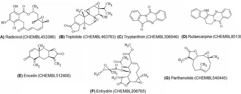
inhibitors < 1μM (Bento et al., 2014). Moreover, only 7 out of those 9 compounds that meet the Lipinski’s rule of 5 (Figure 2) (Lipinski et al., 2001). Therefore, development of validated SBVS protocols that can identify marginal and potent COX-2 inhibitors to cover phytochemicals as potential lead compounds is required.
The research presented in this paper was aimed to construct and retrospectively validate SBVS protocols to identify marginal to potent COX-2 inhibitors by using data from DUD-E with additional marginal active COX-2 inhibitors from DUD-E as active compounds (Mysinger et al., 2012). The protocols were constructed by employing PLANTS1.2 as the molecular docking software (Korb et al., 2007; Korb et al., 2009) and PyPLIF to identify Protein-Ligand Interaction Fingerprints (PLIF) to COX-2 as the re-scoring functions (Radifar
et al., 2013a; Radifar et al., 2013b). The quality of
the SBVS protocol was subsequently assessed (Cannon et al., 2007; de Graaf et al., 2011; Desaphy et al., 2013; Powers, 2011) and Figure 1. Structures of curcumin, the active substance found in turmeric (Curcuma longa) (A) and resveratrol, a compound mainly found in grapes and red wine (B) (Orlikova et al., 2013; Setyaningsih et al., 2013; Yuniarti et al., 2012)
Figure 2. Structures of phytochemicals published in Journal of Natural Products and stored in ChEMBL_21 database that have IC50 values as COX-2 inhibitors < 1 M and do not violate the
compared to the original SBVS accompanying the release of DUD-E (Mysinger et al., 2012). Very recently, Istyastono (2015) showed that using recursive partition and regression tree (RPART) method with the PLANTS1.2 docking score (ChemPLP score) and the PLIF bitstrings resulted from PyPLIF as the descriptors increased significantly the SBVS predictive ability (Istyastono, 2015; Therneau et al., 2015). This approach could avoid the dependency of the application of PyPLIF towards the reference compound (Istyastono, 2015). By employing the same strategy, this research could increase significantly the predictive ability of the SBVS protocols to identify marginal and potent COX-2 inhibitors with enrichment factor (EF) value of 44.78. The optimized protocol was subsequently employed to virtually screen phytochemicals in figures 1 and 2 as COX-2 inhibitors.
MATERIALS AND METHODS
The crystal structure of COX-2 obtained from the protein data bank (PDB) with PDB id of 3LN1 (Wang et al., 2010a) was used as the
reference structure. Active (435 compounds) and marginal active (1538 compounds) COX-2 inhibitors and the decoys (23150 compounds) from DUD-E (Mysinger et al., 2012) were employed to perform retrospective validations for the SBVS protocol. All calculations and computational simulations were performed on a Linux (Ubuntu 12.04 LTS Precise Pangolin) machine with Intel(R) Xeon(R) CPU E31220 (@
3.10 GHz) as the processors and 8.00 GB of RAM. Computational medicinal chemistry applications employed in this research were SPORES (ten Brink and Exner, 2009), PLANTS1.2 (Korb et al., 2007; Korb et al., 2009), Open Babel 2.2.3 (O’Boyle et al., 2011), PyPLIF 0.1.1 (Radifar et al., 2013a; Radifar et al.,
2013b), and PyMOL 1.2r1 (Lill and Danielson,
2011). Statistical analysis was performed by using R 3.2.3 (R Core Team, 2015).
Computational Methods
Virtual molecular target preparation
The crystal structure of COX-2 with the PDB id of 3LN1 (Wang et al., 2010a) was
downloaded from
http://www.rcsb.org/pdb/explore.do?structur
eId=3ln1. Only chain A of the crystal structure
used further in this research (Mysinger et al., 2012). The module splitpdb in SPORES was subsequently used to split and to convert the splitted files into mol2 files the virtual COX-2 (protein.mol2), the co-crystal ligand celecoxib
(ligand_CEL682_0.mol2), and the water
molecules. The mol2 files were then ready to be employed in molecular docking simulation employing PLANTS1.2 docking software.
Ligands preparation for retrospective virtual screening
and decoys_final.ism. Each compound in the files was then subjected to Open Babel 2.2.3 conversion software to be converted in its three dimensional (3D) format at pH 7.4 as a mol2
file. The settypes module in SPORES was subsequently employed to properly check and assign the mol2 file into a proper mol2 file ready to dock by using PLANTS1.2 docking software.
Automated molecular docking and virtual screening
Similar to previously published procedures (Istyastono and Setyaningsih, 2015; Istyastono et al., 2015a; Setiawati et al., 2014), all
virtual screenings were performed by docking program PLANTS1.2. For each compound, 50 poses were calculated and scored by the ChemPLP scoring function at speed setting 2. The binding pocket of COX-2 was defined by the coordinates of the center of the co-crystal ligand celecoxib and a radius of 5 Å (which is the maximum distance from the center defined by a 5 Å radius around the reference ligand). All other options of PLANTS1.2 were left at their default setting (Anita et al., 2012; Istyastono and Setyaningsih, 2015). Every compound was virtually screened five times independently.
Rescoring using protein-ligand interaction fingerprints calculated by PyPLIF
face-to-face, and hydrophobic interactions) were used to define the PLIF for each docking pose (Radifar et al., 2013b; Setiawati et al., 2014). The
cavity used for the PLIF analysis consisted of a set of amino acid residues in the binding pocket of COX-2 defined in subsection Automated molecular docking and virtual screening.
Optimizing the SBVS predictive ability using RPART
The docking pose with the best ChemPLP score was selected for each virtually screened compound. The results were then ranked based on their ChemPLP score and the enrichment factor of True Positives (TP) at 1% False Positives (FP) value or the EF1% value
was calculated (EF1% = %TP/FP1%) (de Graaf
and Rognan, 2008; Istyastono and Setyaningsih, 2015). The compound was predicted as a COX-2 inhibitor if it showed ChemPLP score ≤ the ChemPLP score of the compound at 1% FP (Istyastono et al., 2015b). Therefore, the
protocol was encoded as the EF1%-ChemPLP
based SBVS. By employing RPART package (R Core Team, 2015; Therneau et al., 2015), decision trees were generated using ChemPLP score resulted from PLANTS1.2 (Korb et al., 2009) and all PLIF bitstrings resulted from PyPLIF (Radifar et al., 2013b) as the descriptors
(Istyastono, 2015). The predictive quality of the best decision tree resulted from RPART method was measured by examining the EF value (de Graaf et al., 2011), the balance accuracy (Cannon et al., 2007; Therneau et al., 2015) and F-measure value (Desaphy et al., 2013). The predictive quality of the best decision tree was also compared to the predictive quality of the EF1%-ChemPLP based
SBVS by employing McNemar’s test (Cannon et al., 2007).
Virtual screening on some phytochemicals By employing the best SBVS protocol resulted from the subsection Optimizing the SBVS predictive ability using RPART, virtual screening campaigns to predict whether the COX-2 inhibitors (Figures 1 and 2) were also identified as COX-2 inhibitors virtually were performed. The virtual compounds were downloaded in their SMILE formats from the ChEMBL_21 database (Bento et al., 2014).
Subsequently, the virtual compounds were prepared and examined using the best SBVS protocol.
RESULTS AND DISCUSSION
Aimed to construct and evaluate the validation of an SBVS protocol to identify phytochemicals as inhibitors for COX-2, this research employed marginal and potent COX-2 inhibitors organized and stored in DUD-E (Mysinger et al., 2012) as the retrospective compounds for the validation. Similar to the construction of an SBVS protocol to identify
potent ligands for adrenergic β2 receptor
(Istyastono and Setyaningsih, 2015), the SBVS constructed here employed PLANTS1.2 as the molecular docking software (Korb et al., 2007; Korb et al., 2009) and PyPLIF as the PLIF identification software for rescoring the results from PLANTS1.2 (Radifar et al., 2013a; Radifar et al., 2013b). Additionally, the SBVS protocol
constructed here employed the ChemPLP scores from PLANTS1.2 (Korb et al., 2009) and the PLIF bitstrings from PyLIF (Radifar et al., 2013b) obtained from the docking pose with
the best ChemPLP score in each virtually screened compound as descriptors to generate decision tress using RPART package in R (Istyastono, 2015; R Core Team, 2015; Therneau et al., 2015).
Retrospective Validation of the SBVS protocol
The retrospective SBVS campaigns on COX-2 ligands and their decoys have resulted in 3,767,550 docking poses and 1,318,642,500 PLIF bitstrings resulted from 25,117 out of 25,123 screened compounds. Six decoys could not pass the constructed protocol, which were then assigned as True Negatives (TN). The EF1%-ChemPLP based SBVS resulted in
ChemPLP score of -108.028 as the cutoff score. Employing this cutoff score resulted in a confusion matrix presented in Table 1. The EF value of the protocol was 3.55, which was very low compared to the reference (EF value = 12.9) and was not recommended to be employed further (Istyastono et al., 2015b;
Mysinger et al., 2012). Inspired by Istyastono
Figure 3. The decision tree adopted from the best decision tree resulted from the RPART method (Istyastono, 2015; Therneau et al., 2015).
Table I.The confusion matrices and the statistical significances resulted from the validation of the SBVS protocol to identify COX-2 inhibitors
Parameters SBVS
EF1%-ChemPLP based Optimized using RPART
Confusion Matrices
True Positives (TP) 70 668
False Negatives (FN) 1903 1305
True Negatives (TN) 22919 22975
False Positives (FP) 231 175
Statistical Significances
Sensitivity 0.04 0.34
Specificity 0.99 0.99
Enrichment Factor (EF) 3.55 44.78
Balanced Accuracy 0.51 0.67
F-measure 0.06 0.47
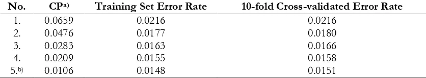
Table II.Decision trees resulted from employing RPART method on the SBVS results to identify marginal COX-2 ligands
No. CPa) Training Set Error Rate 10-fold Cross-validated Error Rate
1. 0.0659 0.0216 0.0216
2. 0.0476 0.0177 0.0180
3. 0.0283 0.0163 0.0166
4. 0.0209 0.0155 0.0158
5.b) 0.0106 0.0148 0.0151
a)Complexity parameter of the decision tree; b)The selected decision tree with the lowest training set
Therneau et al., 2015) was used to generate decision trees using ChemPLP scores and PLIF bitstrings descriptors to optimize the SBVS protocol. The best decision tree (Figure 3) resulted in a confusion matrix with EF value of 44.78. The EF value of the optimized SBVS protocol was higher than the EF1%-ChemPLP
based SBVS (3.55) and the reference (12.9). Moreover the predictive ability of the optimized protocol was statistically better in confidence level of 95% compared to the EF1%-ChemPLP based SBVS (p-value < 0.05)
using McNemar’s test with chi-squared value of 417.23 (Cannon et al., 2007; R Core Team, 2015).
The predictive ability of the optimized protocol was considered as acceptable since it outperformed the reference protocol (Mysinger
et al., 2012). By examining the statistical significances (Table I), although the optimized protocol could be used further in prospective campaigns since the EF value was sufficiently high (de Graaf et al., 2011; Istyastono et al., 2015b), the sensitivity value was still considered
as low (Desaphy et al., 2013). The predictive ability was mainly contributed by the high specificity value. This indicated that if a compound predicted as a COX-2 inhibitor using this optimized in silico screening protocol, it would be high likely as COX-2 inhibitor in vitro. But, if a compound predicted as a non 2 inhibitor, it would still likely be a COX-2 inhibitor in vitro since the sensitivity value was
low caused by the high number of the false negatives (FN; Table 1). This high number of the FN was the limitation of the optimized SBVS protocol that could be improved to increase the predictive ability of the SBVS protocol by employing some more advanced approaches, for example: (i) employing anchor reactions during molecular docking simulations (Yuniarti et al., 2011) or post-docking pose selection (de Graaf et al., 2011; Istyastono et al., 2015b), and/or (ii) employing advanced used of
PLIF bitstrings (Desaphy et al., 2013).
Based on Figure 3, the most important descriptor to identify COX-2 inhibitors was the hydrogen bond interaction to ARG499 (previously reported as ARG513 in the older COX-2 crystal structure (Kurumbail et al., 1996)) with the inhibitors as the acceptor (PLIF bitstring #221). This interaction was identified previously as the anchor interaction of COX-2 inhibitors to COX-2 binding pocket (Kurumbail et al., 1996; Wang et al., 2010a; Yuniarti et al., 2011). This anchor
interaction was identified in the interaction of selective COX-2 inhibitor celecoxib in the COX-2 binding pocket (Wang et al., 2010a).
Alternatives important interactions identified in this research were: (i) hydrogen bond interaction with the residue as the acceptor, which were to GLN178 (PLIF bitstring # 68; Previously reported as GLN192 in the older COX-2 crystal structure (Kurumbail
et al., 1996)), LEU338 (PLIF bitstring #138; Table III. The in silico screening results on some phytochemicals as COX-2 inhibitors
Name IC50 (nM)a)
Decision Tree Parametersb)
TP or FNc)
ChemPLP score PLIF bitstring number
17 18 68 138 221 242
Curcumin 79200 -86.1881 1 0 0 0 0 0 FN Resveratrol 32000 -90.2518 1 0 1 0 0 1 TP Radicicol 27 -49.8668 0 0 0 0 0 0 FN Triptolide 40 -67.6514 0 0 0 0 0 0 FN Tryptanthrin 64 -87.7228 1 0 0 0 0 0 FN Rutaecarpine 300 -86.6826 1 0 0 0 0 0 FN Encelin 400 -66.2878 0 0 0 0 0 0 FN Enhydrin 600 -37.5564 0 0 0 0 0 0 FN Parthenolide 800 -63.2679 0 0 0 0 0 0 FN
a)Ref: (Bento et al., 2014); b)See Figure 3 for more explanation; c)TP and FN stand for True Positive
Previously reported as LEU352 in the older COX-2 crystal structure (Kurumbail et al., 1996)) or PHE504 (PLIF bitstring #242; Previously reported as PHE518 in the older COX-2 crystal structure (Kurumbail et al., 1996)); (ii) hydrogen bond interaction to HIS75 (PLIF bitstring #18; Previously reported as HIS90 in the older COX-2 crystal structure (Kurumbail et al., 1996)) with the residue as the donor; and (iii) edge-to-face aromatic interaction to HIS75 (PLIF bitstring #17).
Celecoxib was also reported having the interaction to GLN178, HIS75 and PHE504 (Wang et al., 2010a). The interaction to LEU352
could categorized as novel interaction, but it was not rare in COX-2 binding since very recently several COX-2 inhibitors could proceed further to the clinical trial phase although the compound did not show interaction to the previously known important residues in the COX-2 binding pocket (Wang et al., 2010a; Wang et al., 2010b). The decision tree
resulted from RPART method was therefore could identify alternative important interactions which in turn could increase the predictive ability of SBVS protocols by decreasing the number of FP and FN. Moreover, the 10-fold cross validation in the construction of decision trees (Table 2) showed that there was no evidence of overfitting of the selected decision tree, and the 1000 times Y-randomization showed that there is no evidence of chance correlation (Cappel et al., 2015; Lim et al., 2009).
Phytochemicals Virtual Screening Employing Optimized Protocol
Nine compounds presented in Figures 1 and 2 were examined using the optimized protocol exclusively identified marginal COX-2 inhibitors. This should be verified by testing more representative numbers of other external marginal and potent COX-2 inhibitors.
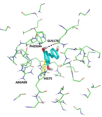
The docking protocol used here is the same as the docking protocol employed by Mumpuni et al. (2015), which re-dock co-crystalized ligand celecoxib to the crystal structure 3LN1 with the root-mean-square
deviation (RMSD) value of 0.525 Ǻ. Since the value was less than 2.0 Ǻ (Mumpuni et al., 2015), the selected pose of resveratrol here (Figure 4) could be considered as the right pose. In the visual inspection on the best pose of resveratrol in COX-2 binding pocket (Figure 4) using PyMOL (Lill and Danielson, 2011) and the examination of Figure 3 and Table 3, resveratrol was predicted as COX-2 inhibitor by binding to HIS75 (edge-to-face aromatic interaction), GLN178 (hydrogen bond), and PHE504 (hydrogen bond). Surprisingly, the selected pose for resveratrol did not bind to ARG499. The decision tree (Figure 3) provided alternative interactions in COX-2 ligand binding (Wang et al., 2010a; Wang et al., 2010b).
Since the first branch of the decision tree involved hydrogen bond to ARG499 (Figure 3), employing this as the anchor interaction in the molecular docking simulation (Wang et al.,
2010a; Yuniarti et al., 2011) could therefore increase the predictive ability of the SBVS protocol.
CONCLUSIONS
The optimized SBVS protocol employing PLANTS1.2 and PyPLIF followed by RPART method to produce decision tree to identify phytochemicals as COX-2 inhibitors has been retrospectively validated using DUD-E with additional marginal active compounds as the active compounds. The protocol resulted in better predictive ability in COX-2 inhibitors identification compared to the original protocol accompanying the release of DUD-E. However the sensitivity value was still considered as low, which was also indicated by predicting correctly only 1 out of 9 phytochemical as COX-2 inhibitors. The improvement could be achieved by employing hydrogen bond to ARG499 as the anchor interaction during the molecular docking simulations using PLANTS1.2 before the PLIF identification using PyPLIF.
ACKNOWLEDGEMENT
This work was financially supported by the Penelitian Hibah Bersaing (No. 010/HB-LIT/III2016) from Directorate of Research and Community Services, Ministry of Research, Technology and Higher Education, the Republic of Indonesia. Dr. Sri Hartati Yuliani, Apt. from Division of Pharmaceutical Technology, Faculty of Pharmacy, Sanata Dharma University is acknowledged for her technical assistance and suggestions in writing this article.
REFERENCES
Anita Y., Radifar M., Kardono L., Hanafi M., Istyastono EP., 2012, Structure-based design of eugenol analogs as potential estrogen receptor antagonists.
Bioinformation, 8, 901–906
Bento AP., Gaulton A., Hersey A., Bellis LJ., Chambers J., Davies M., Krüger FA., Light Y., Mak L., McGlinchey S., Nowotka M., Papadatos G., Santos R., Overington JP., 2014, The ChEMBL bioactivity database: An update. Nucl. Acids Res., 42, 1083–1090
Mitchell, J.B.O., 2007, Support vector inductive logic programming outperforms the naive Bayes classifier and inductive logic programming for the classification of bioactive chemical compounds. J. Comput. Aided Mol. Des., 21, 269–280
Cappel D., Dixon SL., Sherman W., Duan J., 2015, Exploring conformational search protocols for ligand-based virtual screening and 3-D QSAR modeling. J. Comput. Aided Mol. Des., 29, 165–182 Chakraborti AK., Garg SK., Kumar R.,
Motiwala HF., Jadhavar PS., 2010, Progress in COX-2 inhibitors: a journey so far. Curr. Med. Chem., 17, 1563–1593 Cianchi F., Cortesini C., Schiavone N., Perna
F., Magnelli L., Fanti E., Bani D., Messerini L., Fabbroni V., Perigli G., Capaccioli S., Masini E., 2005, The role of cyclooxygenase-2 in mediating the effects of histamine on cell proliferation and vascular endothelial growth factor production in colorectal cancer. Clin. Cancer Res., 11, 6807–6815
Dai Z., Ma X., Kang H., Gao J., Min W., Guan H., Diao Y., Lu W., Wang, X., 2012, Antitumor activity of the selective cyclooxygenase-2 inhibitor, celecoxib, on breast cancer in vitro and in vivo. Cancer Cell Int., 12, 53
de Graaf C., Kooistra AJ., Vischer HF., Katritch V., Kuijer M., Shiroishi M., Iwata S., Shimamura T., Stevens RC., de Esch IJP., Leurs R., 2011, Crystal structure-based virtual screening for fragment-like ligands of the human histamine H1 receptor. J. Med. Chem., 54,
8195–8206
de Graaf C. and Rognan D., 2008, Selective structure-based virtual screening for full and partial agonists of the β2 adrenergic
receptor. J. Med. Chem., 51, 4978–4985 Desaphy J., Raimbaud E., Ducrot P., Rognan
D., 2013, Encoding protein-ligand interaction patterns in fingerprints and graphs. J. Chem. Inf. Model., 53, 623–637 Gautam R., Jachak SM., Kumar V., Mohan
CG., 2011, Synthesis, biological evaluation and molecular docking studies of stellatin derivatives as cyclooxygenase
(COX-1, COX-2) inhibitors and anti-inflammatory agents. Bioorg. Med. Chem. Lett., 21, 1612–1616
Huang N., Shoichet BK., Irwin JJ., 2006, Benchmarking sets for molecular docking. J. Med. Chem., 49, 6789–6801 Istyastono EP., 2015, Employing recursive
partition and regression tree method to increase the quality of structure-based virtual screening in the estrogen receptor alpha ligands identification. Asian J. Pharm. Clin. Res., 8, 21–24
Istyastono EP., Riswanto FDO., Yuliani SH., 2015a, Computer-aided drug
repurposing: a cyclooxygenase-2 inhibitor celecoxib as a ligand for estrogen receptor alpha. Indones. J. Chem., 15, 274–280
Istyastono EP., Kooistra AJ., Vischer H., Kuijer M., Roumen L., Nijmeijer S., Smits R., de Esch I., Leurs R., de Graaf C., 2015b, Structure-based virtual
screening for fragment-like ligands of the g protein-coupled histamine H4 receptor. Med. Chem. Commun., 6, 1003–1017 Istyastono EP. and Setyaningsih D., 2015,
Construction and retrospective validation of structure-based virtual screening protocols to identify potent ligands for human adrenergic β2
receptor, Indones. J. Pharm., 26, 20–28 Jendrossek V., 2013, Targeting apoptosis
pathways by Celecoxib in cancer. Cancer Lett., 332, 313–324
Kaserer T., Temml V., Kuti, Z., Vanek T., Landa P., Schuster D., 2015, Prospective performance evaluation of selected common virtual screening tools. Case study: Cyclooxygenase (COX) 1 and 2.
Eur. J. Med. Chem. 96, 445–457
Korb O., Stützle T., Exner TE., 2007, An ant colony optimization approach to flexible protein–ligand docking. Proc. IEEE Swarm Intell. Symp. 1, 115–134
Korb O., Stützle T., Exner TE., 2009, Empirical scoring functions for advanced protein-ligand docking with PLANTS. J. Chem. Inf. Model., 49, 84–96 Krüger DM. and Evers A., 2010, Comparison
complementarity and enrichment factors.
ChemMedChem, 5, 148–158
Kurumbail R., Stevens A., Gierse J., 1996, Structural basis for selective inhibition of cyclooxygenase-2 by anti-inflammatory agents. Nature, 384, 644–648
Larsson J., Gottfries J., Bohlin L., Backlund A., 2005, Expanding the ChemGPS chemical space with natural products. J. Nat. Prod., 68, 985–991
Lill MA. and Danielson ML., 2011, Computer-aided drug design platform using PyMOL. J. Comput. Aided Mol. Des., 25, 13–19
Lim HD, Istyastono EP, van de Stolpe A, Romeo G, Gobbi S, Schepers M, Lahaye R, Menge WMBP, Zuiderveld OP, Jongejan A, Smits RA, Bakker RA, Haaksma EEJ, Leurs R, de Esch IJP, 2009, Clobenpropit analogs as dual activity ligands for the histamine H3 and
H4 receptors: synthesis, pharmacological
evaluation, and cross-target QSAR studies. Bioorg. Med. Chem., 17, 3987– 3994.
Lipinski CA., Lombardo F., Dominy BW., Feeney PJ., 2001, Experimental and computational approaches to estimate solubility and permeability in drug discovery and development settings. Adv. Drug Deliv. Rev., 46, 3–26
Maggon K., 2005, Best-selling human medicines 2002-2004. Drug Discov. Today, 10, 739–742
Mumpuni E., Nurrochmad A., Jenie UA., Pranowo HD., 2015, Virtual screening and bonding mode elucidation of curcumin analogue in cyclooxygenase-2 enzyme using EE_COX2_V.1.0 protocol. Indones. J. Phar. Sci., 13, 235-241 Mysinger MM., Carchia M., Irwin JJ., Shoichet Vandermeersch T., Hutchison GR., 2011, Open Babel: An open chemical toolbox. J. Cheminform., 3, 33–47
Orlikova B., Legrand N., Panning J., Dicato M., Diederich M., 2013, Anti-inflammatory
and anticancer drugs from nature. Cancer Treat. Res., 159, 123–143
Pany S., Pal A., Sahu PK., 2013, In silico analysis of cyclooxygenase inhibitory activity of some natural molecules. Int. J. Pharm. Pharm. Sci., 5, 7–9
Penning TD., Talley JJ., Bertenshaw SR., Carter JS., Collins PW., Docter S., Graneto MJ., Lee LF., Malecha JW., Miyashiro JM., Rogers RS., Rogier DJ., Yu SS., Anderson GD., Burton EG., Cogburn JN., Gregory SA., Koboldt CM., Perkins WE., Seibert K., Veenhuizen AW., Zhang YY., Isakson PC., 1997, Synthesis and biological evaluation of the 1,5-diarylpyrazole class of cyclooxygenase-2 inhibitors: identification of 4-[5-(4- methylphenyl)-3-(trifluoromethyl)-1H-environment for statistical computing. Vienna. http://www.r-project.org. Radifar M., Yuniarti N., Istyastono EP., 2013a,
PyPLIF-assisted redocking
indomethacin-(R)-alpha-ethyl-ethanolamide into cyclooxygenase-1.
Indones. J. Chem., 13, 283–286.
Radifar M., Yuniarti N., Istyastono EP., 2013b,
PyPLIF: Python-based protein-ligand interaction fingerprinting. Bioinformation, 9, 325–328
Rao PNP., Chen Q., Knaus EE., 2006, Synthesis and structure-activity relationship studies of 1,3-diarylprop-2-yn-1-ones: dual inhibitors of cyclooxygenases and lipoxygenases. J. Med. Chem., 49, 1668–1683
Rowlinson SW., Kiefer JR., Prusakiewicz JJ., Pawlitz JL., Kozak KR., Kalgutkar AS., Stallings WC., Kurumbail RG., Marnett LJ., 2003, A novel mechanism of cyclooxygenase-2 inhibition involving interactions with Ser-530 and Tyr-385. J. Biol. Chem., 278, 45763–45679
“target-specific” drugs? Celecoxib as a case in point. Mol. Interv., 6, 196–198 Setiawati A., Riswanto FDO., Yuliani SH.,
Istyastono EP., 2014, Retrospective validation of a structure-based virtual screening protocol to identify ligands for estrogen receptor alpha and its application to identify the alpha-mangostin binding pose. Indo. J. Chem., 14, 103–108.
Setyaningsih D., Radifar M., Murti YB., Istyastono EP., 2013, Construction of in silico structure-based screening tools to study the oxidative metabolites formation of curcumin by human cytochrome 450 3A4. Indones. J. Pharm., 24, 75–85
ten Brink T. and Exner TE., 2009, Influence of protonation, tautomeric, and stereoisomeric states on protein-ligand docking results. J. Chem. Inf. Model., 49, 1535–1546
Therneau T., Atkinson B., Ripley B., 2015, rpart: Recursive partitioning and regression trees. R package version 4.1-9.
https://CRAN.R-project.org/package=rpart
Wang JL., Carter J., Kiefer JR., Kurumbail RG., Pawlitz JL., Brown D., Hartmann SJ., Graneto MJ., Seibert K., Talley JJ., 2010a,
The novel benzopyran class of selective cyclooxygenase-2 inhibitors-part I: The first clinical candidate. Bioorg. Med. Chem. Lett., 20, 7155–7158
Wang JL., Limburg D., Graneto MJ., Springer
J., Hamper JRB., Liao S., Pawlitz JL., Kurumbail RG., Maziasz T., Talley JJ., Kiefer JR., Carter J., 2010b, The novel
benzopyran class of selective cyclooxygenase-2 inhibitors. Part 2: The second clinical candidate having a shorter and favorable human half-life.
Bioorg. Med. Chem. Lett. 20, 7159–7163 Wilgus TA., Bergdall VK., Tober KL., Hill KJ.,
Mitra S., Flavahan NA., Oberyszyn TM., 2004, The impact of cyclooxygenase-2 mediated inflammation on scarless fetal wound healing. Am. J. Pathol., 165, 753– 761
Wilgus TA., Vodovotz Y., Vittadini E., Clubbs EA., Oberyszyn TM., 2003, Reduction of scar formation in full-thickness wounds with topical celecoxib treatment.
Wound Rep. Reg., 11, 25–34
Willoughby DA., Moore AR., Colville-Nash PR., Gilroy D., 2000, Resolution of inflammation. Int. J. Immunopharmacol., 22, 1131–1135
Yuniarti N., Ikawati Z., Istyastono EP., 2011, The importance of ARG513 as a hydrogen bond anchor to discover COX-2 inhibitors in a virtual screening campaign. Bioinformation, 6, 164–166 Yuniarti N., Nugroho PA., Asyhar A.,