Behavioral/Systems/Cognitive
Conditional Inactivation of Androgen Receptor Gene in the
Nervous System: Effects on Male Behavioral and
Neuroendocrine Responses
Kalina Raskin,
1,2Karel de Gendt,
3Anne Duittoz,
4Philippe Liere,
5Guido Verhoeven,
3Franc¸ois Tronche,
1,2and
Sakina Mhaouty-Kodja
1,21Centre National de la Recherche Scientifique (CNRS) Unite´ Mixte de Recherche (UMR) 7148, “Ge´ne´tique Mole´culaire-Neurophysiologie et
Comportement,” and2Institut de Biologie, Colle`ge de France, 75231 Paris, France,3Laboratory of Experimental Medicine and Endocrinology, Katholieke
Universiteit Leuven, 3000 Leuven, Belgium,4CNRS UMR 6175/Institut National de la Recherche Agronomique, 37380 Nouzilly, France, and5UMR 788 –
Inserm–Universite´ Paris XI, 94276 Kremlin Biceˆtre, France
Testosterone (T) profoundly influences central sexual differentiation and functions. In the brain, T signals either directly through androgen receptor (AR) or indirectly through estrogen receptor (ER) following aromatization into E2 (17--estradiol). As T, through AR, also controls peripheral male sexual differentiation, the relative contribution of central AR in T-mediated regulation of behavioral and neuroendocrine responses still remains unclear. To address this question, we generated, by using Cre-loxP technology, mice selectively lacking AR expression in the nervous system. The mutant male urogenital tract was normally developed, and mice were able to produce offspring. Nonetheless, sexual motivation and performance as well as aggressive behaviors were affected. Only a low percentage of males displayed a complete sexual behavior and offensive attacks. The latency to show masculine behaviors was increased and copulation length prolonged. Erectile activity during mating was also altered. These alterations occurred despite increased levels of T and its metabolites, and an unaffected number of ER␣-immunoreactive cells. Olfactory preference and neuronal activation, mapped by Fos immunoreactivity, following exposure to estrus female-soiled bedding were also normal. At comparable T levels, greater differences in masculine behaviors were observed between gonadectomized control and mutant males.ARinvalidation in the nervous system also
disrupted the somatotropic axis since mutant males exhibited growth retardation and decreased serum levels of insulin-like growth factor I. Our findings show that central AR is required in T-induced regulation of male-typical behaviors and gonadotrope and somato-tropic axes. This genetic model offers a unique opportunity in the understanding of AR’s role in cerebral functions of T.
Introduction
Testosterone (T) exerts permanent (organizational) effects on the developing male nervous system during the perinatal period and transient (activational) roles in adulthood (Phoenix et al., 1959). In rodents, the prenatal and neonatal T surges masculinize neural circuitries, leading to an enhancement of behavioral pat-terns that are exclusively elicited by males. Indeed, a wide range of behaviors (sex, aggression, juvenile play . . .) and neuroendo-crine functions (feedback effect of steroids on gonadotropin re-lease, growth) are sexually dimorphic.
In the brain, T can signal either directly through activation of androgen receptor (AR), or indirectly via stimulation of estrogen receptors (ERs) following conversion into estradiol by aromatase
cytochrome P-450 enzyme (Ar). AR, ER␣, and ERare members
of the nuclear receptor superfamily and regulate target genes at a transcriptional level (Matsumoto et al., 2003). To delineate the relative importance of the direct signaling through AR versus the indirect pathway through ER in androgen-mediated regulation of brain functions, genetic models were studied. Data from
knock-out (KO) mice ubiquitously lackingCyp19 ar(arKO) or
ER␣(ER␣KO) indicated that 17--estradiol (E2) deriving from
neural aromatization of T and ER␣signaling pathway play a key
role in the expression of sexual and aggressive behaviors (Ogawa et al., 1997; Wersinger et al., 1997; Bakker et al., 2002). Unfortu-nately, a better understanding of the real involvement of cerebral
AR functionsin vivowas hampered by the complex phenotype of
Tfm (testicular feminization mutation) mice carrying
spontane-ously mutatedARgene (Lyon and Hawkes, 1970) and ubiquitous
ARKO mice (Sato et al., 2003; Chang et al., 2004; De Gendt et al., 2004). Indeed, genetic (XY) males of both models display a com-plete androgen insensitivity phenotype with female-typical exter-nal appearance, small cryptorchidic azoospermic testes, and low
Received Jan. 19, 2009; revised Feb. 20, 2009; accepted Feb. 20, 2009.
This work was supported by the Centre National de la Recherche Scientifique (CNRS) and Colle`ge de France. We thank Drs. R. Counis and G. Garrel [CNRS Unite´ Mixte de Recherche (UMR) 7079, Paris, France] for providing the monoclonal anti-LH; Dr. Caraty A. [CNRS UMR 6175/Institut National de la Recherche Agronomique (INRA), Nouz-illy France) for anti-GnRH; and Dr. M. Schumacher, A. Pianos, A. Cambourg, and B. Eychenne (UMR 788 –Inserm– Universite´ Paris XI, Kremlin Biceˆtre, France) for intratesticular steroid content measurements. We are also indebted to Dr. O. Rampin (UMR1197 INRA–Universite Paris 11, Jouy-en-Josas, France) for helpful discussions on erection.
Correspondence should be addressed to either Sakina Mhaouty-Kodja or Franc¸ois Tronche, Centre National de la Recherche Scientifique Unite´ Mixte de Recherche 7148, Ge´ne´tique Mole´culaire-Neurophysiologie et Comportement, Colle`ge de France, 11 place Marcelin Berthelot, 75231 Paris Cedex 05, France. E-mail: [email protected] or franc¸[email protected].
DOI:10.1523/JNEUROSCI.0296-09.2009
levels of circulating T. Due to the lack of external genitals, male sexual behavior of Tfm and ARKO mice is limited to coital (mounts and thrusts) behavior as previously described (Ono et al., 1974; Sato et al., 2004). Furthermore, given the critical role of T in several peripheral functions related or not to reproduction, it is difficult to distinguish, in these genetic models, between its central and peripheral mediated effects.
Therefore, to define more precisely the specific role of central AR in T-dependent regulation of male behaviors and
neuroen-docrine functions, we generated mice lackingARin the nervous
system. For this purpose, we crossed floxed AR mice (De Gendt et al., 2004) with transgenic mice expressing Cre recombinase driven by the promoter and the nervous system-specific enhancer of rat nestin (Nes) in neuronal and glial precursor cells (Tronche
et al., 1999). The obtained ARfl/Y, Nes-Cre (ARNesCre) males,
unlike ARKO mice, had a normal development of the urogenital tract and were able to produce offspring with reduced fertility. Nevertheless, they exhibited impaired sexual and aggressive
be-haviors despite high levels of T and its metabolites [5-␣
-dihydrotestosterone (DHT) and E2]. They also showed growth retardation and altered growth hormone (GH)/insulin-like growth factor I (IGF-I) system.
Materials and Methods
Generation of mice with conditional inactivation of AR gene in the nervous system
Mutant ARNesCremice and their control (ARfl/Y) littermates, with a
genetic mixed background (C57BL/6 and 129SvEv), were obtained from crossings of ARNesCremales with ARfl/ARflfemales. Mice were weaned at
21–25 d of age and group housed under a controlled photoperiod (12 h light/12 h dark cycle—lights on at 7:00 A.M.) and temperature (22°C) and givenad libitumaccess to food and water. The presence of Cre transgene was detected by dot blot analysis and ARflallele by PCR as
previously described (De Gendt et al., 2004). Cre-mediated excision of ARflallele was found only in neuronal tissues. The genetic sex was
determined by using specific primers forSryandNDSgenes, located, respectively, on the Y and X chromosomes (Kunieda et al., 1992). All studies were performed on 2- to 5-month-old animals, in accordance with the National Institutes of HealthGuide for the Care and Use of Laboratory Animals.
Western blot analysis
Brain, skeletal muscle, and testis were dissected and stored at⫺80°C. Tissues were homogenized in buffer (2% SDS, 50 mMDTT, 62.5 mMTris, pH 6.8, 10% glycerol) containing a mixture of protease inhibitors (Roche). Samples were then boiled for 5 min and centrifuged at 7500⫻
gfor 10 min at 4°C. Supernatants were collected, and protein concentra-tions were determined using a Bradford assay (Bradford, 1976). Samples (35g of proteins for brain and skeletal muscle and 15g for testis) were then loaded and separated on a 7.5% polyacrylamide gel. After transfer onto a nitrocellulose membrane, the blot was probed with 1:200 diluted polyclonal antibody directed against the N-terminal epitope of AR (Sc816, Santa Cruz Biotechnology) and 1:1000 diluted actin anti-body (Sigma). After incubation with peroxidase-conjugated secondary antibody (GE Healthcare) diluted at 1:5000, signals were visualized by using ECL Plus detection kit (GE Healthcare).
Immunohistochemistry
Animals were killed and transcardially perfused with a solution of 4% paraformaldehyde (PFA) in PBS. Brains were sliced into 30m coronal sections and stored in 0.5% PFA. Pituitary glands were dissected and fixed in 4% PFA for 1 h and washed three times in PBS for 20 min at 4°C. After an overnight incubation in 18% saccharose at 4°C, pituitary glands were sliced (7m). Testes were fixed in Bouins fluid (for 100 ml: 75 ml of saturated picric acid, 25 ml of formaldehyde 37%, 5 ml of glacial acetic acid) overnight at 4°C, transferred to 70% ethanol at 4°C, then processed into paraffin wax and sliced into 5m sections.
Brain and pituitary sections were rinsed in PBS and treated with 1% H2O2in PBS for 30 min, then incubated for 2 h with 5% normal goat
serum (NGS, Sigma-Aldrich) in PBST (PBS/0.1% Triton X-100). Testis sections were first dewaxed after heat-induced antigen retrieval for 5 min in 0.01Mcitrate buffer, pH 6, in a microwave oven. Sections were incu-bated at 4°C for 36 h with 1:200 diluted rabbit anti-AR or anti-ER␣ (Santa Cruz Biotechnology), or overnight with 1:500 diluted anti-c-fos antibody (Abcam). Immunostaining was then performed with biotinyl-ated goat rabbit (1:400, Vector Laboratories) for 2 h. Bound anti-bodies were visualized by 30 min of incubation with the biotin–strepta-vidin complex reagent (ABC kit; Vector Laboratories), followed by color development with 3,3⬘-diaminobenzidine tetrahydrochloride chromo-genic substrate (DAB) from Sigma-Aldrich. For immunofluorescence, pituitary sections were incubated overnight with anti-AR and then 1 h with mouse anti-luteinizing hormone (LH) (1:300, gift from Drs. R. Counis and G. Garrel, CNRS, Paris, France) in PBST with 1% NGS. They were then incubated for 2 h with 1:500 diluted anti-mouse Alexa 488 and anti-rabbit Cy3 secondary antibodies (Invitrogen). For quantification of ER␣immunoreactivity, stained sections were matched on anatomical landmarks and compared across animal groups by tallying the number of labeled cells in each region.
Fertility study
Each control or mutant male (2 months old) was mated with two control females for a period of 4 months. The total number of litters and pups per male was recorded at birth.
Sperm count
Epididymides were dissected and cut into small pieces in PBS and left to incubate at 37°C for 10 min. After 5 min of centrifugation at room temperature at 100⫻g, the supernatant was removed and 10-fold di-luted. Spermatozoa were counted in a hemocytometer with a light microscope.
Measurements of hormones and IGF-I levels and gonadotropin-releasing hormone and T contents
Blood samples were centrifuged at 4500⫻gfor 10 min at 4°C, and sera were stored at⫺20°C until analysis. LH, follicle-stimulating hormone (FSH), T, E2, and IGF-1 levels were measured by using commercially available RIA kits (Biocode-Hycel for LH and FSH, Biosource Interna-tional for T and Diagnostic Systems Laboratories for E2 and IGF-1). Assay sensitivities were 0.14 ng/ml, 0.2 ng/ml, 0.05 ng/ml, 2.2 pg/ml, and 21 ng/ml, and interassay coefficients of variation were 12.2%, 8.5%, 5.5%, 8%, and 6.7% for LH, FSH, T, E2, and IGF-1, respectively.
For gonadotropin-releasing hormone (GnRH) content, the hypothal-amus was dissected on ice from the ventral surface of the brain and rapidly frozen at⫺80°C. Tissue samples were homogenized in 0.1N HCl containing a mixture of protease inhibitors before adding NaOH to bring the pH to 7 as previously described (Moore and Wray, 2000). The ho-mogenates were then centrifuged at 4°C for 10 min, and the supernatants were stored at⫺80°C until RIA analysis. Samples were resuspended in PBS-gelatin buffer, and GnRH concentration was measured in 100l duplicate aliquots as described previously (Caraty et al., 1995). GnRH assay sensitivity was 0.2 pg/ml, and the mean interassay and intra-assay coefficients of variation were 13% and 9%, respectively. The used Gn-RHBDS037 antibody is specific to the C-terminal moiety and also binds pro-GnRH and Hyp9-GnRH.
Intratesticular contents of T and DHT were measured by gas chromatography-mass spectrometry as previously described (Liere et al., 2000; Meffre et al., 2007).
Ten to 15 animals per genotype were used for each assay.
Male-typical behaviors
Analyses were conducted in Plexiglas cages under red-light illumination 2 h after lights off and were videotaped for further analysis.
the receptive female if no sexual behavior was displayed. If a mounting episode occurred during this period, the test was extended for 30 addi-tional min or until the male ejaculated. In the latter case, the subject was no longer tested during the next sessions. For males that exhibited sexual behavior on test 2 or 3, the latencies to mount, intromit, thrust, and ejaculate were cumulative. In experiment 2, each male was tested once in its housing cage for 10 h after female introduction.
Stimulus C57BL/6J females were ovariectomized and implanted with SILASTIC implants filled with 50g of E2-benzoate (Sigma-Aldrich) in 30l of sesame oil. Four to five hours before the tests, females were subcutaneously administered with 1 mg of progesterone (Sigma-Aldrich) in 100l of sesame oil. Female receptivity was screened with sexually experienced males before the beginning of the test. At the end of experiment 2, females were still receptive when put in the presence of sexually experienced males.
Erectile activity.The latency to the first grooming erection, and the time spent in erection after each mount with intromission were mea-sured in experiment 2. Erection was scored when the male stood on its hindlimbs, bent its body forward, head down to reach the penis, and performed genital grooming while displaying hip movements, as previ-ously described (Rampin et al., 2003; Rampin et al., 2006).
Olfactory preference.One week after sexual behavior tests, males were placed for 10 min in cages where three containers filled with clean, male-soiled, or female-soiled bedding were equidistantly placed. Animals were first habituated for 2 consecutive days in the same paradigm with only clean bedding for 10 min. The time spent sniffing each container was recorded. Male-soiled bedding was obtained from animals placed in a cage with clean bedding 24 h before the test. Female-soiled bedding was obtained from five group-housed E2-treated females, injected with pro-gesterone 6 h before bedding collection.
Aggression.Males, individually housed for 2 weeks without bedding changes, were tested in a resident–intruder paradigm in their home cages for 3 consecutive days by using A/J mice (The Jackson Laboratory) as intruders. The A/J mouse strain was chosen on the basis of its low scores of aggression (Le Roy et al., 2000). In fact, we never noted an aggressive behavior from A/J mice toward resident animals. Each test was per-formed with a different intruder. The test was stopped immediately after the first offensive attack (defined by biting and wrestling) or after 10 min when no attack occurred. In the latter case, the latency was 600 s. For gonadectomized and T-treated mice, tests lasted 10 min. The latency to the first aggressive act (tail rattling, biting, lunge, offensive attack), the total aggression duration, and the number of offensive attacks and lunge and bites aggression bouts displayed toward the intruder mice were scored for each resident animal.
Elevated plus maze
Animals (10 per genotype) were tested for their behavior in the elevated plus maze (EPM). Mice were brought into the test room at least 1 h before the testing onset. Analysis was performed for 10 min under controlled light conditions (500 lux) and started 4 h before dark. At the beginning of the test, each mouse was placed in the central area. The time spent as well as the number of entries into the open arms were registered.
Locomotor activity
Locomotor activity of animals was analyzed in a computed circular cor-ridor as previously described (Salomon et al., 2006). Briefly, mice were introduced in a circular corridor made of two concentric cylinders crossed by four diametrically opposite infrared beams (Imetronic). The locomotor activity was counted when animals interrupted two successive beams and thus had traveled a quarter of the circular corridor. In the device, the lighting was programmed on the usual dark:light cycle. Spon-taneous activity was recorded for 14 h.
Since aggressive behavior, anxiety state, and locomotor activity were comparable between naive and sexually experienced males, we presented only the results obtained on sexually experienced mice.
T and DHT treatments
Naive males were castrated at 8 weeks of age and implanted with SILAS-TIC tubes containing 10 mg of either T or DHT (Sigma-Aldrich). Two weeks later, animals were tested in the behavioral tests.
Statistics
The percentages of animals showing behaviors were compared by Fisher exact tests. All other data are expressed as means⫾SEM. Analysis of olfactory preference and aggression was performed by a two-way ANOVA for repeated measures for the main effects of genotype and bedding or genotype and test day, respectively, as factors, followed by Bonferroni tests aspost hoccomparisons. Data of nonrepeated measure-ments were analyzed by one-way ANOVA or Student’sttest for unpaired data.pvalue⬍0.05 was considered significant.
Results
Generation and characterization of conditional mutant mice lackingARin the nervous system
To selectively inactivate AR gene in the nervous system, we
crossed females carrying floxedARgene allele (De Gendt et al.,
2004) with transgenic male mice expressing Cre recombinase un-der the control of the rat Nes promoter and neural-specific en-hancer (Tronche et al., 1999).
Analysis by Western blotting indicated the presence of 110 kDa AR protein in the testis and skeletal muscle of both control
(ARfl/Y) and ARNesCremice (Fig. 1A). Nuclear AR protein was
indeed present in testicular Sertoli cells, peritubular myoid cells,
and Leydig cells as well as in LH-positive gonadotrope cells of
both genotypes (Fig. 1B,C). In the brain, AR signal was detected
in control but not in mutant animals (Fig. 1A). Detailed
immu-nohistochemical studies revealed a high nuclear AR expression in
several brain regions of control mice (Fig. 1D). Similar
distribu-tion and density of nuclear AR expression were observed in the
brain of AR⫹/Y and AR⫹/Y mice carrying Nes-Cre transgene
(data not shown). In ARNesCreanimals, AR protein was
com-pletely lost in the CA1 region of the hippocampus, cortex, medial amygdala (MA), cortical amygdala, lateral septum, and bed
nu-cleus of stria terminalis (BNST) (Fig. 1D). In the hypothalamus,
AR expression also disappeared in many regions, including the medial preoptic area (MPOA), ventromedial hypothalamus, and
arcuate nucleus (Fig. 1D); only a few scattered cells retained
N-terminal AR labeling in the anterior and lateral hypothalamus.
Altogether, these results indicate thatARwas selectively
dis-rupted in the brain of ARNesCremales, including regions
impor-tant in the regulation of neuroendocrine and behavioral func-tions related to reproduction, such as mating and aggression (MA, lateral septum, BNST, MPOA, and ventromedial hypothal-amus), and areas involved in learning and memory (cortex and hippocampus).
Effects of nervous systemARdisruption on male
neuroendocrine functions
Elevated ranges of T, DHT, E2, and LH in ARNesCremales Comparison of phenotypic and genetic sex indicated a normal
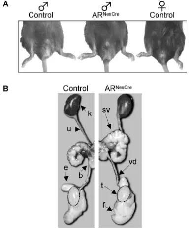
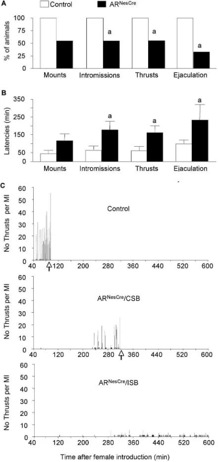
sex ratio of 1:1 male/female for ARNesCremice. In addition, adult
ARNesCremales showed normal development of external genitals
(Fig. 2A) and urogenital tract (Fig. 2B). This clearly indicates that conditional AR gene inactivation in the nervous system did not interfere with male sexual differentiation of the urogenital
sys-tem. Despite a slight reduction of testicular weight (0.29⫾0.01%
of body weight vs 0.35⫾0.01%;p⬍0.05), mutant males had,
respectively, 2.6-fold and twofold higher gonadal contents of T
(70.9⫾16.9 ng/g vs 26.7⫾3.3 ng/g;p⬍0.05) and its metabolite
DHT (8.4⫾1.3 ng/g vs 3.7⫾0.9 ng/g;p⬍0.01) than controls.
Circulating levels of T were also significantly enhanced by
four-fold in ARNesCremice (3.9⫾0.6 ng/ml vs 1.3⫾0.1 ng/ml;p⬍
0.01), which is in good correlation with the increased weight of
the androgen-sensitive seminal vesicles (0.65⫾0.02% of body
metabolite of T, were also significantly augmented by 42% in ARNesCremice (7.8⫾0.7 pg/ml vs 5.5⫾0.3 pg/ml;p⬍0.01).
In males, the testicular synthesis of T is under the control of the hypothalamic GnRH and pituitary LH. The liberated T acts,
in turn, to reduce both GnRH and LH release. In mutant animals,
LH levels were also increased by twofold (2.0⫾0.2 ng/ml vs
0.98⫾0.06 ng/ml;p⬍0.001) while FSH levels were at the normal
range (38.2⫾6.00 ng/ml for mutants vs 36.4⫾6.0 ng/ml for
controls). No significant differences were observed in the hypo-thalamic GnRH content between control and mutant animals
(499.6⫾78.5 pg/animal for controls vs 426.1⫾36.6 pg/animal
for mutants).
When tested in continuous mating, ARNesCremales were able
to produce offspring, but 45% of them were hypofertile with a significantly decreased litter size and total number of pups (Table
1). ARNesCremales had also consistently lower (2.4-fold decrease)
spermatozoa number in the epididymides (Table 1).
Growth retardation and reduced serum IGF-I levels in ARNesCremales
Mutant males exhibited a significant decrease of body weight
(⫺16%,p⬍0.05) (Fig. 3A) and length (⫺6.5%) at 3– 4 months
of age (94.0⫾1.2 mm vs 99.8⫾1.0 mm;p⬍0.001). Analysis of
body composition indicated that fat, muscle, and bone masses,
Figure 1. ARgene disruption is specific to the nervous system in ARNesCremice.A, Western blotting of the 110 kDa AR and 42 kDa actin proteins from brain (b), skeletal muscle (m), and testicular (t) extracts of control and ARNesCremice.B, Immunostaining of AR protein in testicular sections. AR staining was found in Leydig (L), Sertoli (S), and myoid peritubular (MPT) cells of control and ARNesCremice.C, Immunofluorescent detection of AR protein (red) and LHsubunit (green) in pituitary sections.D, Immunostaining of AR protein in coronal brain sections. In control brain, AR protein was detected in the CA1 region of hippocampus (D1), II/III/V layers of the sensorimotor cortex (D2), MA and cortical amygdala (CA) (D3), lateral septum (LS) (D4), BNST and different hypothalamic regions, including the MPOA (D5), and arcuate nucleus (Arc) and ventromedial hypothalamus (VMH) (D6). The corresponding ARNesCresections show no specific AR immunostaining.
Figure 2. Male phenotype of genetic (XY) ARNesCremice.A, External sexual development of ARNesCremales was compared with control male and female littermates at 3– 4 months of age. Anogenital distance was similar between control (10.8⫾0.6 mm) and mutant (11.1⫾1.2 mm)males(n⫽7–10pergenotype).B,UrogenitaltractofcontrolandARNesCremales.k,Kidney;sv, seminal vesicle; vd, vas deferens; t, testis; b, bladder; f, fat tissue; e, epididymis; u, ureter.
Table 1. Four month fertility test and epididymal sperm count
ARNesCre
Control Fertile Hypofertile
Fertility
Total number of pups 43.2⫾3.0 (6) 42.6⫾1.3 (5) 22.5⫾4.2* (4) Total number of litters 5.8⫾0.2 (6) 6.0⫾0.4 (5) 4.5⫾0.9 (4) Litter size 8.0⫾0.4 (6) 6.8⫾0.4* (5) 5.1⫾0.6* (4) Sperm count
No. of spermatozoa (⫻106/ml) 14.1
⫾1.1 (26) 5.8⫾0.6* (21)
expressed as percentages of total body weight, were not
signifi-cantly different between control and ARNesCremice (Fig. 3B). To
investigate whether the somatotropic axis was altered in mutant males, we measured serum levels of IGF-I, the key mediator of GH actions. We thus found that it was reduced by twofold in ARNesCre males in comparison with their control littermates
(255.6⫾26.8 ng/ml vs 525.8⫾19.7 ng/ml;p⬍0.0001).
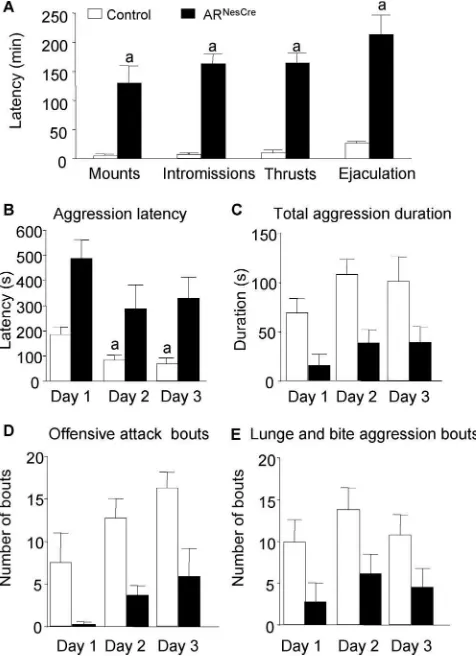
Nervous systemARdisruption impaired masculine behaviors
Sexual behavior
We investigated the effects of nervous systemARgene
inactiva-tion on the expression of male sexual behavior by using two pro-tocols. In 30 min tests (experiment 1), 55% of control males exhibited a complete sexual behavior with mounting, intromis-sions, pelvic thrusts, and ejaculation (supplemental Table 1, available at www.jneurosci.org as supplemental material). In
contrast, none of the tested ARNesCremales showed male sexual
behaviors, even mounting attempts (supplemental Table 1, avail-able at www.jneurosci.org as supplemental material). Since ARNesCre males were able to produce offspring in continuous
fertility test, we measured sexual behavior of another group of naive control and mutant males for 10 h from the introduction of receptive females (experiment 2). In these experimental condi-tions, 100% of control animals mounted, intromitted, performed
pelvic thrusts, and ejaculated (Fig. 4A). In contrast, only 55% of
ARNesCremales exhibited mounting, intromissions, and thrusts
with 33% reaching ejaculation while the 45% remaining mutant males did not show any sexual behavior attempt within the
10 h of the test (Fig. 4A). In addition, the latencies to the first
mount, intromission, pelvic thrusting, and ejaculation were
significantly greater for mutant animals (Fig. 4B,C). The
per-formance of mutant mice was also altered since they exhibited
a prolonged mating behavior (Fig. 4C, Table 2). Even those
that showed a complete sexual behavior reached ejaculation much more slowly than their control littermates. This was
associated with a reduced number of mounts with
intromis-sions and thrusts (Fig. 4C, Table 2).
Erectile activity
We investigated whether the disrupted performance of ARNesCre
mice could be related to an erectile dysfunction. In a first attempt to answer this question, we measured the intromission ratio, a
parameter generally used to evaluate the efficiency of erectionin
copula(Agmo, 1997; Cruz et al., 1999), and the thrusting train, defined as the number of thrusts per second during an
intromis-Figure 3. Growth of ARNesCremice.A, Control and ARNesCremales from the same litters ( n⫽ 15–19 per genotype) were weekly weighed. The growth curves are significantly different (p⬍ 0.05).B, Body composition of live adult (3 months old) males (n⫽5 per genotype) was analyzed by using a Piximus densitometer (Lunar Corporation). Muscle, fat, and bone masses are given as percentages of total body weight.
Figure 4. Male sexual behavior of control and ARNesCremice measured in a 10 h test.A, Percentage of males showing mounts, mounts with intromissions, thrusts with intromissions, and ejaculation.B, Latency to the first mount (without intromission), intromission, thrust, and ejaculation for mice that displayed complete sexual behavior (n⫽9 –11 per genotype;a
sion act. Both the intromission ratio
(Ta-ble 2) and the thrusting train (0.99⫾0.05
thrusts/s vs 1.22⫾0.05;p⬍0.01) were
significantly decreased in ARNesCremice,
thereby suggesting a reduced erectile
activ-ity of ARNesCre males. We thus scored
grooming erection during mating. For both control and mutant males, the first grooming penile erection followed the first intromission. However, a significantly
in-creased latency was observed for ARNesCre
mice (182.3⫾ 47.7 min vs 66.7 ⫾22.8
min;p⬍0.05). Furthermore, the mean length of grooming
erec-tion was significantly reduced for ARNesCremice (9.0⫾0.6 s vs
14.0⫾1.7 s;p⬍0.05).
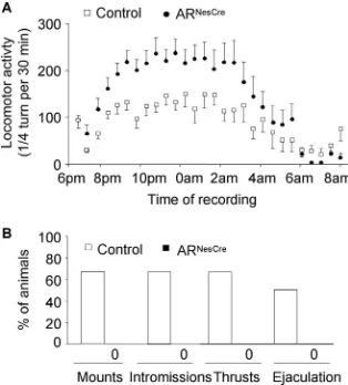
Aggressive behavior
We also tested the effects ofARgene inactivation in the nervous
system on aggression, another sexually dimorphic behavior, by using the resident–intruder paradigm over 3 consecutive days. All resident males exhibited anogenital chemoinvestigation to-ward intruder mice with a comparable mean latency to the first
sniff (Fig. 5A). However, the first contact with intruder mice
clearly enhanced the aggressive behavior of control males, while it had no effect on mutant males. First, if only 30% of control males
and 40% of ARNesCremice attacked on day 1, this proportion
reached 80 –90% on days 2 and 3 among the control group, while
it remained unchanged for mutant mice (Fig. 5B). Second,
con-trol mice that exhibited an aggressive behavior on day 1 attacked
faster the following days (Fig. 5C), whereas no difference in
la-tency to attack was observed for ARNesCremice (Fig. 5C).
Unaltered olfactory preference and neuronal activity after exposure to female pheromones
In rodents, masculine behaviors are activated by olfactory cues. Pheromonal cues are transmitted from the main olfactory epithe-lium and vomeronasal organ to, respectively, the main and acces-sory olfactory bulbs, then to chemosenacces-sory responsive nuclei in MA, septum, BNST, and MPOA, where they are processed in
behavioral responses. We thus tested whether ARNesCre males
show partner preference when given a choice between clean, male-soiled, and female-soiled bedding. The total time spent sniffing containers was similar between sexually experienced
control and ARNesCremice (188.5⫾10.1 s for control vs 185.5⫾
10.8 s for mutant). There were no differences between both ge-notypes in the time spent chemoinvestigating either male or
female-soiled bedding (Fig. 6A). However, all males spent
signif-icantly more time chemoinvestigating soiled beddings than the clean bedding, with a significant preference for bedding soiled by
estrous females (Fig. 6A).
The expression of the immediate early gene c-fos, a marker of
neuronal activity, was further examined in chemosensory regions involved in the expression of sexual behavior following exposure of males to clean or female-soiled bedding. Exposure to female olfactory cues increased Fos immunoreactivity in both the MA and MPOA with no gross differences between control and ARNesCremice (Fig. 6B,C). Similar results were obtained in the
BNST (data not shown).
Characterization of other behaviors
Assessment of anxiety-related behavior in the EPM test did not reveal any differences in the total number of entries into the open
arms (13⫾2 entries for controls vs 10⫾1 entries for mutants) or
in the time spent in the open arms (107.7⫾20.0 s for controls vs
108.1⫾29.0 s for mutants) between genotypes. Recording of
locomotion for 14 h showed a progressive increase of activity, which was maximal between 9:00 P.M. (2 h after lights off) and
3:00 A.M. of the dark phase for both control and ARNesCremice
with a significantly higher activity (a mean of 62% above control,
p⬍0.05) observed for ARNesCremice during the dark phase (Fig.
7A). When measured in gonadectomized and T-treated mice,
locomotor activity was comparable between both genotypes
(supplemental Fig. 1B, available at www.jneurosci.org as
supple-mental material). This strongly suggests that the increased
activ-ity of intact ARNesCremice was caused by their higher levels of E2.
These results indicate that the observed differences in male-typical behaviors among the mouse genotypes could not be at-tributed to an increased anxiety-related behavior or decreased
locomotion of ARNesCremales.
Effects of the NesCre transgene and DHT treatment
To make sure that the observed alterations were specifically
linked toARgene disruption in the nervous system, we first
ex-Table 2. Comparison of male sexual behavior in the 10 h test
ARNesCre
Control CSB ISB NSB
Sexual behavior length (min) 55.6⫾10.2 (11) 166.7⫾59.7* (3) ⬎600.0 (2) 0 (4) Number of mounts without intromissions 9.7⫾1.8 (11) 17.3⫾8.8 (3) 106.5 (2) 0 (4) Number of mounts with intromissions 34.1⫾8.3 (11) 20.3⫾9.2 (3) 23.5 (2) 0 (4) Number of thrusts with intromissions 508.5⫾63.5 (11) 219.0⫾82.8* (3) 219.0 (2) 0 (4) Intromission ratio (MI/M⫹MI) 0.76⫾0.03 (11) 0.55⫾0.11* (3) 0.19 (2) 0 (4)
Male sexual behavior of 11 control and 9 ARNesCre
mice was tested in experiment 2. The number of animals that exhibited complete sexual behavior (CSB), incomplete sexual behavior (ISB), or no sexual behavior (NSB) is given in parentheses.I, Intromissions;M, mounts without intromission; MI, mounts with intromission. *p⬍0.05 versus control.
Figure 5. Aggressive behavior of mice in the resident–intruder paradigm over 3 consecutive days.A, Latency to anogenital chemoinvestigation of intruders with no significant effect of genotype (F(1,40)⫽0.62,p⫽0.44).B, Percentage of males showing aggressive bouts on days 1, 2, and 3 (a
p⬍0.05 vs control mice).C, Latency to attack intruder mice. For residents that did not show aggressive behavior on days 2 and 3 of the test, the latency was 600 and 1200 s, respectively. There was a significant effect of genotype (F(1,24)⫽8.55,p⬍0.05).n⫽20 per genotype;post hocanalysis showed significant decreased latency for control mice to attack at days 2 or 3 versus day 1 (a
amined whether the used transgene, by itself, could have any influence on male neuroendocrine and behavioral responses. As shown in supplemental Table 2 (available at www.jneurosci.org as supplemental material), the Nes-Cre transgene alone, in the
absence of anyARinvalidation, had no effects on the studied
phenotypes. Second, we assessed the effects of the nonaromatiz-able DHT on sexual behavior of gonadectomized control and ARNesCremice. DHT reinstated sexual behavior in 86% of mice in
the control group (Fig. 7B). In contrast, none of tested ARNesCre
mice showed sexual behavior during the 10 h test (Fig. 7B).
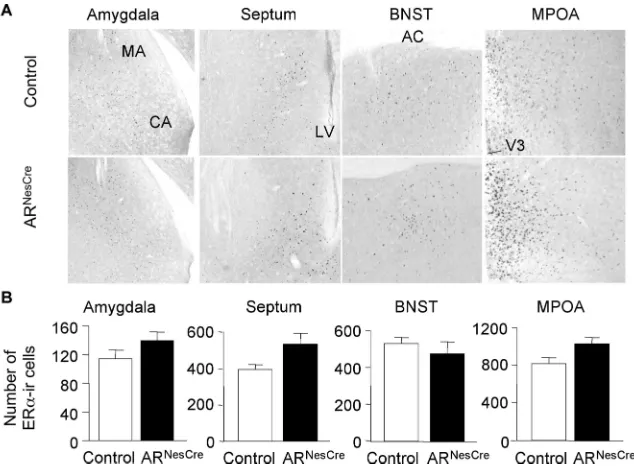
Quantification of ER␣-immunoreactive neurons
Several studies highlighted the importance of ER␣ signaling
pathway in T-induced masculine behaviors (Ogawa et al., 1997; Wersinger et al., 1997). We thus compared the distribution and
density of ER␣-immunoreactive (IR) neurons between control
and ARNesCremice to check whether this ER␣signaling pathway
is altered or not in ARNesCremice. High nuclear ER␣
immuno-staining was detected in brain regions known to express this re-ceptor, such as the MA, septum, BNST, and MPOA of both
ge-notypes (Fig. 8A). The average number of ER␣-IR cells per unit
area in brain areas involved in sexual and aggressive behaviors (MA, septum, BNST, and MPOA) was not statistically different
between control and ARNesCremice (Fig. 8B), thereby indicating
that ER␣was not decreased in ARNesCremice. Rather, an
in-creased intensity of ER␣immunoreactivity was observed in the
septum and MPOA of mutant mice (Fig. 8A).
Sexual and aggressive behaviors of gonadectomized and T-treated mice
To assess, at comparable levels of T, the effects ofARinvalidation
on male-typical behaviors, control and mutant males were gon-adectomized and supplemented with T. As observed for intact
animals, 100% of control versus 60% of ARNesCremales exhibited
sexual behavior. Again, an altered motivation, as evidenced by
the increased latency to initiate sexual behavior (Fig. 9A), and
decreased performance (supplemental Table 3, available at www.jneurosci.org as supplemental material) were observed for ARNesCremales. Mutant males also exhibited a greater latency to
initiate aggressive behavior toward intruder mice (Fig. 9B). A
detailed analysis of aggressive behavior indicated that ARNesCre
males spent less time in aggressive acts (Fig. 9C) and showed less
offensive and nonoffensive bouts (Fig. 9D,E). In addition, 33%
of control but none of ARNesCremales attempted mounts toward
the intruders.
When compared with data obtained in gonadally intact mice, greater differences were observed between the two genotypes. Indeed, at T levels comparable to those found in gonadally intact ARNesCremice, as assessed by seminal vesicle weights
(supple-mental Fig. 1A, available at www.jneurosci.org as supplemental
material), no changes in the phenotype were observed for mutant males, while an amelioration of sexual and aggressive behaviors was seen for control littermates. The latency to initiate both
be-haviors (Figs. 4B, 9A) and mating length (Table 2; supplemental
Table 3, available at www.jneurosci.org as supplemental
mate-rial) were decreased for control males (p⬍0.05). Furthermore,
100% of gonadectomized and T-treated control mice exhibited aggressive behavior since the first day in contrast to the low per-centage (30%) obtained for gonadally intact males.
Figure 6. Olfactory preference and neuronal activation.A, Time spent chemoinvestigating clean, male-soiled, and female-soiled-bedding by control and ARNesCremice (n
⫽20 per ge-notype). A significant effect of bedding (F(2,76)⫽143.58,p⬍0.0001) but not of genotype was found;post hocanalysis showed differences in the time spent sniffing the three beddings (ap
⬍ 0.001 vs clean bedding;bp
⬍0.001 vs male-soiled bedding).B,C, Representative c-fos immu-nostaining in the medial amygdala (B) and medial preoptic area (C) of control and ARNesCre males exposed to clean or female-soiled beddings for 1 h. CA, Cortical amygdala; AC, anterior commissure; V3, third ventricle.
Discussion
The present study addressed the role of ce-rebral AR in T-mediated regulation of male neuroendocrine and behavioral pro-cesses. To this aim, we generated, for the
first time, mice selectively lackingARgene
in the nervous system by using Cre-loxP technology, without interfering with pe-ripheral AR functions.
Requirement of cerebralARin the
regulation of male neuroendocrine functions
ARdisruption in the nervous system
af-fected the negative feedback exerted by T on the hypothalamus–pituitary– gonad (HPG) axis since high levels of LH and in-tratesticular and circulating T were found
in ARNesCremales. Neither the presence of
AR in gonadotropes nor the unaffected
hy-pothalamic ER␣ expression were
suffi-cient to maintain normal LH and T levels
in ARNesCre mice. This is in accordance
with previously reported data in ER␣KO
males, which suggested that cerebral AR plays the primary physiological role in the
steroid feedback on LH secretion (Wersinger et al., 1999). Pitu-itary LH synthesis and liberation are under the positive control of hypothalamic GnRH decapeptide. Our results strongly suggest that T through AR controls liberation rather than synthesis of GnRH. It remains to be determined whether this control is ex-erted directly or indirectly on GnRH neurons. We did not detect AR protein in GnRH neurons of control mice (K. Raskin and S. Mhaouty-Kodja, unpublished observations). However, AR was found to be expressed in the hypothalamic KiSS-1 neurons (Smith et al., 2005), which send projections to GnRH neurons (Kinoshita et al., 2005; Clarkson and Herbison, 2006) and seem to regulate pulsatile GnRH and gonadotropin secretion (Mes-sager et al., 2005). Since the Nes-Cre transgene used in the present
study drivesARablation in the whole nervous system, it is
possi-ble that an altered KiSS-1 expression underlies the observed al-teration of T-mediated negative feedback on HPG axis.
The reduced testis weight and sperm production of ARNesCre
mice were secondary toARgene invalidation in the nervous
sys-tem. These alterations could be related to decreased levels of IGF-I, which plays a critical role in testis growth and spermato-genesis process (Baker et al., 1996; Froment et al., 2004). How-ever, we cannot exclude possible deleterious effects of high levels of T and its metabolites.
Another consequence of centralARgene invalidation was the
alteration of the sexually dimorphic somatotropic axis. A tran-sient decrease in body weight was described in ubiquitous ARKO mice, but it was reversed since 12 weeks of age due to the in-creased adipose tissue mass and dein-creased lipolytic activity (Sato
et al., 2003; Fan et al., 2005). In ARNesCremales, growth
retarda-tion was persistent until 20 –30 weeks of age. This adds another
argument in favor of the selectivity ofARinvalidation in the
nervous system and strongly suggests that the mechanisms un-derlying the lipolytic effect of AR reside primarily in peripheral tissues. Together, these data point out the major role of central AR signaling pathway in the organizational and/or activational
effects of T on somatostatin and GHRH (GH-releasing hormone) neurons (Chowen et al., 2004).
Requirement of centralARin the expression of
masculine behaviors
By using phenotypically male mice, we evaluated precisely the
effects of centralARinvalidation on different components of
sexual behavior. We thus report that both sexual motivation and
performance were disrupted byARgene inactivation in the
ner-vous system. The prolonged mating length probably results from a weak reinforcement of sexual motivation due to the reduced intromission behavior. The latter deficiency could be attributed,
at least partly, to the disrupted penile erection of ARNesCremice.
Androgens, through AR activation, are known to play a major role in the masculinization of the spinal nucleus of the bulbocav-ernosus (SNB) (Johansen et al., 2004). Further experiments will
examine whetherARgene disruption in the nervous system
af-fects AR expression in the SNB and consequently innervation of the striated muscles bulbocavernosus and levator ani attached to the penis base.
ARNesCremice showed different extents of intromission and fertility alteration, but had all reduced sperm number. As it was previously reported that the fewer were the male preejaculatory intromissions, the lower was the sperm number recovered in female tract (Toner and Adler, 1986), we thus suggest that mu-tants with the lowest intromission behavior may correspond to the hypofertile group.
ARNesCremice exhibited also a low and heterogeneous
aggres-sive behavior in resident–intruder tests. It is of interest that
dis-rupted sexual and aggressive behaviors of ARNesCremice were
associated with normal (1) discrimination between male and fe-male nonvolatile olfactory cues, (2) chemoinvestigating behavior toward intruder mice, and (3) induction of neuronal activity, mapped by Fos immunoreactivity, in chemosensory responsive regions (MA, BNST, and MPOA) following exposure to female pheromonal cues. This leads us to suggest that deficiency in mas-culine behaviors is caused by an altered neuronal function in
chemosensory responsive regions, downstream of the olfactory bulb. To verify this hypothesis, further studies will address vola-tile odors.
Previous studies in Tfm mice reported a minor contribution of AR in the display of aggressive and coital behaviors (Scordalakes and Rissman, 2004; Bodo and Rissman, 2007) and a major role in partner preference (Bodo and Rissman, 2007). This discrepancy with our results could rely on differences between
ubiquitous and nervous system-specificARmutations. In
olfac-tory preference tests similar to that performed in the present
study, ER␣KO mice were unable to discriminate between
male-and female-soiled bedding (Wersinger male-and Rissman, 2000a). The
similarity between Tfm and ER␣KO mice in terms of lack of
partner preference (Wersinger and Rissman, 2000a; Bodo and Rissman, 2007) leads us to suggest that brain masculinization by T-mediated activation of ER signaling pathway during the
peri-natal period is not comparable between Tfm and ARNesCremice.
Indeed, although indirect evidence supported the conclusion of unaffected T production in newborn testes (Goldstein and Wil-son, 1972), it is still unknown whether circulating T levels are in
the normal range in perinatal Tfm mice. In ARNesCremice,
mas-culine behaviors were altered despite high levels of T and its
me-tabolite E2. At comparable levels of T, sexual and aggressive be-haviors of gonadectomized and T-treated control mice were greatly ameliorated. The inability of DHT to reinstate sexual
be-havior in gonadectomized ARNesCremice strongly suggests that
the remaining sexual behavior of intact ARNesCremice is probably
due to the only activation of ER␣signaling pathway. It is,
how-ever, important to note that ER␣was not sufficient to
compen-sate for the absence of AR. In line with our data, administration of E2 with DHT to castrated males, but not E2 alone, induced the same levels of masculine sexual behavior as those observed after treatment with T or in intact males (Cooke et al., 2003; Burns-Cusato et al., 2004; Arteaga-Silva et al., 2007). Nevertheless, as it has been previously shown that androgens upregulate aromatase expression (Yamada et al., 1993; Roselli et al., 1997), it would be interesting to examine whether brain aromatase expression is
affected in ARNesCremice.
In conclusion, we report here a pertinent genetic model that allows exploring the specific involvement of central AR in T-induced effects in the nervous system. Our results support the idea that cerebral AR and ER signaling pathways play comple-mentary roles in the masculinization of the nervous system dur-ing the perinatal period and/or the regulation of neuroendocrine and behavioral processes at the adult stage. The relative contri-bution of each of cerebral AR or ER in T-induced effects might differ between brain areas. Indeed, as strongly suggested in the present study, regulation of the somatotropic axis by T seems to be exclusively dependent on AR activation, while both AR and ER signaling pathways are required for the expression of masculine behaviors in mice. A precise comparison of the involvement of each of these signaling pathways in the latter responses needs further generation of conditional ERKO mutants in the nervous system.
References
Agmo A (1997) Male rat sexual behavior. Brain Res Brain Res Protoc 1:203–209.
Arteaga-Silva M, Rodríguez-Dorantes M, Baig S, Morales-Montor J (2007) Effects of castration and hormone replacement on male sexual behavior and pattern of expression in the brain of sex-steroid receptors in BALB/c AnN mice. Comp Biochem Physiol A Mol Integr Physiol 147:607– 615. Baker J, Hardy MP, Zhou J, Bondy C, Lupu F, Bellve´ AR, Efstratiadis A
(1996) Effects of an Igf1 gene null mutation on mouse reproduction. Mol Endocrinol 10:903–918.
Bakker J, Honda S, Harada N, Balthazart J (2002) Sexual partner preference requires a functional aromatase (cyp19) gene in male mice. Horm Behav 42:158 –171.
Bodo C, Rissman EF (2007) Androgen receptor is essential for sexual differ-entiation of responses to olfactory cues in mice. Eur J Neurosci 25:2182–2190.
Bradford MM (1976) A rapid and sensitive method for the quantitation of microgram quantities of protein utilizing the principle of protein-dye binding. Anal Biochem 72:248 –254.
Burns-Cusato M, Scordalakes EM, Rissman EF (2004) Of mice and missing data: what we know (and need to learn) about male sexual behavior. Physiol Behav 83:217–232.
Caraty A, Antoine C, Delaleu B, Locatelli A, Bouchard P, Gautron JP, Evans NP, Karsch FJ, Padmanabhan V (1995) Nature and bioactivity of gonadotropin-releasing hormone (GnRH) secreted during the GnRH surge. Endocrinology 136:3452–3460.
Chang C, Chen YT, Yeh SD, Xu Q, Wang RS, Guillou F, Lardy H, Yeh S (2004) Infertility with defective spermatogenesis and hypotestosteron-emia in male mice lacking the androgen receptor in Sertoli cells. Proc Natl Acad Sci U S A 101:6876 – 6881.
Chowen JA, Frago LM, Argente J (2004) The regulation of GH secretion by sex steroids. Eur J Endocrinol 151 [Suppl 3]:U95–U100.
Clarkson J, Herbison AE (2006) Postnatal development of kisspeptin neu-rons in mouse hypothalamus; sexual dimorphism and projections to
Figure 9. Male sexual and aggressive behaviors of gonadectomized and T-treated mice.A, Latency to the first mount, mount with intromission, thrust, and ejaculation in the 10 h test (n⫽6 per genotype;a
p⬍0.05 vs control mice).B, Latency to the first aggressive behavioral act in the 10 min resident–intruder paradigm with a significant effect of genotype (F(1,28)⫽ 10.99,p⫽0.0051);post hocanalysis showed a significantly decreased latency for control mice to attack at days 2 or 3 versus day 1 (a
p⬍0.05).C, The total aggression duration was decreased in mutant mice (F(1,28)⫽20.13,p⫽0.0005).D, The number of offensive attacks was lower in ARNesCremice (
F(1,28)⫽16.71,p⫽0.0011).E, The number of lunges and bites was reduced for ARNesCremales (
gonadotropin-releasing hormone neurons. Endocrinology 147:5817–5825.
Cooke BM, Breedlove SM, Jordan CL (2003) Both estrogen receptors and androgen receptors contribute to testosterone-induced changes in the morphology of the medial amygdala and sexual arousal in male rats. Horm Behav 43:336 –346.
Cruz MR, Liu YC, Manzo J, Pacheco P, Sachs BD (1999) Peripheral nerves mediating penile erection in the rat. J Auton Nerv Syst 76:15–27. De Gendt K, Swinnen JV, Saunders PT, Schoonjans L, Dewerchin M, Devos
A, Tan K, Atanassova N, Claessens F, Le´cureuil C, Heyns W, Carmeliet P, Guillou F, Sharpe RM, Verhoeven G (2004) A Sertoli cell-selective knockout of the androgen receptor causes spermatogenic arrest in meio-sis. Proc Natl Acad Sci U S A 101:1327–1332.
Fan W, Yanase T, Nomura M, Okabe T, Goto K, Sato T, Kawano H, Kato S, Nawata H (2005) Androgen receptor null male mice develop late-onset obesity caused by decreased energy expenditure and lipolytic activity but show normal insulin sensitivity with high adiponectin secretion. Diabetes 54:1000 –1008.
Froment P, Staub C, Hembert S, Pisselet C, Magistrini M, Delaleu B, Seurin D, Levine JE, Johnson L, Binoux M, Monget P (2004) Reproductive abnor-malities in human insulin-like growth factor-binding protein-1 trans-genic male mice. Endocrinology 145:2080 –2091.
Goldstein JL, Wilson JD (1972) Studies on the pathogenesis of the pseudohermaphroditism in the mouse with testicular feminization. J Clin Invest 51:1647–1658.
Johansen JA, Jordan CL, Breedlove SM (2004) Steroid hormone masculin-ization of neural structure in rats: a tale of two nuclei. Physiol Behav 83:271–277.
Kinoshita M, Tsukamura H, Adachi S, Matsui H, Uenoyama Y, Iwata K, Yamada S, Inoue K, Ohtaki T, Matsumoto H, Maeda K (2005) Involve-ment of central metastin in the regulation of preovulatory luteinizing hormone surge and estrous cyclicity in female rats. Endocrinology 146:4431– 4436.
Kunieda T, Xian M, Kobayashi E, Imamichi T, Moriwaki K, Toyoda Y (1992) Sexing of mouse preimplantation embryos by detection of Y chromosome-specific sequences using polymerase chain reaction. Biol Reprod 46:692– 697.
Le Roy I, Pothion S, Mortaud S, Chabert C, Nicolas L, Cherfouh A, Rouber-toux PL (2000) Loss of aggression, after transfer onto a C57BL/6J back-ground, in mice carrying a targeted disruption of the neuronal nitric oxide synthase gene. Behav Genet 30:367–373.
Liere P, Akwa Y, Weill-Engerer S, Eychenne B, Pianos A, Robel P, Sjo¨vall J, Schumacher M, Baulieu EE (2000) Validation of an analytical procedure to measure trace amounts of neurosteroids in brain tissue by gas chromatography-mass spectrometry. J Chromatogr B Biomed Sci Appl 739:301–312.
Lyon MF, Hawkes SG (1970) X-linked gene for testicular feminization in the mouse. Nature 227:1217–1219.
Matsumoto T, Takeyama K, Sato T, Kato Sb (2003) Androgen receptor functions from reverse genetic models. J Steroid Biochem Mol Biol 85:95–99.
Meffre D, Pianos A, Liere P, Eychenne B, Cambourg A, Schumacher M, Stein DG, Guennoun R (2007) Steroid profiling in brain and plasma of male and pseudopregnant female rats after traumatic brain injury: analysis by gas chromatography/mass spectrometry. Endocrinology 148:2505–2517. Messager S, Chatzidaki EE, Ma D, Hendrick AG, Zahn D, Dixon J, Thresher RR, Malinge I, Lomet D, Carlton MB, Colledge WH, Caraty A, Aparicio SA (2005) Kisspeptin directly stimulates gonadotropin-releasing hor-mone release via G protein-coupled receptor 54. Proc Natl Acad Sci U S A 102:1761–1766.
Moore JP Jr, Wray S (2000) Luteinizing hormone-releasing hormone (LHRH) biosynthesis and secretion in embryonic LHRH. Endocrinology 141:4486 – 4495.
Ogawa S, Lubahn DB, Korach KS, Pfaff DW (1997) Behavioral effects of estrogen receptor gene disruption in male mice. Proc Natl Acad Sci U S A 94:1476 –1481.
Ono S, Geller LN, Lai EV (1974) TfM mutation and masculinization versus feminization of the mouse central nervous system. Cell 3:235–242. Phoenix CH, Goy RW, Gerall AA, Young WC (1959) Organizing action of
prenatally administered testosterone propionate on the tissues mediating mating behavior in the female guinea pig. Endocrinology 65:369 –382. Rampin O, Je´roˆme N, Suaudeau C (2003) Proerectile effects of
apomor-phine in mice. Life Sci 72:2329 –2336.
Rampin O, Je´roˆme N, Briant C, Boue´ F, Maurin Y (2006) Are oestrus odours species specific? Behav Brain Res 172:169 –172.
Roselli CE, Abdelgadir SE, Resko JA (1997) Regulation of aromatase gene expression in the adult rat brain. Brain Res Bull 44:351–357.
Salomon L, Lanteri C, Glowinski J, Tassin JP (2006) Behavioral sensitization to amphetamine results from an uncoupling between noradrenergic and serotonergic neurons. Proc Natl Acad Sci U S A 103:7476 –7481. Sato T, Matsumoto T, Yamada T, Watanabe T, Kawano H, Kato S (2003)
Late onset of obesity in male androgen receptor-deficient (AR KO) mice. Biochem Biophys Res Commun 300:167–171.
Sato T, Matsumoto T, Kawano H, Watanabe T, Uematsu Y, Sekine K, Fukuda T, Aihara K, Krust A, Yamada T, Nakamichi Y, Yamamoto Y, Nakamura T, Yoshimura K, Yoshizawa T, Metzger D, Chambon P, Kato S (2004) Brain masculinization requires androgen receptor function. Proc Natl Acad Sci U S A 101:1673–1678.
Scordalakes EM, Rissman EF (2004) Aggression and arginine vasopressin immunoreactivity regulation by androgen receptor and estrogen receptor alpha. Genes Brain Behav 3:20 –26.
Smith JT, Dungan HM, Stoll EA, Gottsch ML, Braun RE, Eacker SM, Clifton DK, Steiner RA (2005) Differential regulation of KiSS-1 mRNA expres-sion by sex steroids in the brain of the male mouse. Endocrinology 146:2976 –2984.
Toner JP, Adler NT (1986) The pre-ejaculatory behavior of male and female rats affects the number of sperm in the vagina and uterus. Physiol Behav 36:363–367.
Tronche F, Kellendonk C, Kretz O, Gass P, Anlag K, Orban PC, Bock R, Klein R, Schu¨tz G (1999) Disruption of the glucocorticoid receptor gene in the nervous system results in reduced anxiety. Nat Genet 23:99 –103. Wersinger SR, Rissman EF (2000a) Oestrogen receptor alpha is essential for
female-directed chemo-investigatory behaviour but is not required for the pheromone-induced luteinizing hormone surge in male mice. J Neu-roendocrinol 12:103–110.
Wersinger SR, Rissman EF (2000b) Dopamine activates masculine sexual behavior independent of the estrogen receptor ␣. J Neurosci 20:4248 – 4254.
Wersinger SR, Sannen K, Villalba C, Lubahn DB, Rissman EF, De Vries GJ (1997) Masculine sexual behavior is disrupted in male and female mice lacking a functional estrogen receptor alpha gene. Horm Behav 32:176 –183.
Wersinger SR, Haisenleder DJ, Lubahn DB, Rissman EF (1999) Steroid feedback on gonadotropin release and pituitary gonadotropin subunit mRNA in mice lacking a functional estrogen receptor alpha. Endocrine 11:137–143.