doi: 10.3389/fpls.2015.00233
Edited by:
Martin Weih, Swedish University of Agricultural Sciences, Sweden
Reviewed by:
Tanya Waldie, Sainsbury Laboratory Cambridge University, UK Alicia Moreno-Cortés, Universidad Politécnica de Madrid, Spain
*Correspondence:
Thomas Teichmann, Plant Cell Biology, Georg-August-Universität Göttingen, Julia-Lermontowa-Weg 3, Göttingen, Germany [email protected]
Specialty section:
This article was submitted to Plant Biotechnology, a section of the journal Frontiers in Plant Science
Received:28 January 2015
Accepted:23 March 2015
Published:09 April 2015
Citation:
Teichmann T and Muhr M (2015) Shaping plant architecture. Front. Plant Sci. 6:233. doi:10.3389/fpls.2015.00233
Shaping plant architecture
Thomas Teichmann* andMerlin Muhr
Plant Cell Biology, Georg-August-Universität Göttingen, Göttingen, Germany
Plants exhibit phenotypical plasticity. Their general body plan is genetically determined, but plant architecture and branching patterns are variable and can be adjusted to the prevailing environmental conditions. The modular design of the plant facilitates such morphological adaptations. The prerequisite for the formation of a branch is the initiation of an axillary meristem. Here, we review the current knowledge about this process. After its establishment, the meristem can develop into a bud which can either become dormant or grow out and form a branch. Many endogenous factors, such as photoassimilate availability, and exogenous factors like nutrient availability or shading, have to be integrated in the decision whether a branch is formed. The underlying regulatory network is complex and involves phytohormones and transcription factors. The hormone auxin is derived from the shoot apex and inhibits bud outgrowth indirectly in a process termed apical dominance. Strigolactones appear to modulate apical dominance by modification of auxin fluxes. Furthermore, the transcription factor BRANCHED1 plays a central role. The exact interplay of all these factors still remains obscure and there are alternative models. We discuss recent findings in the field along with the major models. Plant architecture is economically significant because it affects important traits of crop and ornamental plants, as well as trees cultivated in forestry or on short rotation coppices. As a consequence, plant architecture has been modified during plant domestication. Research revealed that only few key genes have been the target of selection during plant domestication and in breeding programs. Here, we discuss such findings on the basis of various examples. Architectural ideotypes that provide advantages for crop plant management and yield are described. We also outline the potential of breeding and biotechnological approaches to further modify and improve plant architecture for economic needs.
Keywords: axillary meristem, branching, apical dominance, auxin, cytokinins, strigolactone, ideal plant architec-ture, poplar
Introduction
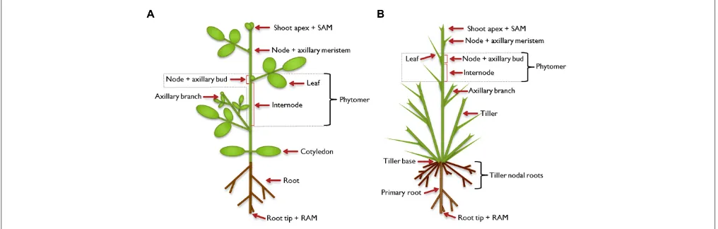
of an internode and a node with one or more attached axillary leaves (Figure 1). In the leaf axils, secondary, lateral meris-tems are established and allow the formation of higher order morphological structures. The axillary meristems may develop a bud that can extend to form a branch, which constitutes a sec-ondary growth axis. Branches are built up in the same way as the primary growth axis, and higher order branching can occur, lead-ing to a complex structure. The architecture of a mature plant is therefore determined by the number and activity of axillary meristems and the growth characteristics of the branches that develop from axillary buds (Kerstetter and Hake, 1997; Sussex and Kerk, 2001;McSteen and Leyser, 2005;Schmitz and Theres, 2005; Bennett and Leyser, 2006; De Smet and Juergens, 2007; Janssen et al., 2014).
Axillary Meristem Initiation
Axillary meristems are the origin of lateral branches. They are formed in the center of the boundary zone at the adaxial side of the leaf base. The boundary zone separates the shoot api-cal meristem (SAM) from the developing leaf primordium. This zone is not just a border but fulfills an important function in meristem maintenance and organ development (Zadnikova and Simon, 2014). It is characterized by small cells, stiff cell walls and a low cell division rate. A key factor during establish-ment of the boundary zone is the transcription factor LATERAL ORGAN BOUNDARIES1 (LOB1) that induces the expression of BAS1, encoding a protein that has brassinosteroid inacti-vating activity (Bell et al., 2012). Brassinosteroids are plant steroid hormones that influence cell expansion and cell divi-sion (reviewed inHardtke, 2007;Fridman and Savaldi-Goldstein, 2013). The LOB1-mediated decrease of brassinosteroid activ-ity causes a reduction of cell size and cell division rate
in the boundary zone compared to neighboring zones (Bell et al., 2012; Gendron et al., 2012). This effect is enhanced by the outward orientation of the auxin efflux carrier PIN-FORMED1 (PIN1) causing depletion of the plant growth hor-mone auxin in the boundary zone. During initial outgrowth of the leaf primordium, PIN1 is oriented toward the pri-mordium. However, as the boundary zone develops, PIN1 is reoriented toward the SAM (Wang et al., 2014a,b). This reorientation depends on the kinase PINOID (PID) that con-trols basal-apical localization of PIN1 (Furutani et al., 2004). The importance of PIN reorientation and the role of PID in development of a functional boundary zone can be seen in
pin1 and pid mutants that exhibit defects in axillary meris-tem formation (Wang et al., 2014a,b). Artificial increase of auxin in the developing boundary zone by localized expression of the auxin biosynthesis gene iaaM in transgenicArabidopsis
resulted in the lack of axillary meristems in a portion of the leaf axils (Wang et al., 2014a,b). On the contrary, bound-ary zone specific expression of a stabilized version of the AUX/IAA protein BODENLOS to reduce auxin signaling in this area resulted in the formation of axillary buds in the axils of cotyledons which was never observed in wild type plants (Wang et al., 2014a). Therefore, a local auxin minimum in the boundary zone appears to be important for axillary meristem formation.
Another gene having an effect on shoot lateral organ development isRPS10B, which was found in a suppressor screen of the more axillary branching2-1(max2-1) mutant. It encodes the ribosomal protein S10e.Stirnberg et al.(2012a) discuss that in the mutant, levels of proteins, which are important for the regulation of auxin distribution and therefore auxin-mediated organ boundary patterning, may be imbalanced. Especially pro-teins with a high turnover rate, such as the Aux/IAA repressors involved in auxin signaling, may be affected by the ribosomal
FIGURE 1 | Illustration of plant architecture.Typical architecture of a dicot plant(A)and a monocot plant(B). The shoot apical meristem (SAM) establishes the shoot as the primary growth axis of the plant by continuously initiating phytomers, the basic modules of the plant shoot. A phytomer consists of an internode and a node with its attached leaf. In the leaf axils, axillary (secondary) meristems are formed in dicot and some monocot plants, which develop into an axillary bud and have the potential to continue growth to form an axillary branch.
rps10b-1mutation (Stirnberg et al., 2012a). In the same suppres-sor screen,FAR-RED ELONGATED HYPOCOTYL3(FHY3) was found. The authors discuss this gene to be potentially involved in the regulation of auxin homeostasis, too (Stirnberg et al., 2012b). Therefore, there appear to be many factors controlling the precise spatiotemporal auxin distribution during meristem devel-opment. In addition to auxin,Wang et al.(2014b) also discuss a role of cytokinin during AM initiation. They report a cytokinin pulse following and being dependent on the establishment of an auxin minimum in the boundary zone of the leaf axil and pro-vide hints for the importance of cytokinin signaling during the establishment of the axillary meristem.
Tissue markers of the boundary zone are the Arabidopsis
NAM-ATAF1/2-CUC2 (NAC) transcription factors CUP
SHAPED COTYLEDONS1, 2, and 3 (CUC1, 2, and 3;Spinelli et al., 2011) that have redundant functions in meristem forma-tion. In tomato,GOBLET(GOB)was identified as an ortholog of theCUCgenes (Busch et al., 2011). Expression of these genes is a prerequisite for development of the SAM and the consecutive formation of the boundary zone.CUCgenes are down-regulated by brassinosteroids. Thus, low brassinosteroid activity in the boundary zone not only reduces cell expansion and division as described above, but also allows the induction ofCUCgenes (Bell et al., 2012;Gendron et al., 2012).
The most pronounced difference between the SAM, the neigh-boring boundary zone and the developing leaf primordium is that cells in the SAM are kept in an indeterminate, non-differentiated state while cells of the boundary zone and the primordium differentiate. Meristematic identity of the SAM cells is retained by activity of the homeobox class I KNOX gene
SHOOT MERISTEMLESS (STM; Long et al., 1996; Long and Barton, 2000). As soon as cells start to differentiate, STM is down-regulated by the MYB transcription factor AS1 and the LATERAL ORGAN BOUNDARY DOMAIN (LBD) transcrip-tion factor AS2 (Ikezaki et al., 2010). Interestingly, during an early phase of boundary zone development,STM continues to be transcribed in all cells of the boundary zone, albeit at a low level (Long and Barton, 2000). This indicates that, for a restricted time period, cells of the boundary zone keep the capac-ity to return to a meristematic stage. During this developmental phase, the axillary meristem is initiated (Grbic and Bleecker, 2000). A molecular marker of de novo axillary meristem for-mation is the focused and strong expression of STM in the center of the boundary zone. InArabidopsis, this focused STM
expression depends on the presence of the GRAS transcrip-tion factor LATERAL SUPPRESSOR (LAS;Greb et al., 2003). Orthologs of LASareLS in tomato (Schumacher et al., 1999) andMONOCULM1(MOC1) in rice (Li et al., 2003). Knockout mutants of LAS fail to develop axillary meristems during the vegetative stage (Greb et al., 2003). Keller et al. (2006) sug-gested that “LAS is required for reacquisition of indetermi-nate cell fate in axillary cells in the course of AM organiza-tion.”
Axillary meristem initiation and development is modulated by several factors that have partially redundant functions. In addition to LAS, the MYB factors REGULATOR OF AXILLARY MERISTEMS1 (RAX1) inArabidopsis(Keller et al., 2006), as well
as BLIND (BL) and POTATO LEAF (C) in tomato (Schmitz et al., 2002; Busch et al., 2011), influence axillary meristem develop-ment. Another factor is a basic helix-loop-helix (bHLH) protein called REGULATOR OF AXILLARY MERISTEM FORMATION (ROX) inArabidopsis(Yang et al., 2012), LAX PANICLE1 (LAX1) in rice (Komatsu et al., 2001, 2003) and BARREN STALK1 (BA1) in maize (Ritter et al., 2002;Gallavotti et al., 2004).
For the ontogenetic origin of axillary meristems, two theo-ries have been discussed (Sussex and Kerk, 2001). The de novo
meristem formation theory is based on the fact that in some plant species, e.g., Arabidopsis, axillary meristems cannot be detected after leaf initiation by anatomical studies. In contrast, the detached or reserve meristem theory describes the situation in plants like tomato where meristematic cells from the SAM persist in the axils of newly built leaves and then, later during development, form axillary meristems (reviewed inBennett and Leyser, 2006). However, the studies on LAS, RAX1, and ROX1 show that similar key factors control meristem initiation in plant species that seem to have contrasting mechanisms of meristem development. This indicates that axillary meristems in plants are generally formed by the same process. The fact that the boundary zone that just separated from the SAM continues to showSTM
expression argues for the detached meristem hypothesis. Cells of the boundary zone seem to be kept in a stage that is not fully determinate and, as a consequence, the axillary meristem can be initiated from this pool of cells. In conclusion, these data provide evidence that also in plants likeArabidopsis, where the meris-tem appears at later stages of development, the merismeris-tem is not formedde novobut built as a detached meristem (Leyser, 2003; Bennett and Leyser, 2006).
Axillary meristems strictly form on the adaxial side of leaf bases. This may be the reason why the transcription fac-tor REVOLUTA (REV), that determines adaxiality, has been described as a further axillary meristem initiation factor (Otsuga et al., 2001). However, its effect on axillary meristem formation may be secondary and the primary function of REV is the con-trol of radial patterning (Emery et al., 2003;Bennett and Leyser, 2006).
Activity of Apical Meristems and
Control of Bud Outgrowth
Snow(1925) could show that maintenance of apical dominance needs a signal that moves downward from a dominant shoot apex and, in addition, another signal may be transported upward into the dormant bud to suppress outgrowth. Thimann and Skoog (1933) identified the plant hormone auxin as the downward sig-nal. Auxin, mainly synthesized in expanding young leaves of the plant apex (Ljung et al., 2001), is transported basipetally in the stem. Removal of the apical auxin source by decapita-tion abolishes apical dominance, while applicadecapita-tion of auxin to the apex of these decapitated plants can restore apical dom-inance (Thimann and Skoog, 1933). However, the inhibitory effect of auxin is not direct. It was shown that external auxin application to axillary buds does not prevent their outgrowth and experiments with radiolabeled auxin revealed that apex-derived auxin does not enter the dormant bud. Additionally, auxin transport appears to be too slow to mediate a direct effect (Hall and Hillman, 1975;Morris, 1977;Everat-Bourbouloux and Bonnemain, 1980; Booker et al., 2003). As a consequence of these studies, a long distance second messenger was postulated. According to this model, such a second messenger relays the
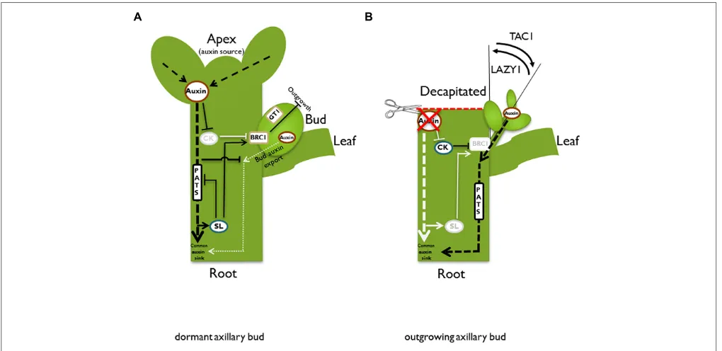
downward auxin signal upward into the dormant bud. There are two good candidates for this messenger: cytokinins and strigolactones. Cytokinin is produced in roots and the stem and transported acropetally in the xylem (Nordstrom et al., 2004). Manipulations of plant cytokinin content show clear effects on bud outgrowth control, e.g., application of cytokinin to axil-lary buds releases dormancy even in plants that have an intact apex (Sachs and Thimann, 1964). Thus, with respect to bud out-growth control, cytokinins act antagonistically to auxin. Most likely, the readout of auxin-cytokinin crosstalk generates part of the signaling chain that controls dormancy. The question of how auxin influences cytokinin as a second messenger was addressed byNordstrom et al.(2004), who found that auxin can dampen cytokinin biosynthesis (Figure 2A). Basipetally transported auxin from the plant apex decreases expression of the cytokinin biosyn-thesis gene ISOPENTENYLTRANSFERASE (IPT) in the stem (Tanaka et al., 2006). In addition, it was shown for pea stems that auxin induces the cytokinin oxidase genePsCKX2 (Shimizu-Sato et al., 2009). Cytokinin oxidases inactivate cytokinin and, thus, lower the pool of active cytokinin (Werner et al., 2001).
FIGURE 2 | Schematic illustration of different pathways and models in the control of bud outgrowth.In an intact plant(A), the apex is a strong auxin source. Auxin is transported basipetally in the polar auxin transport stream (PATS). According to the second messenger model, auxin promotes strigolactone (SL) and represses cytokinin (CK) biosynthesis, respectively. Both hormones have adverse effects on bud outgrowth, most likely acting via the transcription factorBRANCHED1/TEOSINTE BRANCHED1(BRC1/TB1). Auxin indirectly promotesBRC1/TB1expression, which suppresses bud outgrowth. GRASSY TILLERS1(GT1) is a putative downstream target forTB1in monocots. According to the auxin transport canalization model, the axillary bud is also an auxin source and as a prerequisite for vascular tissue formation and bud outgrowth, it has to establish its own auxin export. However, it competes with the shoot apex for the stem as a shared auxin sink. This competition is enhanced by SL, which reduces plasma membrane accumulation of thePIN1 auxin efflux carrier and therefore inhibits the PATS in the main stem. High auxin
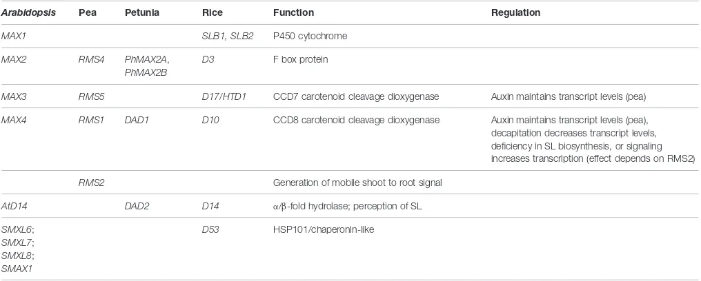
TABLE 1 | Genes involved in strigolactone biosynthesis and signaling.
Arabidopsis Pea Petunia Rice Function Regulation
MAX1 SLB1, SLB2 P450 cytochrome
MAX2 RMS4 PhMAX2A,
PhMAX2B
D3 F box protein
MAX3 RMS5 D17/HTD1 CCD7 carotenoid cleavage dioxygenase Auxin maintains transcript levels (pea)
MAX4 RMS1 DAD1 D10 CCD8 carotenoid cleavage dioxygenase Auxin maintains transcript levels (pea), decapitation decreases transcript levels, deficiency in SL biosynthesis, or signaling increases transcription (effect depends on RMS2)
RMS2 Generation of mobile shoot to root signal
AtD14 DAD2 D14 α/β-fold hydrolase; perception of SL
SMXL6; SMXL7; SMXL8; SMAX1
D53 HSP101/chaperonin-like
The information in the table was taken fromBeveridge et al.(1997, 2000),Foo et al.(2005),Johnson et al.(2006),Braun et al.(2012),Drummond et al.(2012), and Cardoso et al.(2014).
As a consequence of decreased biosynthesis and increased degra-dation, the cytokinin content is lowered in the stem and bud dormancy is maintained. In contrast, the decrease of auxin in the stem after removal of the main auxin biosynthesis site by decap-itation will lead to increased cytokinin biosynthesis (Bangerth, 1994; Figure 2B). In pea, the PsIPT1 and PsIPT2 genes are induced in the nodal stem near the axillary buds after decapita-tion. Consistently, increased cytokinin levels could be detected in excised nodal stems (Tanaka et al., 2006). Cytokinin may then be transported into the adjacent buds. Indeed, it was shown in pea that the zeatin riboside content increased in axillary buds after decapitation (Turnbull et al., 1997).
In addition to damages to the apex, other environmental impacts such as nutrient availability (e.g., nitrogen or phosphorus concentrations in the soil) or planting density-related shading, also profoundly change plant architecture (Casal et al., 1986; Lopez-Bucio et al., 2002;Yoneyama et al., 2013;de Jong et al., 2014). The developmental response of the plant shoot to nutrient supply most likely involves a long distance, graft-transmittable signal from the root. Root tips are a main biosynthesis site of cytokinin (Miyawaki et al., 2004;Nordstrom et al., 2004) and it is tempting to speculate that changes in the cytokinin export from the root to the shoot via the xylem stream provide the postulated long distance signal for root–shoot communication. However, Faiss et al.(1997) showed in grafting experiments that transgenic roots overproducing cytokinin could not induce bud outgrowth in wild type scions, which made cytokinin unlikely to be the elusive signal. Analyses of branching mutants in Arabidopsis
(more axillary branching -max), pea (ramosus -rms), petunia (decreased apical dominance-dad), and rice (dwarf –d) finally led to the discovery of the shoot branching hormone strigolac-tone (SL;Gomez-Roldan et al., 2008;Umehara et al., 2008) which features the required characteristics of the sought-after long dis-tance signal in branching control: it inhibits shoot branching (Gomez-Roldan et al., 2008; Umehara et al., 2008), it can be
transmitted from wild type roots to mutant shoots via grafting and complements the branching phenotype (Beveridge et al., 1997;Sorefan et al., 2003;Booker et al., 2005;Beveridge, 2006). Acropetal SL transport was shown to occur in the xylem (Kohlen et al., 2011) and the biosynthesis is increased by auxin (Sorefan et al., 2003;Figure 2A).
After the discovery of SLs as branching hormones, much effort was put in unraveling their biosynthesis and signaling path-ways. SL biosynthesis starts from carotenoid precursors via the action of the all-trans/9-cis-β-carotene isomerase D27 in rice and AtD27 inArabidopsis(Lin et al., 2009;Waters et al., 2012). Subsequent processing is carried out by the carotenoid cleavage monooxygenases CCD7 and CCD8. These enzymes are known in several species and named MAX3 and MAX4 inArabidopsis
(Sorefan et al., 2003;Booker et al., 2004), RMS5 and RMS1 in pea (Sorefan et al., 2003;Johnson et al., 2006), D17 and D10 in rice (Ishikawa et al., 2005;Arite et al., 2007;Alder et al., 2012), and DAD1 in petunia (Snowden et al., 2005).
Successful complementation ofArabidopsis maxmutants with putativeMAX orthologs from willow and poplar and response of willow buds to the SL analog GR24 indicate that SLs are also synthesized and perceived in woody plants such as trees (Ward et al., 2013;Czarnecki et al., 2014).
In addition to CCD7 and CCD8, the cytochrome P450 monooxygenase MAX1 is involved in downstream SL biosyn-thesis inArabidopsis(Stirnberg et al., 2002;Booker et al., 2005;
Table 1). An overview about all aforementioned SL biosynthesis genes can be found inTable 1.
has been discussed as a possible mobile SL precursor (Seto and Yamaguchi, 2014). This hypothesis is based on the observation that amax1rootstock can complement the branching phenotype of amax4scion (Booker et al., 2005). Recently,Abe et al.(2014) analyzed the MAX1 reaction in SL biosynthesis in detail. They demonstrated that carlactone is converted to carlactonic acid by the action of MAX1 inArabidopsis. However, subsequent reac-tions to generate bioactive SLs remain to be elucidated. The same authors reported hints for the interaction of a carlactonic acid methyl ester with the putative SL receptor AtD14 (see SL signal-ing discussion below;Abe et al., 2014). Therefore, we are close to fully understanding the biosynthesis pathway of at least one bioactive SL. However, there are multiple other bioactive vari-ants of SLs. The detailed reactions leading to this diversity as well as possible alternative biosynthesis pathways remain to be discovered.
Grafting experiments also revealed a class of SL response mutants that could not be complemented by wild type root-stocks (Beveridge et al., 1996;Stirnberg et al., 2007), indicating a role in SL perception and signaling rather than biosynthe-sis. An example is the Arabidopsis max2 mutant (Stirnberg et al., 2007) that encodes an F-box protein involved in SL sig-naling. Its counterparts in rice and pea were described pre-viously (Ishikawa et al., 2005; Johnson et al., 2006; Table 1). Within 6 years after the first description of SLs as branch-ing hormones, further key components of SL signalbranch-ing have been identified and a tentative scaffold of the signal transduc-tion pathway has been assembled (Bennett and Leyser, 2014; Waldie et al., 2014). The α/β hydrolase D14 is most likely a receptor for SL. d14 mutants in Arabidopsis, petunia and rice are insensitive to treatment with the SL analog GR24 and show an increased branching phenotype. Also, D14 exhibits high and specific affinity to GR24 (Kagiyama et al., 2013). In the presence of GR24, the petunia D14 ortholog DAD2 inter-acts with PhMAX2 (Hamiaux et al., 2012). This indicates that in analogy to other plant hormone signaling pathways, D14 may interact with the F-box protein MAX2 upon SL binding, leading to ubiquitination-mediated degradation of a SL signal-ing repressor (Bennett and Leyser, 2014). Sincemax2mutants show pleiotropic effects, it is likely that MAX2 interacts with several pathways and may mediate degradation of different tar-get proteins. Indeed, three different candidate repressors for the strigoalactone signaling pathway have been identified: DELLA proteins (Nakamura et al., 2013), BES1 (Wang et al., 2013), and D53 in rice (Jiang et al., 2013;Zhou et al., 2013). While inter-action of MAX2 with DELLA proteins and BES1 may point to cross talk with the gibberellic acid and brassinosteroid pathway, respectively, D53 emerges as the genuine SL pathway repres-sor (reviewed in Bennett and Leyser, 2014). Dominant gain of function mutations in D53 prevent SL-mediated degrada-tion of the protein and shut off SL signaling. Moreover, rice D53 interacts with D3, which is the rice ortholog of MAX2, andd3 mutants are suppressed by knockdowns of D53 (Jiang et al., 2013;Zhou et al., 2013). A possibleArabidopsisortholog of D53 is SUPPRESSOR OF MORE AXILLARY GROWTH2 LIKE 7 (SMXL7;Stanga et al., 2013; Bennett and Leyser, 2014;
Table 1). Interestingly, the basic principle of SL signaling is
similar to auxin, jasmonic acid, and gibberellic acid signaling (reviewed in McSteen and Zhao, 2008). Briefly, binding of the hormone to a receptor activates an F-box protein-containing SCF E3 ligase complex, which mediates ubiquitination and sub-sequent degradation of a transcriptional repressor. Ultimately, this leads to changes in transcription of a specific set of genes (Hagen and Guilfoyle, 2002; Hartweck, 2008;Memelink, 2009).
Summarized, cytokinin and SL were shown to regulate bud outgrowth, but the mechanism of bud dormancy control and the reciprocal effect of these plant hormones had to be integrated into a model.Prusinkiewicz et al.(2009) andBalla et al.(2011) suggested combining the second messenger model with a model introduced byLi and Bangerth(1999). Their model of “autocor-relative inhibition” is based on the auxin canalization hypothesis bySachs(1981) and discusses a competition of buds for estab-lishment of a polar auxin transport stream (PATS). The auxin canalization hypothesis (reviewed in Domagalska and Leyser, 2011) suggests a feed forward mechanism to explain the establish-ment of polar auxin transport routes that induce the developestablish-ment of vascular tissues. Starting from an auxin source that provides a high auxin concentration, competent cells will transport auxin away from the source and establish an auxin gradient across the tissue. From this initial auxin flow, continuous transport will build up, keeping a high auxin concentration in the transport competent cells and subsequently increasing the expression and polarization of auxin carriers in these cells. As a consequence, auxin transport will further strengthen in a feed forward loop, which sustains and enhances transport competence in files of specific cells. Along these transport routes, vascular tissue will differentiate.
Research on the PIN auxin efflux carrier proteins provided experimental support for the canalization model. Biosynthesis and plasma membrane localization of PIN proteins are elevated by auxin (Paciorek et al., 2005) and the expression of PIN pro-teins precedes vascular development (Sauer et al., 2006;Scarpella et al., 2006;Wenzel et al., 2007). This model can be adapted for a hypothesis on the mechanisms that control apical dominance. As an initial auxin gradient is a prerequisite for the develop-ment of a PATS, only buds that achieve to build up an auxin gradient between the bud as an auxin source and the stem as a common auxin sink have the ability to establish a PATS and grow out. Usually, the actively growing apex is the main auxin source (Figure 2A). According to the auxin canalization model, apical dominance is therefore exerted by the apex through sat-uration of the auxin transport capacity of the stem, acting as an auxin sink. As a consequence, axillary buds are prevented from successfully establishing an initial auxin flux. Hence, they remain dormant.
reduced back to normal levels, preventing further dormant buds from growing out.
Both models, the second messenger model and the model of autocorrelative inhibition/auxin canalization, are complemen-tary. Cytokinin and SL, respectively, influence sink strength of the stem through changes in auxin biosynthesis and modification of PATS. As a consequence of decapitation, the inhibitory effect of auxin on cytokinin biosynthesis is dampened and increased cytokinin levels might enhance local auxin biosynthesis in the bud, increasing its auxin source strength. At the same time, the sink capacity of the stem may be enhanced by a cytokinin-mediated induction of the PATS in the stem by increased syn-thesis and polarization of PIN auxin efflux carriers. Indeed, such increased expression and polarization was shown for PsPIN1 in axillary buds after external cytokinin application (Kalousek et al., 2010). Furthermore,Marhavy et al.(2014) postulated a role for cytokinin in modulating AtPIN1 abundance and polarization during lateral root organogenesis.
In contrast to cytokinin, SL appears to decrease the amount of the PIN auxin efflux carrier at the membrane and, thus, lower auxin transport capacity in the stem. This was observed in stems of Arabidopsis SL-pathway mutants, which showed increased AtPIN1 levels as well as an increased auxin transport (Bennett et al., 2006; Prusinkiewicz et al., 2009). According to the auxin transport canalization model, SLs will, therefore, aggra-vate the establishment of auxin export from axillary buds, lead-ing to increased apical dominance (Prusinkiewicz et al., 2009). Decapitation triggers down-regulation of SL biosynthesis gene
CCD8transcript levels (Foo et al., 2005), most likely resulting in reduced SL biosynthesis. Such a reduction of SL levels would cause a release from their antagonistic effect on PIN polarization. As a result, an increased auxin flux to the root would occur and, thus, further increase the sink capacity of the stem. Summarized, high cytokinin and low SL levels may increase source strength of the bud and increase sink capacity of the stem, and, thus, facilitate the successful establishment of an auxin gradient. This gradient would allow an initial auxin flow from the bud to the stem and the establishment of vascular tissue as a prerequisite for bud out-growth (Figures 2A,B). AlreadySorokin and Thimann (1964) observed that a vascular connection between axillary buds and the main stem coincides or precedes bud outgrowth.
A drawback of the hypotheses on apical dominance control by auxin is the discrepancy between auxin transport velocity and bud outgrowth kinetics after decapitation. In decapitated pea plants, buds start to grow out before auxin concentrations in the associated nodal stem are diminished due to removal of the apical auxin source (Morris et al., 2005). Thus, an alternative primary messenger is discussed.Mason et al.(2014) reported that after decapitation, sucrose concentrations in axillary buds increased. Moreover, buds could be released from dormancy by sucrose treatment and inhibition of sucrose transport by girdling pre-vented outgrowth of buds. Importantly, the measured speed of sucrose transport is sufficient to relay the signal from the shoot apex to a dormant axillary bud in time before first signs of bud outgrowth occur.Mason et al.(2014) therefore suggest that the primary signal after decapitation is sucrose and that auxin con-trols the number of buds that will grow out. The observation
that the branching suppressorBRANCHED1 (BRC1) is down-regulated after sucrose treatment provides further arguments for this “nutritive hypothesis,” whose general concept was postulated earlier (reviewed inPhillips, 1975).
BRANCHED1 is a Key Factor in Bud
Outgrowth Control
BRANCHED1 (BRC1) is a TB1 CYCLOIDEA PCF (TCP) type transcription factor (Aguilar-Martinez et al., 2007; Finlayson, 2007). Proteins of this group are either assigned to class I which contains PCF-like proteins or class II which consists of CYCLOIDEA/TB1-like proteins. It has been suggested that class I TCP factors increase cell division rates, while class II TCP factors inhibit cell cycle progression (Martin-Trillo and Cubas, 2010). The protein group takes its name from the TCP domain which is a highly conserved 59 amino acid basic helix-loop-helix structure that mediates DNA binding, protein–protein interaction, and nuclear targeting. Class II TCP transcription factors that regulate axillary meristem activity have been identi-fied in several plant species (Doebley et al., 1997;Takeda et al., 2003; Kebrom et al., 2006, 2010;Aguilar-Martinez et al., 2007; Finlayson, 2007;Minakuchi et al., 2010;Martin-Trillo et al., 2011; Braun et al., 2012). Even slight expression changes of these fac-tors profoundly modify plant architecture, as it was described for TB1 levels in maize compared to its anticipated ancestor teosinte (Doebley et al., 1997). Orthologs of maizeTB1were identified in other monocots like rice (FINE CULM1/OsTB1) and sorghum (SbTB1; Takeda et al., 2003; Kebrom et al., 2006). Aguilar-Martinez et al.(2007) andFinlayson(2007) described theTB1
orthologs BRANCHED1 (BRC1 = TCP18) and BRANCHED2
(BRC2=TCP12) in the dicot speciesArabidopsis. The fact that
Arabidopsis contains two BRC paralogs is due to duplications of theArabidopsisgenome (Franzke et al., 2011;Vanneste et al., 2014). With respect to axillary branching,BRC1seems to be the major regulator, whileBRC2shows a comparably low expression andbrc2 knockout lines exhibit weaker phenotypes compared tobrc1plants (Aguilar-Martinez et al., 2007; Finlayson, 2007).
BRANCHED1 genes were also identified in tomato (SLBRC1a
andb;Martin-Trillo et al., 2011) and pea (PsBRC1;Braun et al., 2012). In accordance withBRC1being a suppressor of branch-ing,brc1knockout mutants have more rosette branches. While in wild typeArabidopsisplants less than 40% of buds grow out, almost 100% of rosette buds elongate and form a branch inbrc1
plants (Aguilar-Martinez et al., 2007). In addition, leaf axils of cotyledons inbrc1plants sometimes develop axillary meristems that form buds and grow out. In contrast, leaf axils of cotyledons never develop axillary buds in wild type plants. This indicates that
BRC1not only controls bud outgrowth, but also regulates axil-lary meristem initiation. Leaf axils of cauline branches (shoots of the inflorescence) are not affected inbrc1knockout lines. Thus, BRC1 specifically controls axillary meristem initiation and bud outgrowth in rosette leaf axils.
situhybridization experiments,BRC1expression is high in dor-mant rosette leaf buds and low in elongating, i.e., growing buds (Aguilar-Martinez et al., 2007; Finlayson, 2007). In addition, a gradient ofBRC1expression exists along the apical to basal axis in rosette leaf buds ofArabidopsisgrown under long day conditions. Young buds near the shoot apex exhibit low BRC1 expression levels and older buds at the base of the rosette contain high amounts of BRC1 transcript (Finlayson, 2007). This coincides with the basipetal wave of axillary bud initiation and outgrowth in Arabidopsis after onset of flowering, i.e., buds with lower basalBRC1levels grow out earlier (Hempel and Feldman, 1994). In other investigated tissues than buds,BRC1 transcript levels are very low or non-detectable (Aguilar-Martinez et al., 2007; Finlayson, 2007), emphasizing its specific role in the regulation of bud outgrowth.
In order to investigate its subcellular localization, Aguilar-Martinez et al.(2007) expressed BRC1 as GFP fusion under the control of the constitutively and ubiquitously active 35S pro-moter and showed that BRC1 is localized in the nucleus. With these p35S:GFP:BRC1 plants, they observed a severely stunted growth phenotype (Aguilar-Martinez et al., 2007), which is prob-ably the result of misexpression ofBRC1at the shoot apex, further underlining its role as a growth repressor. Taken together, these observations indicate that in dicots, BRC1 acts as a transcrip-tional regulator that inhibits cell division in axillary buds. It was suggested that a final target of the signaling chain that involves TB1/BRC1 may be factors like PCNA that regulate the cell cycle (Müller and Leyser, 2011).
Expression of maizeTB1in wheat from its native maize pro-moter (Lewis et al., 2008) or OsTB1 in rice using the strong and constitutive rice actin promoter (Takeda et al., 2003) did not decrease plant growth but specifically affected outgrowth of axillary buds. Investigations by Guo et al. (2013) indicate that in monocots, TB1 may have a different mode of action than in dicots and may explain why rice OsTB1 overproducers do not show growth depression. Guo et al. (2013) identified the MADS box factor OsMADS57 that functions to increase tillering. Tillers are axillary branches that originate from the shoot base of monocots (Figure 1B). OsMADS57 is a tran-scriptional repressor that down-regulates expression of the SL receptor DWARF14. TB1/BRC1 in turn directly interacts with the OsMADS57 protein and, thereby, inactivates OsMADS57. As a consequence DWARF14 expression is de-repressed and SL perception is increased. Thus, in monocots TB1/BRC1 may not repress progression of the cell cycle, but control outgrowth of axillary buds by enhancement of SL signaling.
BRANCHED1 is a Central Integrator of
Endogenous and Environmental
Factors that Modulate Branching
Endogenous Factors/Hormonal Regulation In order to investigate a possible influence of auxin on BRC1, Aguilar-Martinez et al. (2007) and Finlayson (2007) analyzed
BRC1 expression in rosette buds of 35S:YUCCA plants that
exhibit increased apical dominance due to auxin overproduc-tion.Aguilar-Martinez et al.(2007) reported no effect of increased auxin levels on BRC1 expression in these plants. However, Finlayson (2007) determined BRC1 expression in upper and lower buds separately and found a significant increase in upper buds of 35S:YUCCA plants compared to wild type plants. Therefore, auxin seems at least partially to play a role in influ-encingBRC1expression. Direct application of cytokinin on buds reducedBRC1 transcript levels in pea (Braun et al., 2012; Dun et al., 2012). Also in rice, cytokinin application decreasedFINE CULM1(FC1) expression (Minakuchi et al., 2010). In accordance with these observationsArabidopsis altered meristem program1
(amp1) mutants, which show increased cytokinin levels, exhibit slightly decreasedBRC1expression and more branches than wild type plants (Aguilar-Martinez et al., 2007).
The strongest effect on BRC1transcript levels was observed in max1, max3, max4 SL biosynthesis mutants. The down-regulation ofBRC1expression inArabidopsis maxmutants indi-cates that SLs regulateBRC1transcriptionally (Aguilar-Martinez et al., 2007). Data in favor of the hypothesis that BRC1 acts downstream of SLs has also been obtained from investigations in pea. Studies showed thatPsBRC1transcript levels are upreg-ulated by SL application and down-regupreg-ulated in SL synthesis and signaling mutants (Braun et al., 2012; Dun et al., 2013). In turn, rice fc1 knockout mutants did not respond to SL (Minakuchi et al., 2010) and also in Arabidopsis, GR24 treat-ment did not repress the increased branching phenotype of the
Atbrc1 mutant (Brewer et al., 2009). In contrast, overexpres-sion of FC1 could not suppress the branchiness of SL mutants (Minakuchi et al., 2010) and FC1 expression remains high in buds of SL mutants (Arite et al., 2007). These results appear to be contradicting and may be explained by other branch-ing pathways in which SLs are involved (e.g., modulation of auxin transport, see auxin canalization model) as well as the fact that BRC1 is not solely regulated by SLs. BRC1 was pro-posed to be a central integrator of different branching pathways (Aguilar-Martinez et al., 2007).
Summarized, there appears to be an effect of the three main branching hormones auxin, cytokinin and SL on BRC1
(Figure 2AandTable 2), and further pathways seem to play a role (Rameau et al., 2015).
Exogenous Factors/Shading
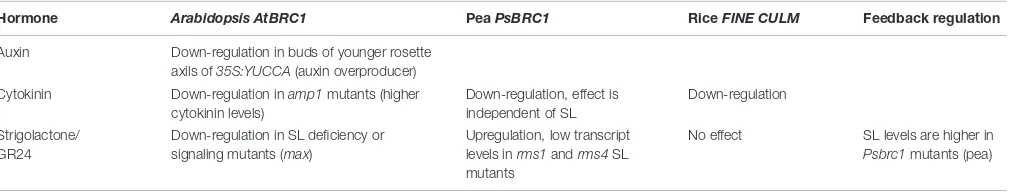
TABLE 2 | Regulation ofBRC1/TB1expression.
Hormone Arabidopsis AtBRC1 PeaPsBRC1 RiceFINE CULM Feedback regulation
Auxin Down-regulation in buds of younger rosette axils of35S:YUCCA(auxin overproducer) Cytokinin Down-regulation inamp1mutants (higher
cytokinin levels)
Down-regulation, effect is independent of SL
Down-regulation
Strigolactone/ GR24
Down-regulation in SL deficiency or signaling mutants (max)
Upregulation, low transcript levels inrms1andrms4SL mutants
No effect SL levels are higher in Psbrc1mutants (pea)
The information in the table was taken fromAguilar-Martinez et al.(2007),Finlayson(2007),Minakuchi et al.(2010),Braun et al.(2012), andDun et al.(2012).
The photoreceptor phytochrome can adopt two different conformations: Pr and Pfr. Upon absorption of red light, Pr (inactive) is converted to Pfr (active) which shuttles to the nucleus and controls gene expression through interaction with PHYTOCHROME INTERACTING FACTOR (PIF) or PIF3-like (PIL; Leivar and Monte, 2014) Within the family of five phytochromes inArabidopsis, mainly phyB was shown to con-trol red light responses of plant architecture (Finlayson et al., 2010;Reddy and Finlayson, 2014). In sorghum, low R/FR ratios or knockout of phyB prevented bud outgrowth, which was cor-related with highTB1transcript levels in axillary buds (Kebrom et al., 2006). It was hypothesized that phyB suppressesTB1/BRC1
and that the high FR proportion of light in a dense plan-tation will convert active phyB Pfr to inactive phyB Pr and thus, suppress bud outgrowth via increasedTB1/BRC1 expres-sion. Similarly, knockout of phyB increases TB1/BRC1 levels and therefore, reduces bud outgrowth. The observation that the
Arabidopsisknockout mutantbrc1-2 does not show branching suppression under low R/FR conditions supports the hypothesis thatTB1/BRC1plays a central role in branching suppression dur-ing shade avoidance (Gonzalez-Grandio et al., 2013). A putative downstream target of TB1 during the shade avoidance response in maize is the HOMEODOMAIN-LEUCINE ZIPPER (HD-ZIP) protein GRASSY TILLERS1 (GT1;Whipple et al., 2011).GT1is expressed in leaf primordia of axillary buds and in provascular tissue below the axillary bud. Interestingly, signals of GFP-tagged GT1 were observed in cells of the axillary meristem, indicating non-cell-autonomous activity of GT1. Comparable totb1 loss-of-function mutants,gt1-1knockout mutants exhibit an increased branching phenotype. The significantly reducedGT1expression intb1mutants indicates that TB1 and GT1 act in the same path-way. SinceTB1expression is not changed ingt1 mutants, it is likely that TB1 acts upstream ofGT1and regulates its expres-sion (Figure 2A). Light conditions with a low R/FR ratio induce the expression of GT1, indicating that suppressed branching during the shade avoidance syndrome is due to TB1-mediated upregulation ofGT1expression.
Plants that suffer from suboptimal nutrient supply also exhibit decreased branching comparable to plants that compete for light. However, in contrast to the shade avoidance syndrome, dur-ing nutrient deprivation resources are not allocated to the shoot but instead to the root to facilitate enhanced nutrient uptake from the soil. Nutrient-induced changes in shoot/root ratio and root development are most obvious with plants grown under phosphate deficiency (Forde and Lorenzo, 2001; Lopez-Bucio
et al., 2002). Branching in these plants is suppressed and many lateral roots develop near the soil surface, which was termed “topsoil foraging” (Peret et al., 2014). These changes in root morphology increase phosphate uptake from soil layers that are enriched in phosphate (Peret et al., 2011; Niu et al., 2013; Hunter et al., 2014).
Kohlen et al.(2011) quantified the number of shoot branches ofArabidopsiswild type and SL biosynthesis (max1,max4) and signaling (max2) mutants under phosphate sufficient and phos-phate deficient conditions. Branching of wild type plants was significantly reduced under phosphate deficiency while none of the max mutants responded to low phosphate. The observed difference in branching suppression correlated with the SL con-tent of the xylem sap. The strigolactone orobanchol could be detected in root exudates and xylem sap of wild typeArabidopsis
and showed an increase in concentration whenArabidopsiswas grown on phosphate deficient substrate. In contrast, the root exu-date of the SL biosynthesis mutantsmax1andmax4that were unresponsive to phosphate deficiency did not show an increase in SL content under phosphate-limiting conditions (Kohlen et al., 2011). Phosphate starvation increased SL synthesis also in tomato (Lopez-Raez et al., 2008), sorghum (Yoneyama et al., 2007), and rice (Umehara et al., 2010).Umehara et al.(2010) showed that the rice SL biosynthesis genesD17(MAX3inArabidopsis) andD10
(MAX4inArabidopsis) are induced by low phosphate conditions. Similarly to phosphate, also the nitrogen supply influ-ences plant architecture. Low nitrogen suppresses branching and changes the root/shoot ratio toward higher root biomass proportions (Forde and Lorenzo, 2001; Euring et al., 2012). Quantification of SLs in roots and root exudates of sorghum and pea plants grown under low nitrogen conditions showed that nitrogen deficiency increased SL levels in these plants (Yoneyama et al., 2007;Foo et al., 2013), which points to SL-mediated sup-pression of bud outgrowth under nitrogen limitation. Vice versa, optimal nitrogen supply decreases SL production, which may lead to increased branching (Yoneyama et al., 2013).
the canalization hypothesis (Sachs, 1981), this weak sink strength will prevent establishment of a PATS from axillary buds and, thus, consolidate bud dormancy. Analyses ofArabidopsismutants showed that intact auxin signaling and SL biosynthesis are both required for increased supply of auxin from the shoot apex lead-ing to suppression of branchlead-ing under nitrogen starvation (de Jong et al., 2014).
In conclusion, phosphate and nitrogen supply of the plant clearly affect plant architecture and SLs are involved in the plant responses to nutrient supply. However, the mechanism leading to a change in branching may vary in different plant species.
The nutrient- or shading-induced changes in plant architec-ture exemplify that plants can adapt their branching patterns to the prevailing environmental conditions. This demonstrates that plant architecture closely correlates with plant growth and sur-vival. Likewise, crop plant performance is determined by branch-ing characteristics and it is not surprisbranch-ing that durbranch-ing domesti-cation of crop plants, certain architectural traits were a major target for selection of improved cultivars. Especially monocot crops like rice, sorghum, maize, and wheat are of great impor-tance for world nutrition. The architectural diversity of monocot plants allowed the selection of specific architectural traits from a broad natural gene pool during domestication.
Branching Relevant Genes Selected
during Domestication and Plant
Breeding
Monocot crop plants belong to the grasses which have been assigned to two major clades, consisting of subfamilies (Barker et al., 2001). Cereals of the first clade, which are important for world nutrition, belong to the subfamily Ehrhartoideae (including rice) and the Pooideae (including oat, wheat, barley, rye). Within the separate, second major clade is the subfamily of the Panicoideae with maize, sorghum, and millets. Grasses exhibit two types of vegetative branching patterns (Doust, 2007), depending on the position of branch development with respect to the plant main axis. Tillers are typical for many grasses and determine their characteristic growth habit (Figure 1B). Tillers are branches that originate from nodes near the plant basis. These branches reach a similar height like the main stem and have the capacity to form adventitious roots. Axillary branches that initiate at upper positions of the culm (the main stem of grasses) are similar to branches of dicot plants. Grasses of the two major phylogenetic clades can be classified according to these branching patterns. Plants of the Ehrhartoideae and the Pooideae develop many tillers and no axillary branches while members of the Panicoideae produce tillers and, in addition, initiate axillary meristems that can grow into axillary branches (Doust, 2007).
The architectural traits selected during the domestication of crop plants include the extent of vegetative shoot and inflores-cence branching, branch angle, as well as internode elongation. Inflorescence branching and genes involved in stem elongation like the DELLA genes (Peng et al., 1999;Sasaki et al., 2002) have been covered in recent reviews (Fernandez et al., 2009;Teo et al.,
2014;Zhang and Yuan, 2014). Here, we will therefore focus on vegetative branching and branch angle.
Changes in vegetative branching phenotypes during plant domestication are most evident in monocot crop plants and the molecular bases of these changes have been thoroughly stud-ied. During domestication of panicoid grasses, plant lines have been selected that show a decrease in both tillering and axil-lary branching. Modern cultivars of domesticated maize plants develop ideally only one female inflorescence (ear) and a high proportion of fixed carbon is allocated to the developing ear. Only the main stem terminates in a single male inflorescence (tassel). In contrast, wild forms of Zea mayssubsp.mays(Zea mays subsp. parviglumis and Zea mays subsp. mexicana, col-lectively named teosinte) develop many axillary branches at the main stem which produce female inflorescences from secondary axillary meristems. Each branch terminates in a male inflores-cence. Doebley et al.(1997) discovered that one of the quanti-tative trait loci that determine maize architectural changes dur-ing domestication carries the TEOSINTE BRANCHED1 (TB1) gene. Small changes in expression strength of TB1 seem to be sufficient to cause the significant differences in branching patterns between teosinte and maize (Doebley et al., 1997). Maize was domesticated in Mesoamerica (Holst et al., 2007; Piperno et al., 2007;Pohl et al., 2007), while the other monocot crops belonging to the Panicoideae, pearl millet and sorghum, were selected in Sub-Saharan Africa (Remigereau et al., 2011). Interestingly, comparative QTL mapping revealed that also in pearl millet, TB1 was the molecular target of domestication (Remigereau et al., 2011). Polymorphism analyses comparing cultivated pearl millet with the wild form Pennisetum glaucum
showed that the nucleotide diversity of the TB1gene dramat-ically dropped in a region upstream of the transcription start site. This analysis indicates that nucleotide changes important for the reduced branching of pearl millet occurred within the promoter region of the TB1 gene (Remigereau et al., 2011). Such decreases of polymorphism restricted to single genes are characteristic of domestication events in contrast to evolution-ary bottle necks that result in a reduction of polymorphism on the whole genome scale. Summarized, the studies in maize and pearl millet indicate that changes in the promoter activ-ity and expression level of the domestication target gene TB1
may be causal for the reduced branching of some monocot crops.
et al.(2010), respectively, carry a mutation in themiR156 com-plementary site. Thus, in both lines, SPL14 mRNA is resistant tomiR156-mediated degradation and accumulates to a higher RNA level than in the rice cultivars Nipponbare and Taichung Native 1 which were used as reference lines in map based cloning.
IPA1/OsSPL14encodes the transcription factor SQUAMOSA PROMOTER BINDING PROTEIN-LIKE 14. A DNA motif that is bound by IPA1/OsSPL14 was found in theOsTB1promoter (Lu et al., 2013). The fact that a transgenic rice line that produces a
miR156resistantIPA1/SPL14mRNA exhibits higherOsTB1 tran-script levels indicates that IPA1/SPL14 positively regulatesOsTB1
expression. As described above, TB1 is an important target during domestication and increased expression ofOsTB1leads to sup-pression of bud outgrowth, which most likely causes the observed low tillering phenotype of the analyzed rice lines with character-istics of IPA. However, low tillering is not the only characteristic of IPA. TheO. japonicalines ST-12 and Shaoniejing also exhibit taller and stronger culms. This observation points to a pleiotropic action of IPA1/SPL14. In addition to bud outgrowth suppression caused by higher expression of OsTB1, increased plant height and higher grain number per panicle may be mediated by induc-tion of DENSE AND ERECT PANICLE1(DEP1; Huang et al., 2009) through IPA1/SPL14 (Lu et al., 2013). Besides from cul-tivars with an altered miR156 – IPA1/SPL14 pathway which were selected by classical breeding during crop domestication, a biotechnological approach, in whichmiR156was overexpressed in switchgrass, was successful. The overexpressing lines exhib-ited increased tillering and also the biomass quantity and quality were improved, which is beneficial for the use of switchgrass as a resource of bioenergy (Fu et al., 2012).
Another example for tillering-relevant genes are
STRIGOLACTONE BIOSYNTHESIS 1 and 2 (SLB1 and
SLB2).Cardoso et al.(2014) identified these closely related genes by QTL mapping in rice. They are present in the low-tillering cultivar Azucena (Japonica subspecies), while they are absent from the high-tillering cultivar Bala (Indicasubspecies) due to a genomic rearrangement. Both genes show high orthology to the
Arabidopsis SL biosynthesis geneMAX1and are functional in
Arabidopsis, since they can rescue themax1mutant phenotype (Cardoso et al., 2014). More recently, they were shown to catalyze the oxidation and subsequent hydroxylation of carlactone to yield the SL orobanchol (Zhang et al., 2014). Consistently, the cultivar Bala exudes low SL levels from roots (Cardoso et al., 2014). A generally reduced SL production would explain the high tillering phenotype and indicate that SLs are also important regulators of the architecture of crop plants, besides from the factors discussed above.
The initial reason for the QTL mapping, however, was not plant architecture. SLs are exuded by roots into the rhizosphere, where they promote arbuscular mycorrhiza, especially under phosphate starvation conditions. Root parasitic plants, such as
Striga, appear to exploit this mechanism and use SLs as germi-nation cues (reviewed inBouwmeester et al., 2007). SLs therefore induce germination of Striga seeds, which is in line with the finding that the rice cultivar Azucena, exhibiting high SL exu-dation, is more susceptible toStriga infection (Cardoso et al.,
2014). Therefore, SLs also play an important role in plant resis-tance in addition to their function in the control of rice tillering. Thus, they are a potent target for breeding efforts for improving agronomical traits in crop plants. Furthermore, a recent publi-cation indicates that SL genes may also be a quantitative trait in trees used on short rotation plantations. The willow ortholog of
MAX4co-localizes with a QTL for shoot resprouting after coppic-ing (Salmon et al., 2014). Thus, manipulation of the SL pathway for improvement of crop plants may specifically be useful for fast growing trees like willow and poplar which are cultivated on short rotation coppices. These trees are grown for 3–5 years and, after harvesting, the plants are allowed to resprout from the stool to start the next rotation.
In addition to the degree of tillering, the angle between tiller and culm determines the suitability of rice varieties for rice farm-ing (Wang and Li, 2005). Tillers of the wild riceOryza rufipogon
grow in a horizontal orientation during the vegetative phase. This horizontal growth habit suppresses competing weeds, but the horizontal tillers have high space requirements and are not suit-able for cultivation of rice in dense stands. Thus, rice varieties with a more compact growth due to a smaller tiller angle were selected during domestication.
Yu et al. (2007) isolated TILLER ANGLE CONTROL1
(TAC1) by map-based cloning in an attempt to character-ize a quantitative trait locus that decreases the tiller angle in rice. They used a mapping population obtained from a rice variety with almost zero tiller angle (straight tillers, compact growth) and a line with spread out tillers. The rice variety with compact growth carries a mutation in the 3′UTR ofTAC1
which leads to aberrant splicing. The resulting mRNA con-tains a mutated 3′UTR that leads to decreased stability. Yu
et al.(2007) could show that high levels ofTAC1mRNA cor-respond to a large tiller angle and low expression levels to a smaller tiller angle, respectively. Analysis of 152 rice accessions (wild type, O. japonica and O. indica cultivars) revealed that all lines with low tiller angle carry the identicaltac1 mutation that leads to aberrant splicing of thetac1 transcript (Yu et al., 2007).
TAC1shows sequence similarity toLAZY1, a gene that is also involved in tiller angle determination. In contrast totac1, a loss of function inlazy1results in wider tiller angles. This effect on tiller angle is caused by a modified gravitropic response of the mutant. In the lazy1mutant, the apical-basal polar auxin transport is increased, while lateral auxin transport is decreased. This results in abnormal auxin distribution leading to a weaker gravitropic response. Therefore,LAZY1controls gravitropism by regulating polar auxin transport (Li et al., 2007).
In conclusion, TAC1 and LAZY1 have opposite functions with respect to branch angle control (Figure 2B). The most obvious difference on the sequence level between TAC1 and LAZY1 is an EAR like domain at the C-terminus that is only present in LAZY1 (Dardick et al., 2013). Phylogenetic analyses and studies of intron–exon structure indicate thatLAZY1 is, from an evo-lutionary perspective, the older gene and TAC1evolved from
repressor through the EAR domain. TAC1, which lacks the EAR domain, may compete with LAZY1 and diminish repression by LAZY1 (Dardick et al., 2013).
Other genes that regulate tiller angle are PROSTRATE GROWTH1 (PROG1; Tan et al., 2008) and LOOSE PLANT ARCHITECTURE 1(LPA1;Wu et al., 2013). Both genes encode putative zinc finger transcription factors with C-terminal EAR-like repression domains. The tiller base in the prog1 mutant shows asymmetric growth due to a higher cell number on the lower side of the tiller base. Like lazy1, the lpa1 mutant exhibits reduced shoot gravitropism, possibly caused by a slower sedimentation of amyloplasts in the statocytes (Wu et al., 2013).
In summary, the analyses on TB1, TAC1, LAZY1, PROG1,
and LPA1 in crop plants indicate that with respect to plant architecture, only few key genes have been the target of selection during domestication.
In the studies mentioned above, monocots were investigated. However,TAC1has also been identified as a candidate gene for branch angle control in dicotyledonous species, e.g., in peach trees (Prunus persica; Dardick et al., 2013). In trees, fruit and wood production are influenced by crown architecture. Trees with compact crowns are suited for high density cultivation and allow yield increases compared to lines with a wider crown (Dardick et al., 2013). P. persica varieties that exhibit a com-pact growth habit are called broomy or pillar lines and the associated semidominant mutation has been designated as br. The mutation was mapped as an insertion that introduces a premature stop codon in a gene encoding a protein with sim-ilarity to the monocot TAC1. A knockout of the orthologous gene inArabidopsisresulted in smaller angles between cauline (i.e., inflorescence) branches and the main inflorescence shoot as well as between rosette branches and the stem. The pyramid poplar (Populus nigra‘Italica’) develops a phenotype compara-ble to the broomy or pillar variety of peach. This poplar growth habit may also be caused by a defect in a poplar ortholog of TAC1. In apple, another compact growth phenotype exists which has been designated columnar (co). However, this phe-notype is different from theP. persicabroomy or pillar growth habit. Columnar apple is not only characterized by a compact crown, but also by shorter branches, a thicker stem with shorter internodes and short fruit spurs (Petersen and Krost, 2013). Moreover, thebr mutation is semidominant, whilecois domi-nant. Thecomutation has been mapped to a region of 393 kb with 36 ORFs on chromosome 10 (Petersen and Krost, 2013). However, the exact locus and its molecular function remain to be determined.
In fruit and timber trees, not only the branch angle, but also the degree of branching is economically important. The leaves of branches contribute to the specific leaf area index which significantly affects photosynthesis rate (Broeckx et al., 2012). In contrast to annual plants, trees build two different types of branches. During the growth period, the shoot apex suppresses the outgrowth of buds to a certain extent (apical dominance), leading to so-called paradormancy. However, this state of dormancy can be overcome by several factors (e.g., by decapitation), leading to bud outgrowth. Buds that develop
and grow out in the same season without an intervening dor-mant season form so-called sylleptic branches. However, many species in temperate regions undergo dormancy during winter as an adaptation to adverse environmental conditions. After the growth period in summer, short day length and low tempera-tures prohibit further growth. The resulting stage of dormancy is called ecodormancy. It can still be broken if the growth con-ditions become more favorable. However, after further exposure to short daylength and low temperatures, the tree enters a stage called endodormancy, in which it can survive the harsh con-ditions in winter. Endodormancy can only be broken after a certain chilling requirement, i.e., a certain cumulative time of cold temperatures, is fulfilled. The plant is then reverted into an ecodormancy state, which will be broken when the envi-ronmental conditions become more favorable in spring. Buds formed during the previous growth period will then grow out and produce so-called proleptic branches. The different stages of dormancy described above are reviewed inAllona et al.(2008). Many tree species of the temperate regions form exclusively pro-leptic branches, but some genera like Populus, Prunus, Alnus, Larix,andTsugacan also grow sylleptic branches (Broeckx et al., 2012). This may be advantageous during the establishment phase of trees since all branches that are built during the first growth period are, by definition, sylleptic branches. The additional leaf area of sylleptic branches contributes to carbon fixation and sylleptic branches have a high translocation efficiency of photo-synthates (Scarascia-Mugnozza et al., 1999). Early canopy clo-sure and the resulting suppression of weed growth might also be an important trait for fast growing trees on short rotation plantations.
In perennial plants, apical dominance seems to be controlled in a similar way as in annuals. Studies by Cline and Dong-Il (2002) indicate that auxin is a key player in this process. They compared three poplar clones with significant differences in sylleptic branching. They showed that “branchiness” of the three poplar clones correlates with sensitivity to auxin, which gener-ally suppresses bud outgrowth: the clone with a low degree of sylleptic branching was more sensitive to auxin than the highly branched clone. A hallmark of branching control by apical dom-inance is a gradient of bud outgrowth across the main shoot. This is most evident and has been thoroughly characterized inArabidopsis. Before flowering, bud initiation and outgrowth occurs in an acropetal direction while after the onset of flower-ing, this gradient is reversed and uppermost buds elongate and grow out first (Hempel and Feldman, 1994). Similarly, sylleptic branching occurs in a basipetal direction in poplar. In con-trast, all proleptic branches that form after a period of dormancy start to elongate at a similar time point (Wilson, 2000). This synchronized growth of proleptic branches points to a control mechanism that is different from apical dominance or it is due to a factor that very efficiently breaks apical dominance. Studies byMoreno-Cortes et al.(2012) identified a protein that may play a role in bud outgrowth control. They isolated CsRAV1 from chestnut that encodes a protein with homology toAtRAV1from
Arabidopsisthat has been classified as a transcriptional repres-sor (Ikeda and Ohme-Takagi, 2009). Overexpression ofCsRAV1
the poplar clone that was used in these studies usually does not branch during the first growth period (i.e., it does not form syllep-tic branches), suppression of branching must have been released by constitutive overexpression of CsRAV1. Interestingly,CsRAV1
is highly expressed during winter.Moreno-Cortes et al.(2012) hypothesized that in perennials that grow in temperate regions, RAV1 accumulates during winter and elicits growth of prolep-tic branches from axillary meristems in the following spring. Overexpression ofRAV1, thus, leads to season-independent accu-mulation of RAV1 and causes growth of sylleptic branches from meristems which have not been exposed to a period of winter dormancy.
Conclusion
Apical dominance as a key control mechanism of branching has been a focus of intense research since Thimann and Skoog performed experiments in the 1930s on the role of auxin in sup-pression of branching (Thimann and Skoog, 1933). As it became evident that auxin does not directly suppress bud outgrowth, the second messenger hypothesis was put forward and the search for the elusive branching hormones initiated. Cytokinin was soon classified as one of the second messengers (Turnbull et al., 1997;Müller and Leyser, 2011), but it took until 2008 to iden-tify SL as another branching hormone (Gomez-Roldan et al., 2008;Umehara et al., 2008). Within 6 years after this discovery, canonical SL biosynthesis and signaling pathways were estab-lished (Waldie et al., 2014). Now, SLs are accepted as branch-ing control factors for herbaceous monocots (Umehara et al., 2008) and dicots (Gomez-Roldan et al., 2008). Loss-of-function mutants of SL biosynthesis and signaling show profound changes of plant architecture. Nonetheless, modification of the SL path-way has not yet been used in genetic engineering to improve architecture of crop plants. Also, SL genes have not been a tar-get during monocot domestication, since the architectural trait selected during domestication of crops is low branching (Doust, 2007). However, the identification ofSLB1andSLB2in rice culti-vars (see discussion above) points to a role of SLs in parasitic weed resistance. Therefore, SLs may be an important target in breeding programs.
Analyses of domestication genes in monocot crops led to the identification of a small set of target genes (Doebley et al., 1997; Doust, 2007; Yu et al., 2007; Tan et al., 2008; Ku et al., 2011; Remigereau et al., 2011), of which each pro-foundly influences plant architecture. Originally, researchers proposed that monocot genes controlling plant architecture, e.g.,TB1 and TAC1, are unique to monocots (Doebley et al., 1997; Yu et al., 2007). However, orthologs of TB1 and
TAC1 were soon also identified in dicots (Aguilar-Martinez et al., 2007; Martin-Trillo et al., 2011; Braun et al., 2012; Dardick et al., 2013). Now, with this knowledge, key genes for genetic engineering or for use as genetic markers for classical breeding of monocot and dicot crops are avail-able.
In contrast to herbaceous plants, knowledge on branching control in woody plants generally is scarce. Currently, this
topic is attracting more attention. Recent studies by Ward et al. (2013) and Czarnecki et al. (2014) showed comple-mentation of Arabidopsis max mutants by Salix and Populus MAX orthologs, respectively, pointing to a role of SLs in trees. Also, PpeTAC1 has been characterized as a controlling factor of branch angle in peach (Dardick et al., 2013) and
CsRAV1from chestnut has been shown to play a role in sea-sonal control of proleptic branching (Moreno-Cortes et al., 2012).
Tree breeding is time consuming due to the long gen-eration time of woody plants. Thus, using these key genes in genetic engineering approaches would be more straight-forward to improve productivity. However, transgenic crops and also transgenic trees are not readily accepted by the public in many countries (Kaiser, 2001). Therefore, the generation of transgenic tree cultivars for wood or fruit production appears to be not economically reasonable at the moment. Alternatively, markers like broomy could be employed to assist classical breeding programs. Another pow-erful technique is Targeting Induced Local Lesions in Genomes (TILLING), which can identify desired point mutations in a mutagenized population in an efficient, high-throughput way (McCallum et al., 2000). A variant of this technique, called Ecotilling (Comai et al., 2004), could be used to screen natural populations for desired polymorphisms in order to exploit natural variation for breeding. These methods work without the production of genetically modified organisms (GMOs).
Additionally, targeted genome editing approaches such as CRISPR-Cas9 and related technologies may be used to actively introduce highly specific changes in the genome instead of screening for random changes (reviewed in Sander and Joung, 2014). However, it is still unclear how this and other new methods will be treated by legislature. Although, the resulting engineered plants cannot be dis-tinguished from plants generated by traditional breeding methods, they may be classified as GMOs at least in the European Union, because their production involves trans-genic intermediates (reviewed in Hartung and Schiemann, 2014).
Furthermore, even though the techniques discussed above are very powerful and may not fall under GMO-regulation, they are still limited to modifications of existing sequence within a given species. The introduction of entirely new sequences, allowing the attainment of com-pletely new traits, can only be achieved by introducing foreign DNA, inevitably resulting in GMO by defini-tion. Therefore, transgenic plants are still not entirely dispensable to match the demand for efficient crops and will most likely play a major role in the future in many countries.