*Corresponding author:
E-mail: [email protected]
Available online at http://medpet.journal.ipb.ac.id/
Partial Sequencing of 16S rRNA Gene of Selected
Staphylococcus aureus
Isolates
and its Antibiotic Resistance
H. D. Kusumaningruma,b*, L. Handayanic, & R. Novriantic
aDepartment of Food Science and Technology, Faculty of Agricultural Engineering and Technology,
Bogor Agricultural University
Campus IPB Darmaga, PO. BOX 220, Bogor 16002, Indonesia
bSoutheast Asian Food and Agricultural Science and Technology (SEAFAST) Center, Bogor Agricultural University
SEAFAST Center Building, Jl. Puspa No 1, Campus IPB Darmaga, Bogor 16680, Indonesia
cFood Science Study Program, Graduate School, Bogor Agricultural University, Indonesia (Received 11-04-2016; Reviewed 24-05-2016; Accepted 13-07-2016)
ABSTRACT
The choice of primer used in 16S rRNA sequencing for identification of Staphylococcus species found in food is important. This study aimed to characterize Staphylococcus aureus isolates by partial sequencing based on 16S rRNA gene employing primers 16sF, 63F or 1387R. The isolates were isolated from milk, egg dishes and chicken dishes and selected based on the presence of sea gene that respon-sible for formation of enterotoxin-A. Antibiotic susceptibility of the isolates towards six antibiotics was also tested. The use of 16sF resulted generally in higher identity percentage and query cover-age compared to the sequencing by 63F or 1387R. BLAST results of all isolates, sequenced by 16sF, showed 99% homology to complete genome of four S. aureus strains, with different characteristics on enterotoxin production and antibiotic resistance. Considering that all isolates were carrying sea gene, indicated by the occurence of 120 bp amplicon after PCR amplification using primer SEA1/SEA2, the isolates were most in agreeing to S. aureus subsp. aureus ST288. This study indicated that 4 out of 8 se-lected isolates were resistant towards streptomycin. The 16S rRNA gene sequencing using 16sF is use-ful for identification of S. aureus. However, additional analysis such as PCR employing specific gene target, should give a valuable supplementary information, when specific characteristic is expected.
Keywords: 16S rRNA gene sequencing, Staphylococcus aureus, enterotoxin-A, antibiotic
ABSTRAK
Pemilihan primer yang digunakan dalam sekuensing gen 16S rRNA untuk identifikasi secara akurat terhadap spesies Staphylococcus pada pangan merupakan hal yang penting. Studi ini bertujuan untuk mengkarakterisasi isolat Staphylococcus aureus melalui sekuensing parsial terhadap gen 16S rRNA menggunakan primer 16sF, 63F atau 1387R. Isolat yang digunakan diisolasi dari susu, telur olahan dan ayam olahan yang dipilih berdasarkan keberadaan gen sea yang berperan dalam pemben-tukan enterotoksin-A. Kerentanan isolat terhadap enam antibiotik juga diuji. Penggunaan primer 16sF secara umum menghasilkan persentase homologi dan cakupan yang lebih tinggi dibandingkan dengan sekuensing menggunakan 63F atau 1387R. Hasil BLAST terhadap semua isolat yang dis-ekuensing dengan 16sF menunjukkan 99% homologi terhadap 4 strain S. aureus dengan karakteristik yang berbeda dalam pembentukan enterotoksin maupun kerentanan terhadap antibiotik. Mengingat bahwa semua isolat mempunyai gen sea, yang ditandai dengan kehadiran amplikon 120 bp setelah
amplifikasi menggunakan primer SEA1/SEA2, semua isolat homolog dengan S. aureus subsp. aureus
ST288. Penelitian ini juga menunjukkan bahwa empat dari delapan isolat resisten terhadap strept-omisin. Sekuensing terhadap gen 16S rRNA menggunakan primer 16sF sangat bermanfaat dalam
mengidentifikasi S. aureus, tetapi tambahan analisis PCR terhadap gen spesifik, akan memberikan
informasi yang berharga terutama jika diharapkan ditemukan karakteristik khusus pada bakteri.
INTRODUCTION
Staphylococcal food poisoning is occurred when people consume food contaminated by Staphylococcus aureus that produce enterotoxins. Various staphylococ-cal enterotoxins (SE) incriminated in staphylococstaphylococ-cal food poisoning have been reported. They were SEA, SEB, SEC, SED and SEE, and recently new serological types of SEs (SEG, SEH, SEI, SEJ, SEK, SEL, SEM, SEN,
SEO, SEP, SEQ, SER and SEU) were also identified
(Argudin et al., 2010; Xie et al. 2011, Roussel et al., 2015). Staphylococcal enterotoxins-A (SEA), which is found on most food poisoning by S. aureus, is expressed in the mid-exponential phase, and its gene appears to be transferred by temperate bacteriophage (Argudin et al., 2010). The prevalence and genetic diversity of S. aureus has been investigated in raw and pasteurized milk (Rall et al., 2008), ready-to-eat foods (Huong et al., 2010), milk and food product (Salasia et al., 2011), and street-vend foods (Rohinishree & Negi, 2011). For epidemiological
purposes, the accuracy of identification of Staphylococcus species isolated from various food products is critical. In this regard, molecular characterization is reported to be more accurate (Becker et al., 2004) than phenotypic
identification (Rohinishree & Negi, 2011). The 16S rRNA
gene is extensively used as taxonomic marker molecules
during molecular characterization (Janda & Abott, 2007), particularly during the sequence analysis for differenti -ating species and sub species of bacteria (Rohinishree & Negi, 2011).
Generally, during sequencing analysis, the DNA
target is firstly amplified using a pair primers followed
by the sequencing using a single primer (SenGupta
& Cookson, 2010). Identification based on highly
conserved genes such as 16S rRNA usually uses long
sequences primers (≥ 500 bp to about 1500 bp) (Janda & Abbott, 2007), although species specific shorter sequenc -es can also be applied. For this purpos-es, universal
primers for amplification of 16S rRNA genes are widely
available, such as primers 63F and 1387R (1350bp) (Marchesi et al., 1998) and 27f and 1492r (Frank et al., 2008). Relatively shorter primer, i.e. 16sF and 16sR3 were also used (Lee et al., 2007). Since the choice of
primers will affect the diversity of bacterial species that
will be detected (Fredriksson et al., 2013), in this study
three different primers targeting the 16S rRNA gene, i.e.
16sF, 63F and 1387R, was used separately to sequence DNA of selected S. aureus isolates from milk, egg dishes and chicken dishes. Milk, egg dishes and chicken dishes were reported contaminated by S. aureus in previous study (Handayani et al., 2014).
Furthermore, as information on the spreading of antibiotic resistance strains among S. aureus in food is also important, the antibiotic susceptibility testing of the isolates against antibiotics was also conducted in this study. The antibiotics tested were antibiotics that usually used to control human infections as well as to control and treat infections on farms. Many S. aureus
strains that demonstrated resistance to different antibi
-otics were isolated from hospitals (Schmitz et al., 1999; Brown & Ngeno, 2007; Xie et al., 2011), hospital waste
waters (Thompson et al., 2012), as well as from animal based food product, such as raw milk and dairy prod-ucts (Jamali et al., 2015), poultry retail meat (Teramoto et al., 2016), and goat milk powder (Xing et al., 2016).
MATERIALS AND METHODS
Bacterial Strain and Isolates
The wild-type of S. aureus from raw milk (S1, S4 and S10), egg dishes (TB1), sautéed chicken cuts (UA2 and UA13) and chicken cuts satay (SJ1 and SJ4) were isolated in previous study (Handayani et al., 2014). S. au-reus ATCC 25923 was used as a reference bacterium. All bacteria were grown in a tryptic soy broth or agar (Difco Laboratory, Detroit, MI, U.S.A.), and incubated at 37oC
for 18h-24h. For confirmation, the isolates were spread
onto Baird-Parker Agar (Oxoid Ltd., Hampshire, UK) supplemented with egg-yolk tellurite. Plates were incu-bated at 37°C for 18-24 h, thereafter the colonies were picked and streaked on Mannitol Salt Agar (Oxoid Ltd., Hampshire, UK). Typical colonies were then tested for production of catalase using Staphylase test kit (Oxoid
Ltd., Hampshire, UK) and biochemical identification
using API Staph (bioMérieux Inc., North Carolina, USA) according to manufacturer’s instructions.
DNA Extraction
Genomic DNA was isolated as described previ-ously by Mason et al. (2001) with slight modification, as reported by Handayani et al. (2014), i.e. lysostaphin (10 mg/mL) was substituted by lysozyme (Bio Basic Canada Inc., Ontario, Canada) solution (10 mg/ml). The concentration of genomic DNA was determined by the Spectrophotometer UV -1800 (Shimadzu, Japan) at 260 nm while the quality was assessed based on the ration of the reading at 260/280 nm. The integrity of the DNA was checked by running in 1.5% agarose gel at 75V for 40 min electrophoresis (Bio-Rad Laboratories Pte. Ltd, Singapore).
Detection of 16S rRNA and Sea Gene by PCR
The amplification of the gene encoding 16S
rRNA and sea was performed using primers listed in Table 1 at Thermal Cycler 2720 (Applied Biosystems, California, USA). PCR master mix consisted of 12.5 µL of DreamTaq Green master mix (Thermo Fisher
Scientific, Massachusetts, USA), 1 µL of each primer (10
µM), 2 µL of DNA template, and 8.5 µL nuclease free
water (Thermo Fisher Scientific, Massachusetts, USA).
Cycling parameters were one denaturation cycle for 5
min at 95°C and 30 amplification cycles for denaturation
(1 min at 95°C), annealing (1 min at 55°), extension (1 min at 72°C) and termination for 5 min at 72°C, adopted from Lee et al. (2007). The amplification products were
visualized on 2% agarose gel (Thermo Fisher Scientific, Massachusetts, USA) by electrophoresis (Rad,
Sequence Analysis of 16S rRNA Gene
The genotypic characterization of bacteria isolates was made through partial sequence analysis of 16S rRNA gene, using single primer 16sF, 63F, or 1387R.
The PCR products that were amplified with primers
16sF/16sR3 were sequenced using primer 16sF. The
PCR products amplified with primers 63F/1387R were
sequenced using primer 63F and 1387R separately. The process of DNA sequencing was conducted by BigDye Applied Biosystem sequencer engine model 3730 at Macrogen inc., Singapore. Partial sequence
data obtained, in FASTA format, was then submitted
to the BLAST process at NCBI (National Center for
Biotechnology Information) database for the identifica
-tion of isolates (http://blast.ncbi.nlm.nih.gov/ Blast.cgi).
The BLAST process was conducted using nucleotide
collection searching setting (nr/nt) with Staphylococcus aureus subsp. aureus (taxid:46170) as organism of choice. The sequencing results were then compared to the reported 16S rRNA gene sequences of Staphylococcus species available in the GenBank database. The isolates
were identified based on the highest homology percent -ages to the reported sequences.
Antimicrobial Susceptibility Testing
Antimicrobial susceptibility was tested according to the guidelines of the Clinical Laboratory Standards
Institute (CLSI, 2012) using the disk diffusion tech -nique with commercially available discs (Oxoid). The antimicrobials and concentrations in micrograms tested were gentamicin (10 mcg), streptomycin (10 mcg), kanamycin (30 mcg), chloramphenicol (30 mcg), tetracycline (30 mcg), and oxytetracycline (30 mcg). The inhibition zones, in mm, were measured in duplicate and scored as sensitive, intermediate susceptibility and resistant according to the CLSI recommendations, e.g. >19, 15-18 and <14 for tetracycline; >18, 13-17 and <12 for chloramphenicol; etcetera (CLSI, 2014). Gentamicin
10 µg S ≥15 13–14 ≤12; Kanamycin 30 µg ≥18 14–17 ≤13; Streptomycin 10 µg ≥15 12–14 ≤11; Chloramphenicol 30 µg ≥18 13–17 ≤12; Tetracycline 30 µg ≥19 15–18 ≤14
(CLSI, 2014).
RESULTS
Genotypic Characteristic of Isolates
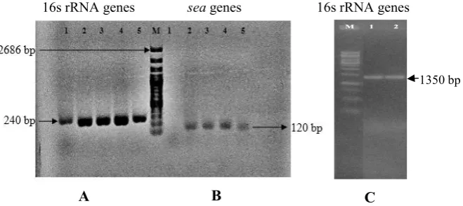
Ten isolates of 78 presumptive S. aureus isolates were positive for S. aureus (Handayani et al., 2014). Eight
of these isolates were reconfirmed in this study carrying
sea gene that responsible for formation of staphylococcal enterotoxin-A (SEA). The presence of sea gene was indi-cated by the occurrence of 120 bp amplicon, after PCR
amplification using primer SEA1/SEA2 (Table 2). The
reference strain, S. aureus ATCC 25923, did not show this gene. In addition to the work of Handayani et al. (2014), all isolates also demonstrated 1350 bp amplicon after
amplification by 63F/1387R primer (universal primer). The amplified PCR products of some isolates are shown
in Figure 1.
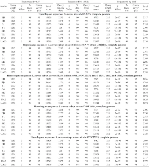
The BLAST results are listed in Table 3. Sequencing by primer 16sF resulted in high identity percentages (almost all achieved 99%) towards the existing genome of S. aureus strains found in database of NCBI GenBank, in comparison to the sequencing by 63F and 1387R. Sequencing by 63F showed similarity to S. aureus strains in a range between 94% and 99%, while by 1387R in a range of 90% to 99%. In addition, using 16sF also result-ed in low E-value. The lower the E-value, or the closer it Table 1. Nucleotide sequences of primers used in PCR analysis
Gene Primer Sequence (5’ → 3’) PCR product of
partial sequence
Temperature annealing (ᵒC)
seaᵃ SEA1 TTGGAAACGGTTAAAACGAA 120 bp 62.1
SEA2 GAACCTTCCCATCAAAAACA 64.2
16S rRNAᵃ 16sF CCGCCTGGGGAGTACG 240 bp 70.1
16sR3 AAGGGTTGCGCTCGTTGC 69.1
16S rRNAᵇ 63F CAGGCCTAACACATGCAAGTC 1350 bp 70.8
(universal) 1387R GGGCGGWGTGTACAAGGC 70.7
Note: ᵃ Lee et al. (2007); ᵇ Marchesi et al. (1998).
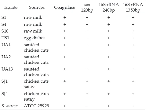
Table 2. Characteristics of S. aureus isolates
Note: + : positive results, - : negative results. Isolate Sources Coagulase sea 120bp
16S rRNA 240bp
16S rRNA 1350bp
S1 raw milk + + + +
S4 raw milk + + + +
S10 raw milk + + + +
TB1 egg dishes + + + +
UA1 sautéed chicken cuts
+ + + +
UA2 sautéed chicken cuts
+ + + +
UA13 sautéed chicken cuts
+ + + +
SJ1 chicken cuts satay
+ + + +
SJ4 chicken cuts satay
+ + + +
is to zero, the match is more significant (Pearson, et al., 2014). The expect value (E) is a parameter that describes the number of hits one can “expect” to see by chance when searching a database of a particular size.
As shown in Table 3, by sequencing using primer 16sF, all isolates demonstrated 99% homology to the sequence of genome of 4 strains of S. aureus. They were S. aureus strain MSSA476, S. aureus subsp. aureus ST772-MRSA-V strain DAR4145, S. aureus subsp. aureus DSM 20231, and S. aureus subsp. aureus ST288 (isolate 10338, 10497, 15532, 16035, 18341, 18412 and 18583). Strain MSSA476, DAR4145 and DSM 20231 do not produce enterotoxin-A, as reported at accession BX571857.1, CP010526.1 and CP011526.1, respectively (NCBI, 2016). On the other hand, S. aureus subsp. aureus ST288 isolates 10338, 10497, 15532, 16035, 18341, 18412 and 18583 are known as enterotoxin-A producer. Complement of the genome for the enterotoxin-A gene (entA) of S. aureus subsp. aureus ST288 isolate 10338, 10497, 15532, 16035, 18341, 18412 and 18583 are shown in Table 4. Since all isolates were detected carrying sea gene (Table 2), the isolates were in agreeing to S. aureus subsp. aureus ST288 that able to form enterotoxin-A. The present study has been indicating that sequencing with 16S rRNA as gene target has been successfully identifying the isolates to
specific strains. Additional PCR analysis employing
SEA1/SEA2 primers increased accuracy of characteriza-tion of the isolates.
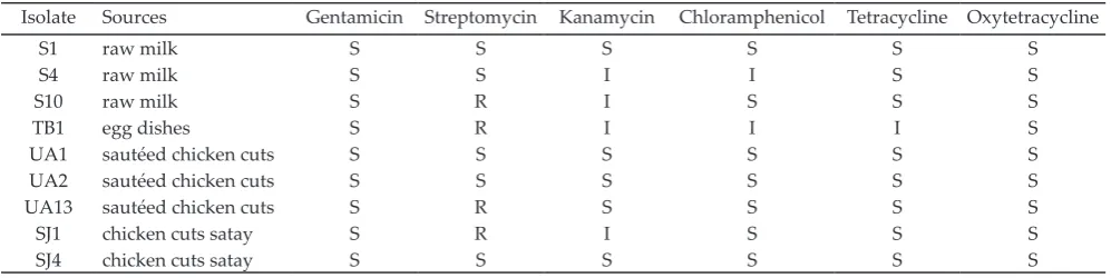
Antibiotic Resistance Among the Isolates
Four isolates showed resistance to streptomycin (Table 5). All isolates, however, were susceptible to gentamycin and oxytetracycline. Interestingly, the resistant strains to streptomycin were isolated from
different food sources, i.e. from milk (S10), egg dishes
(TB1), sautéed chicken cuts (UA13) and chicken cuts satay (SJ1). Next to resistance to streptomycin, isolate TB1 showed also intermediate resistance to kanamycin, chloramphenicol and tetracycline.
DISCUSSION
Among the SEs, SEA is reported as the most com-mon cause of staphylococcal food poisoning worldwide, but the involvement of other SEs has been also found. SEA is considered as the main cause of staphylococcal food poisoning, probably due to its high resistance to proteolytic enzymes (Argudin et al., 2010). In the
pres-ent study, all (eight) isolates were idpres-entified carrying
sea gene, suggesting the potential risk of these strains due to production of SEA. Another study conducted in Indonesia by Salasia et al. (2011), however, did not find this gene in 11 food isolates (i.e. fermented milk prod-uct, sausage, meat ball, cakes and cheese), but found seb, sec, see and seg genes. They found, however, sea gene in 5 isolates of 19 milk isolates.
Regarding the sequencing results, as
pre-dicted, the use of 3 different primers resulted
in various identity percentages. Sequencing by primer 16sF (5’-CCGCCTGGGGAGTACG-3’) re-sulted in higher identity percentages (almost all achieved 99%) towards the existing genome of S. aureus strains in comparison to the sequencing by 63F (5’-CAGGCCTAACACATGCAAGTC-3’) and 1387R (5’-GGGCGGWGTGTACAA GGC-3’). As was presented in Table 3, sequencing by 16sF also resulted in higher query coverage than that achieved by 63F and 1387R, except for isolate SJ4. These results highlighted that 16S rRNA sequencing by short sequence could provide
suf-ficient identification amongst S. aureus strain. The 16sF has been used before to detect 16S rRNA genes of S. aureus isolates from food sample (Lee et al., 2007; Lee & Park 2016).
This study found that all isolates demonstrated 99% homology to the sequence genome of 4 strains of S. aureus by sequencing using primer 16sF. Based on the highest total score of the BLAST results, all isolates showed similarity to the genome of S. aureus strain MSSA476 and S. aureus subsp. aureus ST772-MRSA-V strain DAR4145. Strain MSSA476 is an invasive commu-nity acquired methicillin-susceptible S. aureus (Holden et al. 2004). On the other hand strain DAR4145 is a multidrug resistant strain of ST772-MRSA-V (Steinig et 485
486 487 488
489
490 491 492
1350 bp
A
B
C
16s rRNA genes seagenes 16s rRNA genes
al., 2015). These results suggested that the isolates in this study showed equal likelihood whether they were
cor-responding to the MRSA or the MSSA. This finding is
possible since these strains also occupied many people in Indonesia. Recent epidemiological study showed MRSA carriage rate of 4.3%, and MSSA carriage rate of 1.5% among 1,502 patients in hospitals in Java and Bali (Santosaningsih et al., 2014).
Furthermore, all isolates which were sequenced by 16sF also showed 99% homology to the sequence genome of S. aureus subsp. aureus ST288, a highly trans-missible methicillin-resistant S. aureus (Vogel et al., 2012) that produce enterotoxin-A. The occurrence of strain
ST228 were notified for long periods. Conceicao et al., (2007) reported that strain ST239-MRSA-III was replaced by both strain ST5-MRSA-II and ST228-MRSA-I between Table 3. Query length, E value, identity percentages, query coverage and total score of the sequencing results towards homologous
sequences found in the NCBI GenBank database after the BLAST process
Isolates
Sequenced by 63F Sequenced by 1387R Sequenced by 16sF
Query length (bp)
E Value
%
Iden-tity
Query cover
(%)
Total score
Query length (bp)
E Value
%
Iden-tity
Query cover
(%)
Total score
Query length (bp)
E Value
%
Iden-tity
Query cover
(%)
Total score
Homologous sequence: S. aureus strain MSSA476, complete genome
SJ1 1263 0 94 91 10820 1232 0 90 90 8793 210 2e-97 99 93 2117
SJ4 1126 0 97 96 10798 1272 0 97 98 12349 216 2e-99 99 94 2161
S1 1573 0 98 82 12853 1509 0 98 82 12853 215 4e-101 99 95 2195
S4 1251 0 98 92 11906 938 0 99 90 9075 217 4e-101 99 94 2195
S10 1504 0 98 87 13679 1489 0 99 86 13529 215 9e-102 99 95 2206
TB1 1514 0 97 87 13424 1353 0 98 95 13618 212 2e-98 99 95 2139
UA2 1331 0 97 93 12560 1372 0 98 93 13119 217 4e-101 99 94 2195
UA13 1350 0 99 94 13590 1345 0 98 95 13507 212 2e-98 99 95 2139
Homologous sequence: S. aureus subsp. aureus ST772-MRSA-V, strain DAR4145, complete genome
SJ1 1263 0 94 91 10820 1232 0 90 90 8787 210 2e-97 99 93 2117
SJ4 1126 0 97 96 10793 1272 0 97 98 12344 216 2e-99 99 94 2161
S1 1573 0 97 84 13330 1509 0 98 82 12853 215 4e-101 99 95 2195
S4 1251 0 98 92 11912 938 0 99 90 9070 217 4e-101 99 94 2195
S10 1504 0 98 87 13684 1489 0 99 86 13529 215 9-e102 99 95 2206
TB1 1514 0 97 87 13430 1353 0 98 95 13618 212 2e-98 99 95 2139
UA2 1331 0 97 93 12565 1372 0 98 93 13119 217 4e-101 99 94 2195
UA13 1350 0 99 94 13596 1345 0 98 95 13507 212 2e-98 99 95 2139
Homologous sequence: S. aureus subsp. aureus ST288, isolate 10338, 10497, 15532, 16035, 18341, 18412 and 18583, complete genome
SJ1 1263 0 94 91 9009 1232 0 90 90 7329 210 2e-97 99 93 1756
SJ4 1126 0 97 96 8991 1272 0 96 98 10274 216 2e-99 99 94 1793
S1 1573 0 97 84 11092 1509 0 98 82 10699 215 4e-101 99 95 1820
S4 1251 0 98 92 9911 938 0 99 90 7558 217 4e-101 99 94 1820
S10 1504 0 98 87 11388 1489 0 99 86 11262 215 9e-102 99 95 1830
TB1 1514 0 97 87 11175 1353 0 98 95 11336 212 2e-98 99 95 1774
UA2 1331 0 97 93 10455 1372 0 98 93 10921 217 4e-101 99 94 1820
UA13 1350 0 99 94 11314 1345 0 98 95 11244 212 2e-98 99 95 1774
Homologous sequence: S. aureus subsp. aureus DSM 20231, complete genome
SJ1 1263 0 94 91 10820 1232 0 90 90 8794 210 2e-97 99 93 2106
SJ4 1126 0 97 96 10798 1272 0 96 98 12338 216 2e-99 99 94 2150
S1 1573 0 97 84 13319 1509 0 98 82 12848 215 4e-101 99 95 2183
S4 1251 0 99 92 11900 938 0 99 90 9070 217 4e-101 99 94 2183
S10 1504 0 98 87 13673 1489 0 99 86 13524 215 9e-102 99 95 2195
TB1 1514 0 97 87 13418 1353 0 98 95 13612 212 2e-98 99 95 2128
UA2 1331 0 97 93 12554 1372 0 98 93 13114 217 4e-101 99 94 2183
UA13 1350 0 99 94 13585 1345 0 98 95 13502 212 2e-98 99 95 2128
Homologous sequence: S. aureus subsp. aureus strain ATCC25923, complete genome
SJ1 1263 0 94 91 10826 1232 0 90 90 8782 210 10e-96 98 93 2095
SJ4 1126 0 97 96 10804 1272 0 96 98 12338 216 8e-98 98 94 2139
S1 1573 0 97 84 13313 1509 0 98 82 12848 215 2e-99 99 95 2172
S4 1251 0 98 92 11895 938 0 99 90 9058 217 2e-99 99 94 2172
S10 1504 0 98 87 13668 1489 0 99 86 13524 215 4e-100 99 95 2183
TB1 1514 0 97 87 13413 1353 0 98 95 13612 212 10e-97 98 95 2117
UA2 1331 0 97 93 12549 1372 0 98 93 13114 217 2e-99 99 94 2172
1994 and 2004 in Hungary. A study conducted in
hos-pitals in a relative small geographic area in Switzerland
also observed that several MRSA clones (ST5-MRSA-II, ST45-MRSA-IV, ST228-MRSA-I and ST247-MRSA-I) were present over a period of 8 years from 1997 to 2004 (Blanc et al., 2007). More recently, Vogel et al. (2012) compared the whole genome of eight ST228 isolates recovered between 2001 and 2008 that spread over ten
years in a tertiary care hospital in Switzerland. These
reports suggested that the spreading of ST228 strain was
confirmed.
As discused above, when sequenced by 16sF all isolates showed good homology (99%) to some strains
found in the NCBI GenBank data base with different
characteristics on its antibiotic resistance. Next to this characteristic, the coresponding strains also showed
differences on capability to form enterotoxin. Strain
MSSA476 and DAR4145, as well as strain DSM 20231 did not produce enterotoxin-A (NCBI, 2016). On the other hand, S. aureus subsp. aureus ST288 isolate 10338, 10497, 15532, 16035, 18341, 18412 and 18583 are known as enterotoxin-A producers. Therefore, additional
information to confirm the presence of a gene that re -sponsible for formation of enterotoxin-A is considerably
important. Since all selected isolates were confirmed
carrying sea gene, indicated by the presence of 120 bp
amplicon after PCR amplification using primer SEA1
and SEA2 as presented in Table 2, all isolates were most in agreeing with S. aureus subsp. aureus ST288. The sea
gene is 771 bp in size encoding an enterotoxin A pre-cursor of 257 amino acid residues (Huang et al., 1987).
Specific primers SEA1 and SEA2 were frequently used
in PCR analysis to detect the presence of sea gene in S. aureus isolates from food, such as in raw and pasteur-ized milk (Rall et al., 2008); Kérouanton et al., 2007) and ready-to-eat Kimbap (Lee et al., 2007). Specific PCR
primers have commonly been employed to confirm the presence or absence of specific characteristics associated
with target microorganisms such as virulence factors. Next to genotypic characteristic, information on antibiotic resistance amongst S. aureus strains found in food is also important for surveillance and epidemiol-ogy study. This study found that four isolates from
different food sources (from milk, egg dishes, sautéed
chicken cuts and chicken cuts satay) were resistant to streptomycin. Streptomycin resistance among S. aureus isolates was also reported in other study. Jamali et al. (2015) found that amongst S. aureus isolates from raw
milk and dairy products (n=328), 5.8% demonstrated
resistance to streptomycin, 4% to kanamycin, 3.7% to chloramphenicol, and 2.1% to gentamicin. Most isolates were resistant to tetracycline (56.1%) followed by to penicillin (47.3%). The high percentage of resistant isolates to these last two antibiotics could be due to the widespread use of these antibiotics to control and treat infections on dairy farms (Jamali et al., 2015). Moreover, the fact that streptomycin resistant strains were found in milk, egg dishes and chicken dishes indicated possible Table 4. Accession and complement of the genome for the enterotoxin-A gene (entA) of S. aureus subsp. aureus ST288, isolate 10338,
10497, 15532, 16035, 18341, 18412 and 18583
Isolate Accession Complement for the enterotoxin A gene Source*
10388 HE579059.1 1988966..1989738 http://www.ncbi.nlm.nih.gov/nuccore/HE579059 10497 HE579061.1 1988953..1989726 http://www.ncbi.nlm.nih.gov/nuccore/HE579061 15532 HE579063.1 1989266..1990039 http://www.ncbi.nlm.nih.gov/nuccore/HE579063 16035 HE579065.1 1988898..1989671 http://www.ncbi.nlm.nih.gov/nuccore/HE579065 18341 HE579069.1 1988952..1989725 http://www.ncbi.nlm.nih.gov/nuccore/HE579069 18412 HE579071.1 1988718..1989491 http://www.ncbi.nlm.nih.gov/nuccore/HE579071 18583 HE579073.1 1988770..1989543 http://www.ncbi.nlm.nih.gov/nuccore/HE579073
Note: * Last accessed [5 April 2016]
Table 5. Antibiotic susceptibility of S. aureus isolates
Isolate Sources Gentamicin Streptomycin Kanamycin Chloramphenicol Tetracycline Oxytetracycline
S1 raw milk S S S S S S
S4 raw milk S S I I S S
S10 raw milk S R I S S S
TB1 egg dishes S R I I I S
UA1 sautéed chicken cuts S S S S S S
UA2 sautéed chicken cuts S S S S S S
UA13 sautéed chicken cuts S R S S S S
SJ1 chicken cuts satay S R I S S S
SJ4 chicken cuts satay S S S S S S
occurrence of cross contamination from human or food
vendor. Schmitz et al. (1999) found that 21% of the S. au-reus isolates (n=699) collected from different hospitals in Europe were resistant to streptomycin. They also found, however, 23% of the S. aureus isolates were resistant to gentamycin, 29% to tobramycin, and 31% to kanamycin. In more recent study, Onwubiko & Sadiq (2011) also found that 55.8% of S. aureus from clinical isolates in a
tertiary health institution in North-western Nigeria (n=
129) showed resistance to streptomycin, 68.8% to tetra-cycline, 38.1% to chloramphenicol, and 7.6% to genta-mycin. The spreading of S. aureus strains that resistant to antibiotics has become a global concern. Continued surveillance of S. aureus producing enterotoxin-A in milk, egg and poultry food products at genotypic levels is necessary to understand and limit further increases of staphylococcal food poisoning incidences.
CONCLUSION
This study has demonstrated that in order to
in-crease the accuracy of the identification results, next to
the sequencing of S. aureus targeting 16S rRNA gene,
PCR analysis using specific primer is considerably
important. All eight isolates were carrying sea gene, de-tected by PCR analysis, indicating that they can produce staphylococcal enterotoxin-A. Genotypic characteriza-tion of the selected strains by sequencing using 16sF, showed agreeing to the sequence genome of S. aureus subsp. aureus ST288 that also produce enterotoxin-A. This study also found that 4 of 8 selected isolates were resistant to streptomycin.
ACKNOWLEDGEMENT
This work was funded by The Directorate General of Higher Education, Ministry of Education and Culture of Indonesia, through the University Excellent Research (PUPT) Competitive Research Grant 2013-2014.
REFERENCES
Argudín, M. A., M. C. Mendoza, & M. R. Rodicio. 2010. Food poisoning and Sta-phylococcus aureus enterotoxins. Toxins. 2: 1751-1773. http://dx.doi.org/10.3390/toxins2071751 Becker, K., D. Harmsen, A. Mellmann, M. Christian, P.
Schumann, G. Peters, & C. von Eiff. 2004. Development and evaluation of a quality–controlled ribosomal sequence database for 16S ribosomal DNA–based identification of Staphylococcus species. J. Clin. Microbiol. 42: 4988-4995. http://dx.doi.org/10.1128/JCM.42.11.4988-4995.2004 Blanc, D. S., C. Petignat, A. Wenger, G. Kuhn, Y. Vallet, D.
Fracheboud, S. Trachsel, M. Reymond, N. Troillet, H. H.
Siegrist, S. Oeuvray, M. Bes, J. Etienne, J. Bille, P. Fran
-cioli, & G. Zanetti. 2007. Changing molecular epidemiol-ogy of methicillin resistant Staphylococcus aureus in a small geographic area over an eight-year period. J. Clin. Micro-biol. 45: 3729-3736. http://dx.doi.org/10.1128/JCM.00511-07 Brown, P. D. & C. Ngeno. 2007. Antimicrobial resistance in
clinical isolates of Staphylococcus aureus from hospital and community sources in southern Jamaica. Int. J. Infect. Dis. 11: 220-225. http://dx.doi.org/10.1016/j.ijid.2006.04.005 CLSI, Clinical and Laboratory Standards Institute. 2012.
Per-formance Standards for Antimicrobial Disk Susceptibility
Tests; Approved Standard—Eleventh Edition. CLSI docu-ment M02-A11. Wayne, Pennsylvania 19087 USA. CLSI, Clinical and Laboratory Standards Institute. 2014.
Per-formance Standards for Antimicrobial Susceptibility Test-ing, Twenty-Fourth Informational Supplement. CLSI doc-ument M100-S24. Wayne, Pennsylvania 19087 USA. Conceicao, T., M. Aires-de-Sousa, M. Fuzi, A. Toth, J.
Pasz-ti, E. Ungvari, W. B. van Leeuwen, A. van Belkum, H.
Grundmann, & H. de Lencastre. 2007. Replacement of methicillin-resistant Staphylococcus aureus clones in Hun-gary over time: a 10-year surveillance study. Clin. Micro-biol. Infect. 13: 971–979. http://dx.doi.org/10.1111/j.1469-0691.2007.01794.x
Frank, J. A., C. I. Reich, S. Sharma, J. S. Weisbaum, B. A. Wil-son, & G. J. Olsen. 2008. Critical evaluation of two primers commonly used for amplification of bacterial 16S rRNA genes. Appl. Environ. Microbiol. 74: 2461–2470. http:// dx.doi.org/10.1128/AEM.02272-07
Fredriksson, N. J., M. Hermansson, & B. M. Wilén. 2013. The choice of PCR primers has great impact on assessments of bacterial community diversity and dynamics in a wastewa-ter treatment plant. PLoS ONE 8: e76431. http://journals. plos.org/plosone/article?id=10.1371/journal.pone.0076431. [20 December 2015]. http://dx.doi.org/10.1371/journal. pone.0076431
Handayani, L., D. N. Faridah, & H. D. Kusumaningrum. 2014. Staphylococcal enterotoxin A gene-carrying Staphylococcus aureus isolated from foods and its control by crude alka-loid from papaya leaves. J. Food Prot. 77: 1992-1997. http:// dx.doi.org/10.4315/0362-028X.JFP-13-483
Holden M.T., E. J. Feil, J. A. Lindsay, S. J. Peacock, N. P. Day, M. C. Enright, T. J. Foster, C. E. Moore, L. Hurst,
R. Atkin, et al. 2004. Complete genomes of two clini-cal Staphylococcus aureus strains: Evidence for the rapid evolution of virulence and drug resistance. PNAS. 101: 9786-9791. http://www.ncbi.nlm.nih.gov/pmc/articles/ PMC470752/pdf/019786.pdf. [20 October 2015]. http:// dx.doi.org/10.1073/pnas.0402521101
Huang, I. Y., J. L. Hughes, M. S. Bergdoll, & E. J. Schantz. 1987. Complete amino acid sequence of staphylococcal entero-toxin A. J. Biol. Chem. 262: 7006-7013.
Huong, B. T. M., Z. H. Mahmud, S. B. Neogi, A. Kassu, N. V. Nhien, A. Mohammad, M. Yamato, F. Ota, N. T. Lam, H. T. A. Dao, & N. C. Khan. 2010. Toxigenicity and genetic diversity of Staphylococcus aureus isolated from Vietnamese ready-to-eat foods. Food Cont. 21: 166–171. http://dx.doi. org/10.1016/j.foodcont.2009.05.001
Jamali, H., M. Paydar, B. Radmehr, S. Ismail, & A. Dadrasnia. 2015. Prevalence and antimicrobial resistance of Staphylo-coccus aureus isolated from raw milk and dairy products. Food Cont. 54: 383-388. http://dx.doi.org/10.1016/j.food -cont.2015.02.013
Janda, J. M. & S. L. Abott. 2007. 16S rRNA gene sequencing for bacterial identification in the diagnostic laboratory: pluses, perils, and pitfalls. J. Clin. Microbiol. 45: 2761–2764. http:// dx.doi.org/10.1128/JCM.01228-07
Kérouanton, A., J. A. Hennekinne, C. Letertre, L. Petit, O. Chesneau, A. Brisabois, & M. L. D. Buyser. 2007. Char-acterization of Staphylococcus aureus strains associated with food poisoning outbreaks in France. Int. J. Food Mi-crobiol. 115: 369-375. http://dx.doi.org/10.1016/j.ijfoodmi -cro.2006.10.050
Kim B., H. Yi, J. Chun, & C. Cha. 2014. Genome sequence of type strain of Staphylococcus aureus subsp. aureus. Gut Pathogens 6:6. http://gutpathogens.biomedcentral.com/ articles/10.1186/1757-4749-6-6. [20 December 2015]. http:// dx.doi.org/10.1186/1757-4749-6-6
au-reus isolates based on mRNA analysis. J. Microbiol. Bio-technol. 17: 461-467.
Lee Y. D. & J. H. Park. 2016. Phage conversion for β-lactam anti -biotic resistance of Staphylococcus aureus from foods. J. Mi-crobiol. Biotechnol. 26: 263–269. http://dx.doi.org/10.4014/ jmb.1508.08042
Marchesi, J. R., T. Sato, A. J. Weightman, T. A. Martin, J. C. Fry, S. J. Hiom, & W. G. Wade. 1998. Design and evaluation of useful bacterium-specific PCR primers that amplify genes coding for bacterial 16S rRNA. Appl. Environ. Microbiol. 64: 795-799.
Mason, W. J., J. S. Blevins, K. Beenken, N. Wibowo, N. Ojha,
& M. S. Smeltzer. 2001. Multiplex PCR protocol for the diagnosis of staphylococcal infection. J. Clin. Microbiol. 39: 3332-3338. http://dx.doi.org/10.1128/JCM.39.9.3332-3338.2001
NCBI. 2016. National Center for Biotechnology Information. http://www.ncbi.nlm.nih. gov. [5 March 2016].
Onwubiko, N. E. & N. M. Sadiq. 2011. Antibiotic sensitivity pattern of Staphylococcus aureus from clinical isolates in a tertiary health institution in Kano, Northwestern Nigeria. Pan. Afr. Med. J. 8: 4. http://www.ncbi.nlm.nih.gov/pmc/ articles/PMC3201603/pdf/pamj-8-4.pdf. [20 January 2016]. http://dx.doi.org/10.4314/pamj.v8i1.71050
Pearson, W. R. 2014. BLAST and FASTA similarity searching for multiple sequence alignment. Methods Mol. Biol. 1079:75-101. http://dx.doi.org/10.1007/978-1-62703-646-7_5
Rall, V. L. M., F. P. Vieira, R. Rall, R. L. Vieitis, A. Fernandes Jr., J. M. G. Candeias, K. F. G. Cardoso, & J. P. Araujo Jr. 2008. PCR detection of staphylococcal enterotoxin genes in Staphylococcus aureus strains isolated from raw and pas-teurized milk. Vet. Microbiol. 132: 408-414. http://dx.doi. org/10.1016/j.vetmic.2008.05.011
Rohinishree, Y. S. & P. S. Negi. 2011. Detection, identifica -tion and characteriza-tion of staphylococci in street vend foods. Food Nutr. Sci. 2: 304-313. http://dx.doi.org/10.4236/ fns.2011.24044
Roussel, S., B. Felix, N. Vingadassalon, J. Grout, J. Hennekinne, L. Guillier, A. Brisabois, & F. Auvray. 2015. Staphylococcus aureus strains associated with food poisoning outbreaks in France: comparison of different molecular typing meth -ods, including MLVA. Front Microbiol. 6: 882. http://www. ncbi.nlm.nih.gov/pmc/articles/PMC4566840. [2 February 2016]. http://dx.doi.org/10.3389/fmicb.2015.00882
Salasia, S. I. O., S. Tato, N. Sugiyono, D. Ariyanti, & F. Pra-bawati. 2011. Genotypic characterization of Staphylococcus aureus isolated from bovines, humans, and food in Indo-nesia. J. Vet. Sci. 12: 353-361. http://dx.doi.org/10.4142/ jvs.2011.12.4.353
Santosaningsih, D., S. Santoso, N. S. Budayanti, K. Kuntaman,
E.S. Lestari, H. Farida, R. Hapsari, P. Hadi, W. Winarto,
C. Milheiric, K. et al. 2014. Epidemiology of Staphylococ-cus aureus harboring the mecA or Panton-Valentine leuko-cidin genes in hospitals in Java and Bali, Indonesia. Am.
J. Trop. Med. Hyg. 90: 728–734. http://dx.doi.org/10.4269/ ajtmh.13-0734
Schelin J., N. Wallin-Carlquist, M. T. Cohn, R. Lindqvist, G. C. Barker, & P. Radstrom. 2011. The formation of Staphylococ-cus aureus enterotoxin in food environments and advances in risk assessment. Virulence. 2: 580-592. http://dx.doi. org/10.4161/viru.2.6.18122
Schmitz, F. J., A. C. Fluit, M. Gondolf, R. Beyrau, E. Linden
-lauf, J. Verhoef, H. Heinz, & M. E. Jones. 1999. The preva-lence of aminoglycoside resistance and corresponding resistance genes in clinical isolates of staphylococci from 19 European hospitals. J. Antimicrob. Chemother. 43: 253-259. http://dx.doi.org/10.1093/jac/43.2.253
SenGupta, D. J. & B. T. Cookson. 2010. A general approach for improving cycle-sequencing that facilitates a robust one-step combined amplification and sequencing meth -od. J. Mol. Diagn. 12: 272-277. http://dx.doi.org/10.2353/ jmoldx.2010.090134
Steinig, E. J., P. Andersson, S. R. Harris, D. S. Sarovich, A.
Manoharan, P. Coupland, M. T. G. Holden, J. Parkhill, S. D. Bentley, D. A. Robinson, & S. Y. C. Tong. 2015. Single-molecule sequencing reveals the molecular basis of multi-drug-resistance in ST772 methicillin-resistant Staphylococ-cus aureus. BMC Genomics. 16:388. http://bmcgenomics.
biomedcentral.com/articles/ 10.1186/ s12864-015-1599-9. [5 January, 2016].
Teramoto, H., S. Salaheen, & D. Biswas. 2016. Contamination of post-harvest poultry products with multidrug resistant
Staphylococcus aureus in Maryland-Washington DC metro area. Food Cont. 65: 132–135. http://dx.doi.org/10.1016/j. foodcont.2016.01.024
Thompson J. M., A. Gundogdu, H. M. Stratton, & M. Ka -touli. 2012. Antibiotic resistant Staphylococcus aureus in hospital wastewaters and sewage treatment plants with special reference to methicillin-resistant Staphylococcus au-reus (MRSA). J. Appl. Microbiol. 114: 44-54. http://dx.doi. org/10.1111/jam.12037
Vogel V., L. Falquet, S. P. Calderon-Copete, P. Basset, & D. S. Blanc. 2012. Short term evolution of a highly transmissible methicillin-resistant Staphylococcus aureus clone (ST228) in a tertiary care hospital. PLoS One. 7: e38969. http://journals. plos.org/plosone/article/citation?id=10.1371%2Fjournal. pone. 0038969. [20 January 2016].
Xie Y, Y. He, A. Gehring, Y. Hu, Q. Li, S. Tu. 2011. Genotypes and toxin gene profiles of Staphylococcus aureus clinical isolates from China. PLoS ONE. 6: e28276. http://journals. plos.org/plosone/article?id=10.1371/journal.pone. 0028276. [15 February 2016].