P-ISSN : 1978-225X; E-ISSN : 2502-5600 DOI: https://doi.org/10.21157/j.ked.hewan.v11i3.2960
IDENTIFICATION OF GENETIC DIVERSITY CYTOCHROME OXIDASE
SUBUNIT II (COII) MITOCHONDRIAL GENE AS GENETIC MARKER
FOR ANISAKIS SPECIES IN
Euthynnus affinis
Prasetyarti Utami1*, Niken Satuti Nur Handayani2, and Rony Marsyal Kunda3 1Study Program of Biology, Faculty of Mathematics and Natural Science, Open University, Jakarta, Indonesia
2Department of Genetics, Faculty of Biology, Gadjah Mada University, Yogyakarta, Indonesia
3Study Program of Veterinary Science, Faculty of Veterinary Medicine, Gadjah Mada University, Yogyakarta, Indonesia *Corresponding author: [email protected]
ABSTRACT
This study aimed to get specific genetic marker for Anisakis sp. identification on mackerel tuna using gene sequence cytochrome oxidase subunit II (COII) mitochondrial deoxyribonucleic acid (mtDNA) and to identify taxonomicaffiliation between Anisakis sp. from Indonesia and others Anisakis sp. from GenBank database. This study started with sample collections at three fish auctions in Cilacap (Central Java), morphology classification, DNA isolation, and molecular based identification using polymerase chain reaction (PCR) and sequencing methods. Molecular based identification of Anisakisused gene amplification COII mtDNA as a cell target prior to sequence. Morphology characteristic results showed that Anisakisnematodes which infected mackerel tuna classified as type II L3 larvae. Molecular based identification showed significant result, which found 530 bp COII DNA gene fragment similar to target cell. Gene sequencing alignment results of COII Anisakisgene compared with GenBank showed 11 different nucleotide sites that can be used as genetic barcode for Indonesian Anisakis sp. This study showed that Anisakis sp. infected mackerel tuna in Java Sea is Anisakis physeteris and considered as zoonosis.
____________________________________________________________________________________________________________________ Key words: Anisakis, COII gene, genetic diversity, genetic marker, zoonosis
ABSTRAK
Penelitian ini bertujuan mendapatkan penanda genetik spesifik untuk identifikasi spesies Anisakis pada ikan tongkol menggunakan urutan gen cytochrome oxidase subunit II(COII) mitochondrial deoxyribonucleic acid(mtDNA)serta mengetahui afiliasi taksonomi spesies Anisakis asal Indonesia dengan spesies Anisakis lainnya dari database GenBank. Penelitian ini diawali dengan koleksi sampel di tiga Tempat Pelelangan Ikan (TPI) di kabupaten Cilacap Jawa Tengah, karakterisasi morfologi, isolasi DNA, serta dilanjutkan dengan identifikasi berb asis molekuler dengan metode polymerase chain reaction (PCR) dan pengurutan. Identifikasi Anisakis berbasis molekuler dilakukan dengan amplifikasi gen COII mtDNA sebagai gen target, kemudian diurutkan. Hasil karakterisasi morfologi menunjukkan bahwa nematoda Anisakis yang menginfeksi ikan tongkol termasuk larva L3 tipe II. Identifikasi berbasis molekuler memberikan hasil yang baik, dengan ditemukan fragmen DNA gen COII berukuran 530 bp sesuai gen target. Hasil pensejajaran urutan gen COII Anisakis dengan pembanding dari GenBank memperlihatkan adanya 11 situs nukleotida pembeda yang dapat dijadikan penanda genetik (barcode) untuk spesies Anisakis asal Indonesia. Hasil penelitian ini menunjukkan bahwa spesies Anisakis yang menginfeksi ikan tongkol di perairan laut Jawa merupakan spesies Anisakis physeteris dan bersifat zoonosis.
____________________________________________________________________________________________________________________
Kata kunci: Anisakis, gen COII, keragaman genetik, penanda genetik, zoonosis
INTRODUCTION
Health and disease monitoring in specific fish species are important things to do because the disease, particularly due to parasite, holds a key role in aquatic biology (Indaryanto et al., 2015). According to Indaryanto et al. (2015) and Cavallero et al. (2012), parasitic worms were one of large group of parasites that can infect fish and created biological and economic loss. Nematodes from Anisakis genus are one of parasite that can live inside fish and squid during larvae stage and sea mammal during adult stage (Yman, 2003; Chai et al., 2005; Cavallero et al., 2012). Histopathology and parasitology examination also found that Anisakis sp. is the most zooparasite found in sea fish and mammals (Siagian et al., 2010; Cavallero et al., 2011; D’Amelio et al., 2011; Setyobudi et al., 2011).
Recently, there are nine species from Anisakis genus, divided into two types. Type I L3 Anisakis consists of Anisakis simplex sensustricto (s.s), A. pegreffii, A. simplex C, A. typical, A. ziphridarum, and A. nascettii, whereas type II L3 Anisakis consists of A. paggiae, A. physeteris, and A. brevispiculata (Mattiucci
and Nascetti, 2008; Mattiucci et al., 2009). Mattiucci et al. (2011); Arizono et al. (2012), noted there were only A. simplex s.s, A. pegreffii, and A. physeteris cause anisakidosis in human. Furthermore, Koinari et al. (2013) stated that the most Anisakis sp. that can cause anisakidiosis were Anisakis Dujardin genus, 1845.
Anisakis infection in human reported first time more than 50 years ago by Van Thiel (Audicana and
Kennedy, 2008) which described as ‘a very unusual finding’ that is the discovery of sea nematodes in the
middle of intestinal eosinophilic mass taken from one
patient’s abdomen with abdominal pain. Sakanari and
McKerrow (1989), Audicana and Kennedy (2008), and Koinari et al. (2013) described anisakidosis in human was marked by several symptoms, such as sudden abdominal pain, nausea, vomitus, diarrhea, allergic reaction, and in serious case can cause shock in patient. Mackerel fish is intermediate, paratenic, and definitive host for Anisakis nematode that can cause anisakiasis or anisakidosis in human (Umehara et al., 2006; Siagian et al., 2010; Cavallero et al., 2012).
Cavallero et al., 2011). Anisakis larvae study in Indonesia usually used phenotype character. Hutomo et al. (1978) used three fish species from Thousand Islands sea, namely Rastrelliger kanagurta, Decapterus rosselli, and Sardinella sirm which identified as type I L3 Anisakis and type B Terranova. Daulay (2003) analyzed Anisakis larvae infection and its causal factors in sea fish at south coast fish auction, Yogyakarta. Setyobudi et al. (2007) analyzed prevalence, intensity, and distribution of Anisakis larvae in layur fish (Trichiurus sp.) at south coast Purworejo Regency. Utami (2013) identified Anisakis sp. in mackerel, selar, and tengiri swanggi fish at Cilacap fish auction according to its morphology characteristic. The results demonstrated successfully discovered and identified Anisakis sp. larvae are stage III larvae with most number of Anisakis sp. were found in selar fish. Suadi et al. (2007) studied layur fish (Trichiurus sp.) population which landed at Cilacap fish harbor using Anisakis sp. parasite as biological marker. Indaryanto et al. (2015) inventoried parasitic worms in mackerel (Rastrelliger sp.) at sea of Banten Bay and Pelabuhan Ratu. Study results concluded finding of A. typica in mackerel fish without support information.
Berland (1961) and Koinari et al. (2013) stated that Anisakis nematodes could be identified according to its morphology and molecular data. Before establishment of polymerase chain reaction (PCR) and sequencing technique based molecular, ventriculus and digestive tract were used as marker to differentiate species in Anisakis genus (Berland, 1961), and only type I and II Anisakis could be differentiated from its ventriculus length and presence of tail spine (mucron) (Berland, 1961; Mattiucci et al., 2011; Arizono et al., 2012; Sohn et al., 2014).
Recently, PCR is the most popular method used to characterize Anisakis sp. form its different locus, such as ribosomal internal transcribed spa cer (ITS) region (Zhu et al., 1998; D’Amelio et al., 2000; Nadler et al., 2005; Pontes et al., 2005; Abe et al., 2006; Umehara et al., 2006; Zhu et al., 2007; Kijewska et al., 2009), and
cytochrome C oxidase subunit II (COII) mitochondria region (Valentini et al., 2006; Mattiucci et al., 2009, 2011; Cavallero et al., 2011; D’Amelio et al., 2011; Setyobudi et al., 2011).
In Indonesia, there is no reported study about genetic biodiversity of Anisakis sp. in mackerel fish with COII gene as a genetic barcode, hence we conducted this study. In this study, COII gene was amplified using PCR method. Amplicon then purified with chromatography column then sequenced to determine nucleotide orders. Potency of COII gene sequence as genetic barcode of Anisakis sp. species was proven with analyzed genetic biodiversity in each species using MEGA 6.0 version program (Kumar, 2001).
MATERIALS AND METHODS
Sample Collections
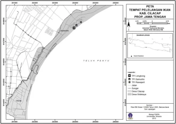
Mackerel tuna fish samples were collected from three fish auctions in Cilacap, Central Java. A total of 65 mackerel tuna fish were collected from three fish auctions in Cilacap Regency, Central Java consist of 20 fish were collected from Lengkong Fish Auction, 25 fish from Samudera Fish Auction, and 20 fish from Rajawarit Fish Auction (Figure 1). The samples collections were done from May until June 2015. Anisakis samples were collected by carried out a necropsy on fish body organ especially abdomen, digestive tract, liver, and muscles. External part observation was done in gill and entire body surface, mainly fin and back. Seven nematodes successfully collected and fixated in RNA buffer later for
morphology and molecular examination at
Biochemistry Laboratory, Faculty of Veterinary Medicine, Gadjah Mada University.
Morpholgy Analysis
Lactophenol solution was drop on Anisakis samples for 24 hours. Then the body length measurement was performed directly, followed by documented using Olympus Bx50 camera with 100x zoom. Body parts
measured were anterior part of cephalic organ and posterior part of caudal organ (Figure 2).
DNA Isolation
Total deoxyribonucleic acid (DNA) from each individual of Anisakis was isolated from body parts using DNA isolation kit (Qiagen) matched with protocol from its manufacture, with combination of RNAase solution treatment. The quality of isolated DNA results were observed using electrophoresis method on 1% agarose gel and buffer 1xTBE on submarine electrophoresis device (Hoefer, USA). Observation was performed using UV-transilluminator with 260 nm wave lengths after provided by gel with bioatlas buffer (Geneaid) functioned as fluorescence. The isolated DNA was then stored at -20 C degree before PCR stage. A total concentration of 50 ng/mL of isolated DNA was used in PCR process.
Primer Design
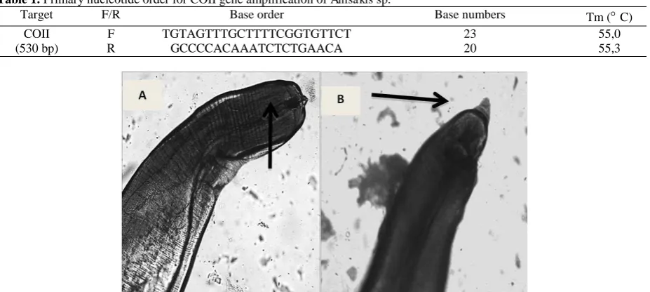
Primer designed was carried out using primer3online program (http://wwwgenome.wi.mit. edu/cgi.bin/primr3.cgi/results_from-primer3) based on mitochondria genome sequence data of Anisakis simplex (access number AB241057.1). Primary base order for COII gene amplification from Anisakis sp. samples was presented in Table 1.
COII Gene Amplification Using PCR Methods In this study, DNA was amplified using PCR machine (Infinigen). DNA amplification was performed in conditions: initial denaturation for 5 minutes at 94 C continued with denaturation at 94 C for 30 seconds, 50 C for primary attachment (annealing), 72 C for 1 minute for elongation; amplification was done for 35 cycles, then post elongation for 5 minutes at 72 C.
PCR products were detected by migrated them on 1% agarosa gel using 1xTBE buffer in submarine electrophoresis device (Hoefer, USA), then observed
using UV-transilluminator with 260 nm wave lengths after gel colored by bioatlas fluorescence (Genaid). DNA marker with the size of 100 bp was used as molecular weight marker. Good PCR result was marked with single band with the nucleotide length is 530 bp.
DNA sequencing
The PCR product was purified using GFX Column purification kit, furthermore it was used as DNA template for DNA sequencing reaction. Each samples was completed two sequencing reactions, using forward primer and reverse primer. The PCR product of COII gene was sequenced. Good sequencing results were marked with single peak spectophotogram results and without noise.
Data analysis
Double alignments of nucleotide sequence of COII gene were analyzed using ClustalW software (Thompson et al., 1994). Results analysis was done based on nucleotide sequencing of COII gen using MEGA 6.0 version program (Kumar, 2001). The genetical length was analyzed with Kimura methods using two parameters (Kumar, 2001). Philogenetic tree was analyzed according to its nucleotide sequence using neighbor joining methods with bootstrap value at 1000x. Comparison data was took from GenBank, there were A. simplex (s.s.) (access number DQ116426), A. pegreffii (access number DQ116428), A. simplex C (access number DQ116429), A. typica (access number
DQ116427), A. ziphidarum (access number
DQ116430), A. physeteris (access number DQ116432), A. brevispiculata (access number DQ116433), A. paggiae (access number DQ116434), and A. nascettii (access number DQ116431).
RESULTS AND DISCUSSION
The observational results of 65 mackerel tuna fishes (Euthynnus affinis) found Anisakis larvae as shown on
Table 1. Primary nucleotide order for COII gene amplification of Anisakis sp.
Target F/R Base order Base numbers Tm (C)
COII (530 bp)
F R
TGTAGTTTGCTTTTCGGTGTTCT GCCCCACAAATCTCTGAACA
23 20
55,0 55,3
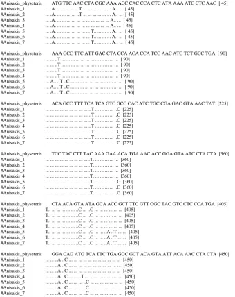
Figure 2. The morphology characterization such as cephalic and caudal length showed positive results of Anisakis sp. (Figure 2).
Mattiucci et al. (2011) and Sohn et al. (2014) stated that stage III Anisakis larvae (L3) has special
feature called drilling tooth “boring tooth” in its anterior tip for perforated and hanged on intestine wall. Morphology identification results revealed that in Anisakis sp. found on mackerel fish shows sharpened area on its anterior and posterior part.
Figure 3. PCR products of Anisakis sp. COII gene on 1% agarose gel. 1Kb= DNA ladder 1000 bp, 1-7= PCR products of Anisakis samples
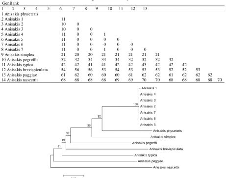
#Anisakis_physeteris ATG TTC AAC CTA CGC AAA ACC CAC CCA CTC ATA AAA ATC CTC AAC [ 45] #Anisakis_1 ... .A. ... ... ... ... ..T ... ... ... ... ... ... A.. ... [ 45]
#Anisakis_2 ... .A. ... ... ... ... ..T ... ... ... ... ... ... A.. ... [ 45] #Anisakis_3 ... .A. ... ... ... ... ... ... ... ... ... ... ... A.. ... [ 45] #Anisakis_4 ... .A. ... ... ... ... ... ... ... ... ... ... ... A.. ... [ 45] #Anisakis_5 ... .A. ... ... ... ... ... ... ... T.. ... ... ... A.. ... [ 45] #Anisakis_6 ... .A. ... ... ... ... ... ... ... T.. ... ... ... A.. ... [ 45] #Anisakis_7 ... .A. ... ... ... ... ... ... ... T.. ... ... ... A.. ... [ 45]
#Anisakis_physeteris AAA GCC TTC ATT GAC CTA CCA ACA CCA TCC AAC ATC TCT GCC TGA [ 90] #Anisakis_1 ... ... ..T ... ... ... ... ... ... ... ... ... ... ... ... [ 90]
#Anisakis_2 ... ... ..T ... ... ... ... ... ... ... ... ... ... ... ... [ 90] #Anisakis_3 ... ... ..T ... ... ... ... ... ... ... ... ... ... ... ... [ 90] #Anisakis_4 ... ... ..T ... ... ... ... ... ... ... ... ... ... ... ... [ 90] #Anisakis_5 ... A.. ..T ..C ... ... ... ... ... ... ... ... ... ... ... [ 90] #Anisakis_6 ... A.. ..T ..C ... ... ... ... ... ... ... ... ... ... ... [ 90] #Anisakis_7 ... A.. ..T ..C ... ... ... ... ... ... ... ... ... ... ... [ 90]
#Anisakis_physeteris ACA GCC TTT TCA TCA GTC GCC CAC ATC TGC CGA GAC GTA AAC TAT [225] #Anisakis_1 ... ... ... ... ... ... ... ... ... ..T ... ... ... ... ..C [225]
#Anisakis_2 ... ... ... ... ... ... ... ... ... ..T ... ... ... ... ..C [225] #Anisakis_3 ... ... ... ... ... ... ... ... ... ..T ... ... ... ... ..C [225] #Anisakis_4 ... ... ... ... ... ... ... ... ... ..T ... ... ... ... ..C [225] #Anisakis_5 ... ... ... ... ... ... ... ... ... ..T ... ... ... ... ..C [225] #Anisakis_6 ... ... ... ... ... ... ... ... ... ..T ... ... ... ... ..C [225] #Anisakis_7 ... ... ... ... ... ... ... ... ... ..T ... ... ... ... ..C [225]
#Anisakis_physeteris TCC TAC CTT TAC AAA GAA ACA TGA AAC ACC GGA GTA ATC CTA CTA [360] #Anisakis_1 ... ... ... ... ... ... ... ... ... .T. ... ... ... ... ... [360]
#Anisakis_2 ... ... ... ... ... ... ... ... ... .T. ... ... ... ... ... [360] #Anisakis_3 ... ... ... ... ... ... ... ... ... .T. ... ... ... ... ... [360] #Anisakis_4 ... ... ... ... ... ... ... ... ... .T. ... ... ... ... ... [360] #Anisakis_5 ... ... ... ... ... ... ... ... ... .T. ... ... ... ... ..G [360] #Anisakis_6 ... ... ... ... ... ... ... ... ... .T. ... ... ... ... ..G [360] #Anisakis_7 ... ... ... ... ... ... ... ... ... .T. ... ... ... ... ..G [360]
#Anisakis_physeteris CTA ACA GTA ATA GCA ACC GCT TTC GTT GGC TAC GTC CTC CCA TGA [405] #Anisakis_1 T.. ... ... ... ... ... ..C ... ..C ... ... ... ... ... ... [405]
#Anisakis_2 T.. ... ... ... ... ... ..C ... ..C ... ... ... ... ... ... [405] #Anisakis_3 T.. ... ... ... ... ... ..C ... ..C ... ... ... ... ... ... [405] #Anisakis_4 T.. ... ... ... ... ... ..C ... ..C ... ... ... ... ... ... [405] #Anisakis_5 T.. ... ... ... ... ... ..C ... ..C ... ... ..A ..T ... ... [405] #Anisakis_6 T.. ... ... ... ... ... ..C ... ..C ... ... ..A ..T ... ... [405] #Anisakis_7 T.. ... ... ... ... ... ..C ... ..C ... ... ..A ..T ... ... [405]
#Anisakis_physeteris GGA CAG ATG TCA TTC TGA GGC GCT ACA GTA ATT ACA AAC CTA CTA [450] #Anisakis_1 ... ... ..A ..C ... ... ... ... ... ... ... ... ... ... ... [450]
#Anisakis_2 ... ... ..A ..C ... ... ... ... ... ... ... ... ... ... ... [450] #Anisakis_3 ... ... ..A ..C ... ... ... ... ... ... ... ... ... ... ... [450] #Anisakis_4 ... ... ..A ..C ... ... ..T ... ... ... ... ... ... ... ... [450] #Anisakis_5 ... ... ..A ..C ... ... ... ..C ... ... ... ... ... ... ... [450] #Anisakis_6 ... ... ..A ..C ... ... ... ..C ... ... ... ... ... ... ... [450] #Anisakis_7 ... ... ..A ..C ... ... ... ..C ... ... ... ... ... ... ... [450]
Boring tooth is observed in anterior part but there is no excretory pore found, whereas in its posterior part there is no mucron but anus is observed.
Based on morphology analysis, Anisakis sp. found in mackerel fish classified as type II L3 Anisakis larvae since there was no mucron at its posterior tip. This result similar with Widjanarko (2015) who discovered Anisakis larvae in karo and mackerel tuna fish which were classified as type II L3 Anisakis larvae. Based on Mattiucci and Nascetti (2009), type II L3 Anisakis nematodes included A. paggiae, A. physeteris, and A. brevispiculata (Mattiucci and Nascetti, 2008; Mattiucci et al., 2009).
A total of 7 isolated DNA samples were amplified for the COII genes by PCR technique which producing amplicons of 540 bp. Subsequently, the PCR products underwent electrophoresis in 1% agarose gel with 1 kb DNA ladder (1st base) (Figure 3). The 540 bp PCR products was obtained after the primer utilized in the PCR was aligned to mitochondiral genome sequence of Anisakis simplex with the access number of (AB241057.1) using BLAST software from the website http://www.ncbi. nlm.nih.gov/tools/primer-blast/index. cgi?LINK_LOC=BlastHome.
The alignment result of the 530 nucleotides of Anisakis showed 11 different sites, which function as
genetic barcode (Figure 4). The 11 nucleotide sites that could be utilized as barcode for Indonesian Anisakis sp. are on the 5th site (A/T), 40th site (A/C), 54th site (T/C), 210th site (T/C), 225th site (C/T), 344th site (T/C), 361st site (T/C), 381st site (C/T), 387th site (C/T), 414th site (A/G), an 417th site (C/A). Mattiucci et al., (2009); Santoro et al. (2010), described that mtDNA COII gene could be used as barcode to distinguish Anisakis at species level. Valentini et al. (2006); Santoro et al. (2010) stated that mtDNA COII gene nucleotides are highly polymorphic among the different Anisakis species. Santoro et al. (2010) reported that COII gene could be function as barcode to identify A. pegreffii that infected barrel-shaped turtles (Celonia mydas), fishes, and cetacean organisms in the Mediterranean Sea.
Table 2 presents the difference of nucleotides number between the Anisakis sample in the research and the species from GenBank. It showed that the difference ranged from zero (0) to 1 nucleotide (nt) among the Anisakis samples in the research and up to 11 when compared to A. physeteris from GenBank.
There was no significant difference among the COII gene sequence and the Anisakis sp. in the research, showing that there is no significant diversity among the samples. There is 1 nucleotide difference between
Table 2. Matrix of the difference between COII gene nucleotide sequence of Anisakis in the research and the Anisakis from GenBank
1 2 3 4 5 6 7 8 9 10 11 12 13 1 Anisakis physeteris
2 Anisakis 1 11
3 Anisakis 2 10 0
4 Anisakis 3 10 0 0
5 Anisakis 4 11 0 0 1
6 Anisakis 5 11 0 0 0 0
7 Anisakis 6 11 0 0 0 0 0
8 Anisakis 7 11 0 0 1 0 0 0
9 Anisakis simplex 21 20 20 21 21 21 21 21
10 Anisakis pegreffii 32 32 34 33 34 32 32 32 32
11 Anisakis typica 42 42 41 41 42 42 43 42 42 42
12 Anisakis brevispiculata 54 56 56 53 54 53 53 53 52 52 53
13 Anisakis paggiae 61 62 60 60 60 61 62 62 61 62 62 62
14 Anisakis nascettii 68 68 68 68 69 69 70 70 68 68 68 68 70
Anisakis 1 Anisakis 4 Anisakis 3 Anisakis 2 Anisakis 7 Anisakis 6 Anisakis 5
Anisakis physeteris Anisakis simplex Anisakis pegreffii
Anisakis brevispiculata Anisakis typica
Anisakis paggiae Anisakis nascettii
Anisakis sample no 3 and 4, as well as Anisakis sample no 4 and 7, while there was no differences (0 nt) among other samples.
It could be concluded that the Anisakis samples found in this research are from the same species because there are no significant nucleotide difference. The diversity percentage of COII gene nucleotide sequence between the Anisakis in the research compared to species from GenBank is presented as phylogenetic tree diagram using neighbor joining method with a bootstrap value of 1000 times. Figure 5 shows the Anisakis phylogenetic tree based on the nucleotide sequence of COII gene.
Figure 5 showed that all of the Anisakis samples studied creates a branch, and closely related to A. physeteris (100%) which have been saved in the GenBank database (Valentini et al., 2006; Mattiucci et al., 2009). This molecular data is consistent and support the previous study by Widjanarko (2015) on the characterization of Anisakis sp. along the coastal port of Sedang, Gunung Kidul, Yogyakarta, based on their morphology characteristics. This research proves that the Anisakis nematodes which infected tuna fishes included type II L3 Anisakis from A. physeteris.
Previously, there was no information on infection from type II L3 Anisakis in Indonesian waters. Mattiucci and Nascetti (2009) also found no type II L3 Anisakis in Asian waters but found some in the Middle East waters, southern ocean of the African continent and South Pacific. The prevalence of individual Anisakis found in this research was 7 nematodes out of 65 tuna fishes studied.
This result indicates that the distribution of type II L3 Anisakis larvae in Java Sea is low. Mattiucci and Nascetti (2008), Mattiucci et al. (2009), and Widjanarko (2015), stated that the low intensity and distribution of type II L3 Anisakis larvae could be influenced by the presence of end phase hosts from the ocean mammals. Gutiérrez-Galindo et al. (2010) reported the infection by type II L3 Anisakis sp. in Micromesistius poutassou and Trachurus trachurus fish in a low prevalence along the Alboran and Catalan waters. Piras et al. (2014) also reported that there was A. physeteris nematode infections in different prevalence in some fish species collected from north Sardinia (North West Mediterranean Sea), with high prevalence among Micromesistius poutassou and M. merluccius, and low prevalence among P. blennoides, S. pilchardus, Sphyraena viridensis, Trachurus mediterraneus, T. Trachurus, and S. pilchardus.
Mattiucci and Nascetti (2008) and Mattiucci et al. (2009) reported that the only end phase host of Type II L3 Anisakis was the sperm whale (Physeteris macrocephalus). In general, sperm whales migrate during mating or birth seasons (Fossette et al., 2014). The presence of Anisakis nematode in marine environment is influenced by several factors such as hosts, nature condition, and the feeding habit of the infected fishes. In general, tuna fishes are sea predators that consume crustaceans, small pelagic fishes, and cephalopods. Widjanarko (2015) studied on tuna fishes
in Gunung Kidul, Yogyakarta, found many large sized crustaceans in the digestive tract of tuna. Piras et al. (2014) and Pozio (2013) stated that Anisakis life cycle includes small crustaceans-planctivore-piscivore-sea mammals-large crustaceans was rarely involved in Anisakis nematode life cycle. Pozio (2013) added that the low prevalence of Anisakis nematode infection in certain fish species could be affected by the number of infected crustaceans.
CONCLUSION
The Anisakis sp. infecting tuna fishes in the three fish markets in Cilacap District, Central Java, was type II L3 Anisakis larvae of A. physeteris (100%) and one of zoonotic organism. There were 32 nucleotide sites in COII mtDNA that could be utilized as genetic barcode for A. physeteris from Indonesia (Java Sea) compared with the A. physeteris from the database (GenBank).
ACKNOWLEDGEMENTS
The author would like to thank LPPM-UT through Hibah Penelitian Lanjut Bidang Ilmu Tahun 2015, 08 April 2015, for funding this research.
REFERENCES
Abe, N., K. Tominaga, and I. Kimata. 2006. Usefulness of PCR-restriction fragment length polymorphism analysis of the internal transcribed spacer region of rDNA for identification of Anisakis simplex complex. Jpn. J. Infect. Dis. 59:60-62.
Arizono, N., M. Yamada, T. Tegoshi, and M. Yoshikawa. 2012.
Anisakis simplex sensu stricto and Anisakis pegreffii: Biological characteristics and pathogenetic potential in human anisakiasis.
Foodborne Pathog. Dis. 9:517-521.
Audicana, T.M., I.J. Ansotegui, L.F. De Corres, and M.W. Kennedy. 2002. Anisakis simplex: Dangerous-dead and alive? Trends
Parasitol. 18:20-25.
Audicana, M.T. and M.W. Kennedy. 2008. Anisakis simplex: From obscure infectious worm to inducer of immune hypersensitivity.
Clin. Microbiol. Rev. 21:360-379.
Berland, B. 1961. Nematodes from some Norwegian marine fishes.
Sarsia. 2:1-50.
Cavallero, S., S.A. Nadler, L. Paggi, N.B. Barros, and S. D’Amelio. 2011. Molecular characterization and phylogeny of anisakid nematodes from cetaceans from south-eastern Atlantic coasts of USA, Gulf of Mexico, and Caribbean Sea. Parasitol. Res. 108:781-792.
Cavallero, S., Ligas, A., Bruschi, F., Smelio, S.D. 2012. Molecular identification of Anisakis spp. from fishes collected in the Tyrrhenian Sea (NW Mediterranean). Vet. Parasitol. 187:563-566.
Chai, J.Y., K.D. Murrell, A.J. Lymbery. 2005. Fish-borne parasitic zoonoses: Status and issues. Int. J. Parasitol. 35:1233-1254. Daulay. 2003. Prevalensi Anisakis pada Delapan Jenis Ikan Laut TPI
Daerah Istimewa Yogyakarta. Thesis. Pasca Sarjana Universitas Gadjah Mada. Yogyakarta.
D’Amelio, S., K.D. Mathiopoulos, C.P. Santos, O.N. Pugachev, S.C.
Webb, M. Picanco, and L. Paggi. 2000. Genetic markers in ribosomal DNA for the identification of members of the genus Anisakis (Nematoda: Ascaridoidea) defined by polymerase-chain-reaction-based restriction fragment length polymorphism.
Int. J. Parasitol. 30:223-226.
D’Amelio, S., S. Cavallero, N.O. Dronen, N.B. Barros, and L. Paggi.
Fossette, S., M.H. Jorgensen, M.V. Jensen, J. Kiszka, M. Berube, N. Bertrand, and M. Vely. 2014. Humpback whale (Megaptera novaeangliae) post breeding dispersal and southward migration in the western Indian ocean. J. Exper. Marine Biol. Eco. 450:6-14. Gutiérrez-Galindo, J.F., A.C. Osanz-Mur, and M.T. Mora-Ventura.
2010. Occurrence and infection dynamics of anisakid larvae in
Scomber scombrus, Trachurus trachurus, Sardina pilchardus, and Engraulis encrasicolus from Tarragona (NE Spain). Food
Control. 21:1550-1555.
Hutomo, M., Burhanuddin, and P. Hadidjaja. 1978. Observations on the incidence and intensity of infection of nematode larvae (famili Anisakidae) in certain marine fishes of waters around Panggang Island, Seribu Islands. Mar. Res. Indonesia. 21:49-60. Indaryanto, F.R., Y. Wardiatno, and R. Tiuria. 2015. Inventarisasi Identification of Anisakis species (Nematoda: Anisakidae) in marine fish hosts from Papua New Guinea. Vet. Parasitol. 193:126-133.
Kumar, S., K. Tamura, J. Dudley, and M. Nei. 2001. Molecular evolutionary genetics analysis (MEGA) software version 6.0.
Mol. Biol. Evol. 24:1596-1599.
Mattiucci, S. and G. Nascetti. 2008. Advances and trends in the molecular systematics of anisakid nematodes, with implications for their evolutionary ecology and host-parasite co-evolutionary processes. Adv. Parasitol. 66:47-148.
Mattiucci, S., M. Paoletti, and S.C. Webb. 2009. Anisakis nascettii. sp. (Nematoda: Anisakidae) from beaked whales of the southern hemisphere: Morphological description, genetic relationships between congeners and ecological data. Syst. Parasitol. 74:199-217. Mattiucci, S., M. Paoletti, F. Borrini, M. Palumbo, R.P. Macarone, V.
Gomes, A. Casati, and G. Nascetti. 2011. First molecular identification of the zoonotic parasite Anisakis pegreffii
(Nematoda: Anisakidae) in a paraffin-embedded granuloma taken from a case of human intestinal anisakiasis in Italy. BMC
Inf. Disease. Doi:10.1186/1471-2334-11-82.
Nadler, S.A., S. D’Amelio, M.D. Dailey, L. Paggi, S. Siu, and J.A.
Sakanari. 2005. Molecular phylogenetics and diagnosis of Anisakis, Pseudoterranova, and Contracaecum from northern Pacific marine mammals. J. Parasitol. 91:1413-1429.
Piras, M,C., T. Tedde, G. Garippa, S. Virgilio, D. Sanna, S. Farjallah, and P Merella. 2014. Molecular and epidemiological data on
Anisakis spp. (Nematoda: Anisakidae) in commercial fish caught off northern Sardinia (western Mediterranean Sea). Vet.
Parasitol. 203:237-240.
Pontes, T., S. D’Amelio, G. Costa, and L. Paggi. 2005. Molecular
characterization of larval anisakid nematodes from marine fishes of Madeira by a PCR-based approach, with evidence for a new Galiero, and G. Nascetti. 2010. Molecular identification and pathology of Anisakis pegreffii (Nematoda: Anisakidae) infection in the Mediterranean loggerhead sea turtle (Caretta caretta). Vet.
Parasitol. 174:65-71.
Setyobudi, E., S. Helmiati, and Soeparno. 2007. Infeksi Anisakis sp. pada layur (Trichiurus sp.) di Pantai Selatan Kabupaten Purworejo. J. Perikanan. 9(1):142-148.
Setyobudi, E., C.H. Jeon, C.H. Lee, K.B. Seong, and J.H. Kim. 2011. Occurrence and identification of Anisakis spp. (Nematoda: Anisakidae) isolated from chum salmon (Oncorhynchus keta) in Korea. Parasitol. Res. 108:585-592.
Siagian, F.E., G.T.Y. Siagian, and A.O. Siagian. 2010. Kelainan yang berhubungan dengan larva Anisakis sp. Majalah Ked. FK. UKI. 27(3):122-129.
Sohn, W.M., J.M Kang, and B.K. Na. 2014. Molecular Analysis of Anisakis type I larvae in marine fish from three different sea areas in Korea. Korean J. Parasitol. 4:383-389.
Suadi, S. Helmiati, and R. Widaningroem. 2007. Population and parasite parameter in hairtail (Trichiurus spp.) landed at PPS Cilacap. J. Fish. Sci. 9(2):226-232.
Thompson, J.D., D.G. Higgins, and T.J. Gibson. 1994. CLUSTAL W: Improving the sensitivity of progressive multiple sequence alignment through sequence weighting, Position-specific gap penalties and weight matrix choice. Nucleic Acid Res. 22:4673-4680.
Umehara, A., Y. Kawakami, T. Matsui, J. Araki, and A. Uchida. 2006. Molecular identification of Anisakis simplex sensu stricto and Anisakis pegreffii (Nematoda: Anisakidae) from fish and cetacean in Japanese waters. Parasitol. Int. 55:267-271. Utami, P. 2013. Identifikasi Anisakis sp. pada beberapa ikan laut di
beberapa tempat pelelangan ikan (TPI) Cilacap. J. Matematika
Sains dan Teknologi. 15(1):21-28.
Valentini, A., S. Mattiucci, P. Bondanelli, S.C. Webb, A.A. Mignucci-Giannone, M.M. Colom-Llavina, and G. Nascetti. 2006. Genetic relationships among Anisakis species (Nematoda: Anisakidae) inferred from mitochondrial cox2 sequences, and comparison with allozyme data. J. Parasitol. 92:156-166. Widjanarko, A. P. 2015. Identifikasi, Prevalensi dan Intensitas
Nematoda Anisakis pada Beberapa Ikan Hasil Tangkapan di Pelabuhan Perikanan Pantai Sedang, Kabupaten Gunung Kidul.
Thesis. Fakultas Perikanan. Universitas Gadjah Mada.
Yogyakarta.
Yman, L. 2003. Spesifik IgE in the diagnosis of parasite-induced allergy. Allergy. 59:14-17.
Zhu, X., R.B. Gasser, M. Podolska, and N.B. Chilton. 1998. Characterisation of anisakid nematodes with zoonotic potential by nuclear ribosomal DNA sequences. Int. J. Parasitol.
28:1911-1921.