R E S E A R C H A R T I C L E
Modulation of Caspase-3 Expression by
Arcangelisia flava
Post Acetaminophen-Induced Hepatotoxicity in Rat’s Liver
Steffi Liem
1, Tina Rostinawati
2, Ronny Lesmana
3, Sri Adi Sumiwi
1, Tiana Milanda
2, Mutakin
4,
Irma Meylani Puspitasari
1, Jutti Levita
1,1Department of Pharmacology and Clinical Pharmacy, Faculty of Pharmacy, Universitas Padjadjaran, Jl. Raya Bandung Sumedang Km. 21, Jatinangor 45363, Indonesia
2Department of Biology Pharmacy, Faculty of Pharmacy, Universitas Padjadjaran, Jl. Raya Bandung Sumedang Km. 21, Jatinangor 45363, Indonesia
3Department of Pharmacology and Therapy, Faculty of Medicine, Universitas Padjadjaran, Jl. Raya Bandung Sumedang Km. 21, Jatinangor 45363, Indonesia
4Department of Pharmaceutical Analysis and Medicinal Chemistry, Faculty of Pharmacy, Universitas Padjadjaran, Jl. Raya Bandung Sumedang Km. 21, Jatinangor 45363, Indonesia
Corresponding author. E-mail: [email protected]
Received date: Dec 12, 2017; Revised date: Apr 4, 2018; Accepted date: Apr 10, 2018
B
ACKGROUND: Acetaminophen, when usedat low doses is a safe drug, but at higher doses it induces apoptosis in hepatoma cells. Arcangelisia
flava that grows widely in Kalimantan Island, Indonesia, contains berberine which is effective in protecting the liver. This work was aimed to study the effect of A. flava extract on the modulation of caspase-3 in acetaminophen-induced hepatotoxicity.
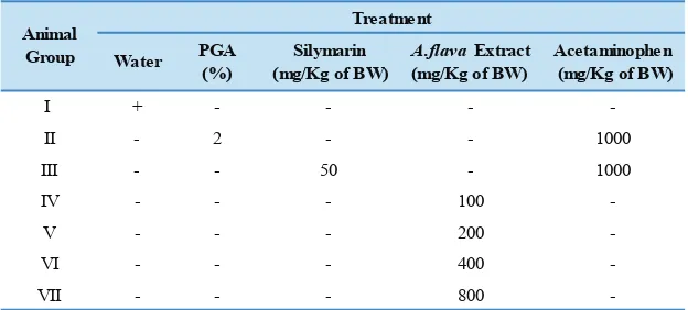
METHODS: Thirty-five Wistar male rats were divided into groups: I the normal control (water); II the negative control (Arabic gum powder or PGA, 2% in suspension); III the positive control (silymarin); IV-VII (A. flava extract 100, 200, 400, and 800 mg/Kg of body weight (BW), respectively) for 14 days. At day 15th, group II-VII were induced with
acetaminophen 1000 mg/Kg of BW per oral for 7 days along with the extracts. At day 22nd, the animals were
measured for their serum glutamic oxaloacetic transaminase (SGOT), serum glutamic pyruvic transaminase (SGPT) and gamma-glutamyl transferase (GGT), histological examination, and Western blotting.
RESULTS: Acetaminophen elevated the SGOT and SGPT (3x compared to normal group), and GGT (5x compared to normal group) of the animals in group II. Pre-treatment with higher doses of A. flava extract (group VI and VII) significantly prevented the biochemical changes induced by acetaminophen. Normal histology of the liver was showed by group I, III, VII, whereas dilated sinusoids, central vein (CV) lesion, and local haemorrage were observed in group II, IV, V and VI. Western blotting showed an inhibition of caspase-3 expression by A. flava extract in dose-dependent manner.
CONCLUSION: Higher dose A. flava extract shows hepatoprotective activity by preventing the elevation of serum transaminases and transferase levels. Eventually, no damage in the acetaminophen-induced rat’s liver was observed. This plant modulates the expression of caspase 3 protein in dose-dependent manner.
KEywORDS: Arcangelisia sp, caspase-3, berberine, hepatoprotective activity, NSAIDs, yellow root
Indones Biomed J. 2018; 10(2): 148-55
Abstract
Introduction
Acetaminophen is widely used in Indonesia since this drug is sold freely in pharmacies and drug stores. When
protein adducts and induces mitochondrial oxidative stress.(1) Cytochrome P450 family 2 subfamily D member 6 (CYP2D6) contributed significantly to the formation of N-acetyl-p-benzoquinone imine (NAPQI), especially at toxic doses of acetaminophen when its plasma concentrations reached 2 mM or more.(2) Both at toxic concentration and therapeutic concentration, NAPQI-glutathione was mostly formed by cytochrome P450 family 3 subfamily A member 4 (CYP3A4).(3) Very high doses of acetaminophen (e.g., 600 mg/Kg) can induce the
mitochondrial permeability transition (MPT) independent of the cyclophilin D (CypD)-regulated MPT pore as a result of extreme oxidative stress.(4) Acetaminophen induce the apoptosis in liver cells and lymphocytes, which is caused by the ectopic expression of human caspase-3 and furthermore promotes the release of cytochrome C into the cytosol of treated cells.(5) The activation of caspase-3 might be characteristic of acetaminophen toxicity in fed mice and was not observed in starved animals due to the lower adenosine triphosphate (ATP) levels, which prevented caspase activation.(6,7)
Arcangelisia flava, that grows widely in Kalimantan Island, Indonesia, is a native plant of South East Asia. This plant has been empirically proven to cure hepatitis in Kalimantan and Sulawesi. Preliminary qualitative tests on wood mill of the stem part and root of this plant detected saponins, flavonoid, and alkaloids in high concentrations (8), while further investigations of this plant had confirmed the presence of palmatine, berberine, jatrorrhizine, dihydroberberine and 20-hydroxyecdysone. (9-11) Berberine prevents inflammation, degeneration and death of liver cells in doxorubicin-induced mice.(12) This compound is effective in protecting the liver from acute CCl4-induced injury. The hepatoprotective mechanisms of berberine may be related to the free radical scavenging and attenuation of oxidative/nitrosative stress, as well as to the inhibition of inflammatory response in the liver.(13,14)
The pharmacological effects of A. gusanlung, a plant
of the same genus, as well as its chemical constituents, had been explored. This plant showed weak cytotoxicity against the SGC 7901 human stomach cancer cell line with an inhibitory concentration (IC)50 value of 85.1 µM (15), whereas A. flava leaves had been proven to exert cytotoxic activity on HeLa, MCF-7 breast cancer cell line, WiDr colon cancer cell line and Vero cell line with the IC50 value of 467±70; 136±17; 213±79, and 1340±288 µg/mL respectively. The activity was selective on MCF-7 and WiDr, but unlikely on HeLa and Vero cell lines.(16) In this work, we investigated the hepato-protective effect
Methods
Materials
A. flava was purchased from Research Institute for Spices and Medicinal Plants (Balittro) Cikampek, Indonesia. The plant was taxonomically identified at Laboratory of Plant Taxonomy, Department of Biology, Faculty of Mathematics and Natural Sciences, Universitas Padjadjaran, Jatinangor, Indonesia, and the voucher specimen (No. 189/ HB/05/2017) was retained in our laboratory for future reference. Silymarin and acetaminophen were obtained from Kimia Farma Bandung, Indonesia. Evogen reagent kits for SGOT, SGPT and GGT from PT Rasani Karya Mandiri, Tangerang, Indonesia, were used.
Animals
Thirty-five healthy male Wistar rats aged 18-20 weeks (200-250 gram of body weight (BW)), were purchased from D’Wistar Experimental Animal Supplier, Jawa Barat, Indonesia. Before starting this study, the protocol was approved by Universitas Padjadjaran Health Research Ethics Committee (No. 1170/UN6.C.10/PN/2017). The rats were acclimatized for 7 days, housed in cages (five animals per cage), and maintained under standard laboratory conditions with dark and light cycles (12/12 hours) according to the ethical standards. Standard feed and drink were given ad libitum. No vitamins were added in their feed.
Extraction of A. flava
Extraction of A. flava stem was performed according to a previous study with a few modifications.(17) One Kg of dry
A. flava stem was ground, weighed, macerated in ethanol 70% for 24 hours, filtered and dried under vacuum. The solvent was evaporated and the residue was re-macerated for 2x24 hours. The yield of extract was found 10.66% to dried stem.
Phytochemical Screening of A. flava Extract
Phytochemical screening was carried out as per the standard methods described by Tiwari and colleagues at the Central Laboratory, Universitas Padjadjaran, Jatinangor, Indonesia.(18)
Experimental Design for Baseline Clinical Biochemistry Parameters
At day 0th, the animals were fasted for 16 hours, and evaluated
for their biochemical parameters (SGOT, SGPT, GGT) using 3 mL of orbital vein blood to obtain the baseline value. Serum was separated by centrifugation at 5,000 rpm for 10 minutes and utilized for the estimation of the biochemical parameters. The animals were then acclimatized for 7 days. At day 8th, the animals were divided into seven groups, five
in each group (19) as shown in Table 1.
Liver/Body weight Ratio, Biochemical Analysis, and Qualitative Histological Study
At day 21st, the animals were re-evaluated for their
clinical biochemistry parameters (SGOT, SGPT, GGT), then they were anaesthetized using overdosage of solid CO2 and sacrificed. The liver was immediately dissected out, weighed, and transferred into Bouin solution for histological examination. The organs were then stained using hematoxyline and eosin, and the histological changes were observed under light-microscope and relevant findings were recorded on 40x magnification. This step was carried out at Laboratory of Animal Biosystem, Department of Biology, Faculty of Mathematics and Natural Science, Universitas Padjadjaran, Jatinangor, Indonesia.
western Blot Analysis
The fresh liver samples were selected randomly from each group and homogenized in radio immunoprecipitation assay buffer. Twenty-five µg of each liver protein were extracted using lysis buffer (0.5% SDS, 0.05 M Tris-HCl, adjust pH to 8.0) and sample buffer (1 M Tris-HCl pH 6.8, 50% gliserol, β-mercaptoethanol, 1% bromophenol blue and distilled water). The samples were heated at 95oC for
5 minutes, loaded into 15% polyacrylamide gel, and run at 150 V for 2 hours. The resolved proteins were transferred
Table 1. Animal groups and the treatment for hepatotoxicity study.
to a polyvinylidene fluoride membrane using 200 mA for 30 minutes. The membranes were then incubated with goat anti-Caspase-3 antibody (1:1000) (Cat. #AAA65015) (R&D System, Minneapolis, USA) and mouse anti-GAPDH antibody (1:1000) (Cat. #AM4300) (Thermo Fisher Scientific Fremont, California, USA). After treatment with the primary antibodies, the membranes were washed and then incubated with secondary antibodies, horseradish peroxidase (HRP)-conjugated chicken anti-goat antibody (1:10000) (Cat. #sc-2953) (Santa Cruz Biotechnology Inc., California, USA) and HRP-conjugated chicken anti-mouse antibody (1:10000) (Cat. #sc-2954) (Santa Cruz Biotechnology Inc.). Each band was visualized by enhanced chemiluminescence (ECL) substrates (Cat. #926-95000) (Li-Cor Biosciences, Lincoln, USA) and the intensity of the band was normalized and measured using Image-J software. This step was performed at the Laboratory of Molecular Physiology, Division of Gene and Protein Analysis, Central Laboratory, Universitas Padjadjaran, Jatinangor, Indonesia.
Statistical Analysis
The obtained data were expressed as mean±SD and the differences between multiple groups were calculated by a one-way analysis of variances (ANOVA) with p-value of
0.05, followed by a nonparametric post hoc Duncan test. The statistical calculations were performed using SPSS for Windows version 16.0 (SPSS Inc., Chicago, USA).
Results
Phytochemical Screening
Animal group N Ratio of liver/body
weight (%) p value
I (normal) 5 3.46±0.14
II (negative) 5 4.65±0.17* 0.000
III (positive) 5 3.45±0.10 0.875
IV (100 mg/Kg of BW) 5 4.47±0.11* 0.000
V (200 mg/Kg of BW) 5 3.68±0.19* 0.030
VI (400 mg/Kg of BW) 5 3.46±0.10 0.294
VII (800 mg/Kg of BW) 5 3.55±0.11 0.875
*indicates significant difference compared to normal group (p<0.05)
Animal group N SGOT p value
I (normal) 5 87.12±7.16
-II (negative) 5 274.58±10.86* 0.000
III (positive) 5 92.32±16.58 0.868
V (200 mg/Kg of BW) 5 147.78±11.45* 0.000
VI (400 mg/Kg of BW) 5 116.46±14.66* 0.000
VII (800 mg/Kg of BW) 5 110.44±21.18 0.098
Animal group N SGPT p value
I (normal) 5 71.80±8.38
-II (negative) 5 230.76±10.19* 0.000
III (positive) 5 84.12±21.23* 0.001
V (200 mg/Kg of BW) 5 123.42±26.19* 0.000
VI (400 mg/Kg of BW) 5 107.75±23.66* 0.000
VII (800 mg/Kg of BW) 5 108.60±10.51* 0.000
Animal group N GGT p value
I (normal) 5 29.54±13.70
-II (negative) 5 174.94±51.07* 0.000
III (positive) 5 49.98±14.82* 0.008
V (200 mg/Kg of BW) 5 82.00±18.06* 0.000
VI (400 mg/Kg of BW) 5 53.74±9.45* 0.000
VII (800 mg/Kg of BW) 5 40.18±23.37 0.351
*indicates significant difference compared to normal group (p<0.05) Table 2. The influence of A. flavastem extract on the ratio of
liver/body weight of the rats.
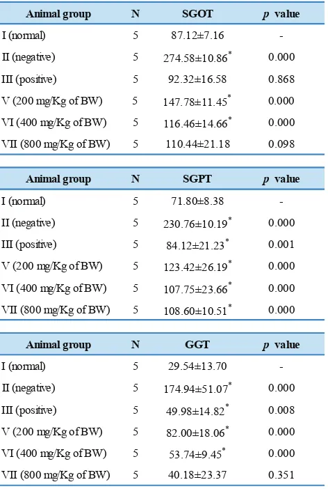
Table 3. The influence of A. flava stem extract on SGOT, SGPT and GGT levels of acetaminophen-induced rats.
Ratio of Liver/Body weight and Biochemical Analysis Table 2 shows the influence of A. flava stem extract on liver/body weight ratio of the acetaminophen-induced rats.
A. flava stem extract improved the liver/body weight ratio of the liver-injured animals.
Liver disease is the most important cause of increased SGPT activity and a common cause of increased SGOT activity. Several factors other than liver disease must be considered in interpreting SGOT and SGPT activities. In most types of liver disease, SGPT activity is higher than that of SGOT. The average SGOT in the blood serum before treatment was 93.66 U/L (normal range 63-175 U/L), whilst the average SGPT level was 55.89 U/L (normal range 19-48 U/L).(20)
Data in Table 3 shows that acetaminophen administration resulted in the elevation of SGOT (3x compared to normal group), SGPT (3x compared to normal group) and GGT (5x compared to normal group) levels of the animals in group II which was treated only with PGA 2%. Pre-treatment with silymarin (group III) and higher doses of A. flava stem extract (group VI and VII) significantly prevented the biochemical changes induced by acetaminophen.
Our results were compared to the previous work on saponins of A. flava. Saponins were able to inhibit 58% of the increasing activity for SGOT and 52% for SGPT after 14 days of saponins administration. This is an indication that saponins of A. flava might have hepatoprotective activity.(8) Influence of A. flava stem extract on SGOT, SGPT and GGT of acetaminophen-induced rats is shown in Figure 1.
Qualitative Histological Study
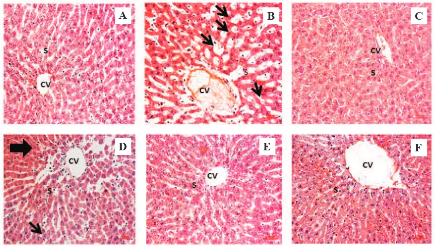
Our study clearly indicated that acetaminophen caused inflammation (Figure 2). Normal histology of the liver defines clear shape of the central vein (CV) and sinusoids, as showed in Figure 2A. In the acetaminophen-treated group, Figure 2B, severe lesion was observed occurs at the CV, the surrounding sinusoids were dilated, Kupffer cells migrated into the CV which indicates inflammation. These liver damages were also observed in group V Figure 2D and group VI Figure 2E, followed by a local haemorrhage (bold arrow) and statosis (thin arrow). Increasing doses of A. flava
were proven could improve the condition of the liver in group VII Figure 2F similarly as was showed in positive control group Figure 2C and normal control Figure 2A.
western Blot Analysis
Figure 1. Influence of A. flava stem extract on SGOT, SGPT and GGT of acetaminophen-induced rats. *indicates significant difference compared to normal group (p<0.05).
A
B
C
D
E
F
Figure 2. Effect of A. flava extract on histological examination of the acetaminophen-induced rat’s liver. A: normal control group; B: negative control group; C: positive control group; D: A. flava extract 200 mg/Kg of BW; E: A. flava extract 400 mg/Kg of BW; F: A. flava extract 800 mg/Kg of BW; CV: Central Vein; S: Sinusoid; Bold arrow: haemorrhage; Thin arrow: steatosis. Magnification 40x.
Discussion
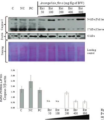
Our study had demonstrated that acetaminophen caused a series of side effects including an increase of liver/body weight ratio, elevation of serum SGOT, SGPT and GGT levels, severe lesion at the CV, the dilation of the surrounding sinusoids, the migration of Kupffer cells into the CV, and furthermore a higher ratio of caspase-3/glyceraldehyde
Control Negative Control Positive Control A. flava extract
400 mg/Kg of BW A. flava extract
200 mg/Kg of BW
A. flava extract 800 mg/Kg of BW
3-phosphate dehydrogenase (GAPDH). These biochemical and pathological alterations in the rat model might resemble acute liver failure in human.
Higher serum transaminases and transferase could be the result of leakage from damaged liver cell membranes post acetaminophen-induced which includes CYP metabolism to a reactive glutathione-depleting metabolite, e.g.,
Arcangelisia flava (mg/Kg of BW) Ext Ext Ext Ext Ext 50 100 200 400 800 C NC PC
Caspase-3
β
-actin
Ponceau Staining
34 kDa (Full length)
17 kDa (Cleaved)
42 kDa
Loading control
Arcangelisia flava (mg/Kg of BW) C NC PC Ext Ext Ext Ext Ext
50 100 200 400 800
3.50
3.00
2.50
2.00
1.50
1.00
0.50
0.00
Ratio Protein Levels Caspase-3
/
β
-actin
Figure 3. The modulation of caspase-3 expression by A. flava extract in rat’s liver. β-actin was used as internal standard. All panels shown above are typical western blot results of this study. C: normal control group; NC: negative control group; PC: positive control group; Ext: A. flava extract (mg/Kg of BW).
Figure 4. Histogram of ratio of caspase-3/ β-actin. C: normal control group; NC: negative control group; PC: positive control group; Ext: A. flava extract (mg/Kg of BW); N/A: not available..
furthermore an increasing of oxidative stress, associated with alterations in calcium homeostasis and initiation of signal transduction responses, causing mitochondrial permeability transition occurring with additional oxidative stress, loss of mitochondrial membrane potential, and loss of the ability of the mitochondria to synthesize ATP, and loss of ATP which leads to necrosis.(1,3,21) Overdose of acetaminophen also caused a temporary moderate caspase activation and generation of caspase-specific cleavage products of cytokeratin 18 in fed CD-1 mice. These effects could be prevented by caspase inhibitors although these inhibitors showed no impact on the overall liver injury.(6) Liver was proven not protected by caspase inhibitor treatment and no morphological evidence for apoptosis was detected. This result indicated that while modest caspase activation due to release of mitochondrial proteins occured, it is insufficient to actually cause significant apoptotic cell death.(22,1)
Previous study had reported that A. flava dose of 250 mg/Kg of BW prevented the elevation of SGOT and SGPT by carbon tetrachloride-induced hepatotoxicity in rat’s liver.(23) Our study confirms that A. flava extract significantly prevent the elevation of SGOT, SGPT and GGT. Moreover, higher doses of A. flava extract indicate equal hepatoprotective activity with silymarin.
Arcangelisia flava (mg/Kg of BW)
Ext Ext Ext Ext
50 100 200 400
C NC PC
Caspase-3
GAPDH
Ponceau Staining
34 kDa (Full length)
17 kDa (Cleaved)
36 kDa
Loading control
C NC PC Ext Ext Ext Ext 50 100 200 400
Arcangelisia flava (mg/Kg of BW)
1.4 1.2 1.0 0.8 0.6 0.4 0.2 0
Ratio Protein Levels Caspase-3/GAPDH
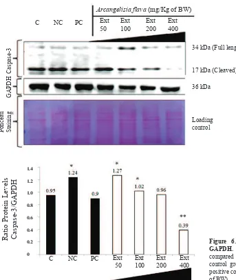
Figure 5. The modulation of caspase-3 expression by A. flava extract in rat’s liver. GAPDH was used as internal standard. C: normal control group; NC: negative control group; PC: positive control group; Ext: A.
flava extract (mg/Kg of BW).
Figure 6. Histogram of ratio of caspase-3/ GAPDH. * and ** indicate significant differences compared to normal group (p<0.05).C: normal
control group; NC: negative control group; PC: positive control group; Ext: A. flava extract (mg/Kg of BW).
will assist in defining the particular substrates and signaling pathways associated with each caspase.(25) In this study,
A. flava extract prevent the elevation of caspase-3 by inhibiting the cleaved of procaspase-3. This may be caused by the bonding between the extract with the active side of hydrophobic S5 site or P5 position of caspase-3.
Furthermore, the ratio of caspase-3/GAPDH was calculated and proved that there was a significant reduction in the particular ratio of higher doses of the extract 200 and 400 mg/Kg of BW (Figure 5-6). Therefore A. flava extract might be potential to be developed as a hepatoprotective agent. This activity is probably caused by secondary metabolites such as flavonoids, alkaloids, phenolics and saponins contained in the extract. Flavonoids and alkaloids in plants had been proven could play an important role in their hepatoprotective effect.(8,12,15,26,27) In
Conclusion
Higher dose A. flava extract shows hepatoprotective activity by preventing the elevation of serum transaminases and transferase levels. Eventually, no damage in the acetaminophen-induced rat’s liver was observed. This plant modulates the expression of caspase 3 protein in dose-dependent manner.
References
1. Jaeschke H, McGill MR, Ramachandran A. Oxidant stress, mitochondria, and cell death mechanisms in drug-induced liver injury: Lessons learned from acetaminophen hepatotoxicity. Drug Metab Rev. 2012; 44: 88-106.
2. Dong H, Haining RL, Thummel KE, Rettie AE, Nelson SD. Involvement of human cytochrome P450 2D6 in the bioactivation of acetaminophen. Drug Metab Dispos. 2000; 28: 1397-400. 3. Laine JE, Auriola S, Pasanen M, Juvonen RO. Acetaminophen
bioactivation by human cytochrome P450 enzymes and animal microsomes. Xenobiotica. 2009; 39: 11-21.
4. LoGuidice A, Boelsterli UA. Acetaminophen overdose-induced liver injury in mice is mediated by peroxynitrite independently of the cyclophilin D-regulated permeability transition. Hepatology. 2011; 54: 969-78.
5. Boulares AH, Ren T. Mechanism of acetaminophen-induced apoptosis in cultured cells: roles of caspase-3, DNA fragmentation factor, and the Ca2+ and Mg2+ endonuclease DNAS1L3. Basic Clin Pharmacol Toxicol. 2004; 94: 19-29.
6. Antoine DJ, Williams DP, Kipar A, Jenkins RE, Regan SL, Sathish JG, et al. High-mobility group box-1 protein and keratin-18, circulating serum proteins informative of acetaminophen-induced necrosis and apoptosis in vivo. Toxicol Sci. 2009; 112: 521-31.
7. Antoine DJ, Williams DP, Kipar A, Laverty H, Park BK. Diet restriction inhibits apoptosis and HMGB1 oxidation and promotes inflammatory cell recruitment during acetaminophen hepatotoxicity. Mol Med. 2010; 16: 479-90.
8. Achmadi SS, Batubara I, Sulistiyani. Saponins of albutra (Arcangelisia flava (L.) Merr) as a hepatoprotector. Technical Report. 2006; 2: 20-30.
9. Verpoorte R, Beek TA, Siwon H, Svendsen AB. Studies on Indonesian medicinal plants. Pharmaceutisch Weekblad. 1982; 4: 87-8.
10. Keawpradub N, Dej-adisai S, Yuenyongsawad S. Antioxidant and cytotoxic activities of Thai medicinal plants named Khaminkhruea: Arcangelisia flava, Coscinium blumeanum and Fibraurea tinctoria. Songklanakarin J Sci Technol. 2005; 27: 456-67.
11. Subeki, Matsuura H, Takahashi K, Yamasaki M, Yamato O, Maede Y, et al. Antibabesial activity of protoberberine alkaloids and 20-hydroxyecdysone from Arcangelisia flava against Babesia gibsoni in culture. J Vet Med Sci. 2005; 67: 223-7.
12. Zhao X, Jie Z, Nannan T, Youran C, Yonghuang L. Protective effects of berberine on doxorubicin-induced hepatotoxicity in mice. Biol Pharm Bull. 2012; 35: 796–800.
13. Feng Y, Siu KY, Ye X, Wang N, Yuen MF, Leung CH, et al. Hepatoprotective effects of berberine on carbon tetrachloride-induced acute hepatotoxicity in rats. Chin Med. 2010; 5: 33. doi: 10.1186/1749-8546-5-33.
14. Domitrović R, Jakovac H, Blagojević G. Hepatoprotective activity of berberine is mediated by inhibition of TNF-α, COX-2, and iNOS expression in CCl(4)-intoxicated mice. Toxicology. 2011; 280: 33-43.
15. Yu LL, Li RT, Ai YB, Liu W, Deng ZS, Zou ZM. Protoberberine isoquinoline alkaloids from Arcangelisia gusanlung. Molecules. 2014; 19: 13332-41.
16. Puspitasari E, Pangaribowo DA, Isparnaning IY, Utami Y. Ethanolic extract of Arcangelisia flava leaves is cytotoxic and selective against breast and colon cancer cell lines. Purwokerto: Proceeding of the 1st University of Muhammadiyah Purwokerto-Pharmacy International Conference; 2005. p. 82-6.
17. Maryani, Marsoedi, Nursyam H, Maftuch. The phytochemistry and the anti-bacterial activity of yellow root (Arcangelisia flava Merr.) against aeromonas hydrophila. J Biol Life Sci. 2013; 4 :180-90. 18. Tiwari P, Kumar B, Kaur M, Kaur G, Kaur H. Phytochemical
screening and extraction: A review. Internationale Pharmaceutica Sciencia. 2011; 1: 98-106.
19. Hasan MN, Khan RA, Nasiruddin M, Khan AA. Ameliorative effect of Nigella sativa oil against paracetamol-induced hepatic and renal damages in rats. J Pharm Res Int. 2016; 13: 1-10.
20. Giknis MLA, Clifford CB. Clinical laboratory parameters for Crl: WI (Han). Wilmington: Charles River; 2008.
21. Hinson JA, Roberts DW, James LP. Mechanisms of acetaminophen-induced liver necrosis. Handb Exp Pharmacol. 2010; 196: 369-405. 22. Gujral JS, Knight TR, Farhood A, Bajt ML, Jaeschke H. Mode of cell death after acetaminophen overdose in mice: apoptosis or oncotic necrosis? Toxicol Sci. 2002; 67: 322-8.
23. Joza N, Kroemer G, Pennigger JM. Genetic analysis of the mammalian cell death machinery. Trends Genet. 2002; 18: 142-9.
24. Wongbutdee J, Sujarit K, Tuchinda P, Thinapong P, Piyachaturawat P. Hepatoprotective effect of Arcangelisia flava Merr extracts in mice. Thai Journal of Physiological Sciences. 2003; 16: 56.
25. Fang B, Boross PI, Tozser J, Weber IT. Structural and kinetic analysis of caspase-3 reveals role for s5 binding site in substrate recognition. J Mol Biol. 2006; 360: 654-66.
26. Yuan HD, Jin GZ, Piao GC. Hepatoprotective effects of an active part from Artemisia sacrorum Ledeb. against acetaminophen-induced toxicity in mice. J Ethnopharmacol. 2010; 127: 528-33.
27. Pattanayak S, Nayak SS, Panda DR, Dinda SC, Shende V, Javad A. Hepatoprotective activity of crude flavonoids extract of Cajanus scarabaeoides (L) in paracetamol intoxicated albino rats. Asian J Pharm Biol Res. 2011; 1: 22-7.
Acknowledgment
This work was facilitated by Academic-Leadership Grant Universitas Padjadjaran Batch-II 2017 No. 872/UN6.3.1/ LT/2017.