483 Cryptococcus neoformans, causes primarily a
chronic infective condition that affects the CNS1-3.Unless, diagnosed early and specific treatment instituted, cryptococcosis is often fatal and can cause severe neurological sequel even after clinical cure. Though once
known to be rare, cryptococcosis has occurred at a high frequency in India in the past two decades 4-9.
Traditionally, diagnosis of cryptococcal meningitis depends on culturing C. neoformans or the
Evaluation of conventional & serological methods for rapid
diagnosis of cryptococcosis
D.C. Saha, Immaculata Xess & Neena Jain
Department of Microbiology, All India Institute of Medical Sciences, New Delhi, India
Received November 1, 2006
Background & objectives: Cryptococcosis is a chronic infective condition affecting the central nervous
system. Unless diagnosed early and specific treatment instituted it can be fatal. There is an urgent need for a rapid and specific diagnostic tool for better management of the patients. Conventional methods such as culture and India ink are specific but cumbersome and time consuming. Serological methods of detection are rapid but have problems of false positivity and cross-reactivity with other micro-organisms. We carried out this study to compare and evaluate the conventional methods with serological methods of detection of cryptococcal meningitis.
Methods: A comparative evaluation of conventional methods (India ink and culture) with LAT (latex
agglutination test) and EIA (enzyme immunoassay) was done in127 CSF samples using culture and EIA as reference separately.
Results: India ink was positive for Cryptococcus in 72.4 per cent of the samples; 56 per cent were
culture positive; LAT positive were 85 per cent and 79.5 per cent were positive by EIA. When culture was positive, all other tests were in agreement to it. However, when culture was negative there was significant difference between the pair of discordance of various diagnostic tests. Culture was 83.46 per cent in agreement to India ink, 76.3 per cent to EIA and 70.8 per cent to LAT. EIA was 92.9 per cent in agreement to India ink and LAT; 6.3 per cent showed false positive by LAT.
Interpretation & conclusions: EIA is valuable in establishing diagnosis when culture is negative for
cryptococcosis. EIA is more specific and has potential advantages over LAT as it gives clear discrimination of positive from negative results. Thus, EIA may be used as a simple, rapid, and reliable serological test for early detection of cryptococcal antigen in clinical samples like CSF in routine laboratories.
demonstration of encapsulated yeasts in India ink preparations from CSF. Direct microscopy and culture are specific but the sensitivity is poor (50-80%)10. A large volume of sample is needed which is often difficult to obtain, especially from paediatric patients. Culture though, is “gold standard” method of diagnosis, is cumbersome, labour intensive and time consuming as the maximum time it takes for the organism to grow sometimes is one month.
Serology, a rapid means of diagnosis, is an indirect and adjunct or complementary procedure to support clinical diagnosis, especially when patient is on treatment. Antigen detection represents the most immediate way to improve methods for cryptococcosis serodiagnosis11-13. Latex agglutination test (LAT) is the most commonly used serological method due to its simplicity in performance14-17. However, it has certain limitations of false positivity18-25, unacceptably high rates of false negativity26,27 and the difficulty of its interpretation in borderline cases.
Enzyme immunoassay (EIA) is another serological tool for detection of capsular polysaccharide antigens of C. neoformans in CSF. This is a rapid test that provides visual and numeric result in less than an hour without pre-treatment of the specimen. It has an advantage of low cross-reactivity with other micro-organisms28-34.
In this study, we performed a comparative evaluation of conventional methods (India ink and culture) with LAT and EIA for detection of cryptococcal antigen in CSF.
Material & Methods
Samples: A total of 127 CSF samples from 81 suspected
cases of cryptococcal meningitis were collected from the routine investigation at Mycology laboratory of the All India Institute of Medical Sciences (AIIMS), New Delhi, from July 2000 to November 2005. The study protocol was approved by the ethics committee of the AIIMS and written consent had been taken by Institute from the patients prior to investigation.
With exception of two patients, the first CSF sample was obtained before starting the antifungal treatment. The serial 48 samples from 22 patients were obtained on different days of infection. All of them were obtained after starting antifungal treatment.
Methodology: CSF samples (1-3 ml) were centrifuged
at 1000 g for 15 min. The pellet was used for direct
microscopy and culture and the supernatant was used for serological tests. Direct microscopy was done by India ink wet mount and culture was done on brain heart infusion (BHI) agar (HiMedia, Mumbai, India) with gentamicin (26 µg/ml). Culture tubes were incubated at 37 and 30°C35.
LAT assays: LAT assays were performed with
CALAS (Meridian Bioscience, Inc., Cincinnati, Ohio). This test utilizes latex particles coated with anti-cryptococcal polyclonal globulin that reacts with the cryptococcal polysaccharide antigen causing a visible agglutination. The test was performed according to manufacturer ’s instructions. CSF specimens were inactivated by placing in boiling water bath for 5 min prior to each test to limit non specific interference.
EIA: EIA was performed using the PREMIER
Cryptococcal antigen kit (Meridian Bioscience, Inc. Cincinnati, Ohio, USA) and the assay was performed as per manufacturer’s instructions. Wells were coated with anticryptococcal polyclonal antibody and the detection system was based upon a monoclonal peroxidase conjugate. Briefly, the test was performed without specimen pre-treatment. A volume of 50 µl CSF was added to antibody coated microtitre plates for 10 min followed by addition of 50 µl of peroxidase-conjugated monoclonal antibody. After 10 min of incubation the microwells were washed (four times) with the wash buffer. The tetramethylbenzidine-urea peroxide substrate was added and incubated for 10 min and stopped with 2N H2SO4. Results were scored visually and spectrophotometrically at 450 nm within 15 min of adding stop solution. The positive and negative cut-off values were 0.150 and 0.10 respectively. Values between 0.1 and 0.15 were considered as indeterminate and were repeated.
Statistical analysis: The comparative evaluation was
done using culture and EIA as a reference separately. Data analysis by Mc Nemar test for each of the diagnostic tests was performed in comparison with culture positive and culture negative.
Results
EIA; 6.3 per cent (n=8) were false positive and 0.78 per cent (n=1) was false negative by LAT. Culture from these samples showed the presence of other organisms like Candida albicans, C. tropicalis, Gram-negative bacteria, Trichosporon and Geotrichum spp. Organisms isolated from false positive LAT: Two samples had Trichosporon (n=2, LAT, titre: 1:6), one sample had Gram-negative bacteria (GNB) and
Geotrichum spp. (n=1, LAT titre: 1:4), one sample had
GNB and C. albicans (n=1, LAT titre: 1:4), one sample had GNB and C. tropicalis (n=1, LAT titre: 1:4) and the rest three samples had GNB (n=3, LAT titre: 1:2). Culture was 83.46 per cent in agreement to India ink, 70.8 per cent to LAT and 76.3 per cent to EIA. EIA was 92.9 per cent in agreement to India ink and LAT. Thirty samples were culture negative but EIA positive. Of these, 30 per cent (n=9) were from patients not on antifungal treatment.
Of the 127 samples, 55 samples were from HIV positive patients and 89 per cent (n=49) were positive by direct microscopy. Of the remaining 6 samples, three were from patients on antifungal treatment, which explains the reason for negative direct microscopy in these samples.
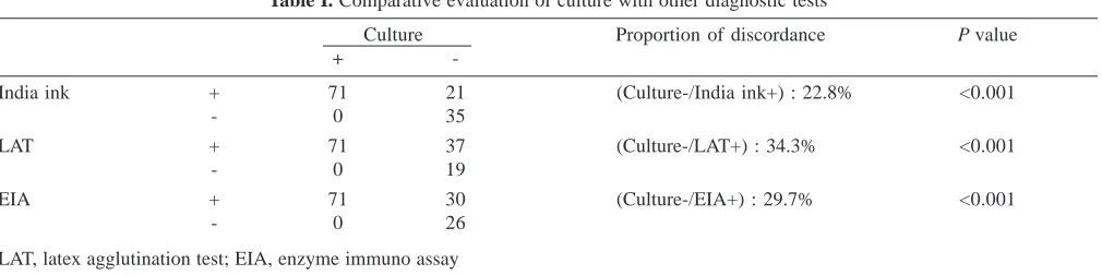
Culture was used as a reference test against other tests for a comparative analysis. The sensitivity of India ink, LAT and EIA was 100 per cent with respect to culture since all samples that tested positive for culture, were positive by other tests while the specificity was 62.5, 33.9, 46.4 per cent respectively. The proportions of discordant pair of culture negative with India ink, LAT and EIA positive were 22.8 per cent, P<0.001 (n=21); 34.3 per cent, P<0.001 (n=37); 29.7 per cent,
P<0.001 (n=30) respectively (Table I).
Mc Nemar test, for each of the diagnostic tests was performed in comparison with culture positive and culture negative, to portray the real scenario for the low specificity obtained in culture negative samples. Culture
was positive in a total of 71 samples (56%) and all diagnostic tests were in agreement with it. Of the 56 culture negative, 37.5 per cent (n=21) were positive and 62.5 per cent (n=35) were negative by India ink; 66 per cent (n=37) were positive and 34 per cent (n=19) were negative by LAT; 53.5 per cent (n=30) were positive and 46.5 per cent (n=26) negative by EIA.
By Mc Nemar test, when culture was negative, there was a significant discordance between the evaluated diagnostic tests. The proportion of discordant pair of LAT positive and India ink negative was 45.7 per cent,
P<0.001 (n=16), proportion of discordance between EIA
positive and India ink negative was 25.7 per cent,
P=0.004 (n=9); and EIA negative and LAT positive was
21.6 per cent, P=0.039 (n=8) and EIA positive and LAT negative was 5.3 per cent (n=1).
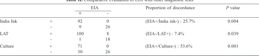
A similar statistical comparative study for all the tests were done for EIA as a reference. The proportion of discordant pair of EIA positive with India ink negative was 25.7 per cent, P=0.004 (n=9); EIA positive and culture negative was 53.6 per cent, P<0.001 (n=30). EIA negative and LAT positive showed a discordance of 7.4 per cent, P=0.039 (n=8) (Table II) while EIA positive and LAT negative was 5.3 per cent (n=1). The sensitivity of India ink, culture and LAT with respect to EIA was 91.1, 70.3, 99 per cent respectively while the specificity was 100 per cent for both India ink and culture and 69.2 per cent for LAT.
The culture was 83.46 per cent in agreement to India ink and 70.8 per cent to LAT and 76.3 per cent to EIA. The EIA was 92.9 per cent in agreement to India ink and LAT.
Discussion
Our results demonstrated that LAT test has higher sensitivity compared to direct microscopic methods. However, a false positivity in 6.3 per cent samples was seen by LAT in presence of other organisms like
Table I. Comparative evaluation of culture with other diagnostic tests
Culture Proportion of discordance P value
+
-India ink + 71 21 (Culture-/India ink+) : 22.8% <0.001
- 0 35
LAT + 71 37 (Culture-/LAT+) : 34.3% <0.001
- 0 19
EIA + 71 30 (Culture-/EIA+) : 29.7% <0.001
- 0 26
C. albicans, C. tropicalis, Gram-negative bacteria, Trichosporon spp. and Geotrichum spp. Two samples
showed cross-reactivity because of Trichosporon spp., and the remaining six samples had Gram-negative bacteria along with C. albicans, C. tropicalis and
Geotrichum spp. All these eight samples were from
HIV negative patients. However, in EIA cross-reaction with these organisms was not seen. Engler & Shea36 have also shown that EIA did not give discrepant results with rheumatoid factor, syneresis fluid, or serum macroglobulins from systemic lupus erythematosis patients while LAT cross-reacted. In another study a false positivity of 1 per cent and false negativity of 1.6 per cent in LAT was reported37. Thus, LAT cannot be relied as a very specific test.
The specificity values also decreased significantly especially in LAT and EIA with reference to culture as it comes negative in low inoculums of organism or if patient is on treatment. Such samples however give positive results with serological methods, as the pick up rate is high. Thus, the tests that show higher number of positives seem less specific when compared to culture. The sensitivity of India ink, LAT and EIA was 100 per cent since all the samples that tested positive for culture, also showed positive by these tests. However, the sensitivity of diagnostic tests varied in respect to EIA, with LAT showing the highest, followed by India ink, and culture. This was because when EIA was positive, India ink and culture were negative in 8.9 and 29.7 per cent samples respectively. The specificity of all the tests in reference to EIA was 100 per cent except in LAT, which showed 8 samples as false positives. The proportion of discordant pair of EIA positive and culture negative was the maximum thus demonstrating a significant difference in sensitivity between conventional and serological methods while that to EIA negative and LAT positive was the least, revealing higher specificity of EIA in comparison to LAT.
A comparative study between LAT and EIA conducted by Gade et al30 demonstrated 99 per cent sensitivity and 97 per cent specificity for EIA. The study resulted in 11 discordant results, 8 of which were identified as positive by the EIA and negative by LAT (EIA+/LAT-). In an earlier study the sensitivities and specificities of commercial kits were shown to be comparable for CSF whereas significant differences in sensitivities were found in serum samples38.
In contrast to the significant discordance in (EIA+/ LAT-) in Gade’s study30, we detected (EIA+/LAT-) in only one sample, as the patient was initially on treatment for 6 wk and then came back with an early relapse and received another course of the treatment for 6 wk. It revealed that EIA could be positive even if patient is on long treatment when LAT demonstrates negative. Scott
et al39 also confirmed the same.
The EIA has potential advantages over LAT as it provides a clear discrimination of positive from negative results (cut-off), and higher sensitivity and specificity12,31. Unlike LAT, no pre-treatment of the specimen is needed30. Moreover the high cost of LAT reagents limits the use of this test on a routine basis in diagnostic laboratories in India. Comparatively the EIA is less expensive (almost half the cost) and can be introduced as a routine diagnostic test.
In conclusion, EIA may be considered as an aid in establishing diagnosis when culture is negative because of low inoculum and treatment. Culture takes a long time, while EIA is rapid. Thus, EIA is a simple, rapid, and reliable test for the early detection of cryptococcal antigen in clinical samples like CSF.
Acknowledgment
Authors thank Drs V. Sreenivas and A.K. Dwivedi for helping us with statistical analysis. The first author (DCS) acknowledges CSIR-UGC for research fellowship.
Table II. Comparative evaluation of EIA with other diagnostic tests
EIA Proportion of discordance P value
+
-India Ink + 92 0 (EIA+/India ink-) : 25.7% 0.004
- 9 26
LAT + 100 8 (EIA-/LAT+) : 7.4% 0.039
- 1 18
Culture + 71 0 (EIA+/Culture-) : 53.6% 0.001
References
1. Dupont B. Cryptococcosis. Rev Prat 1989; 39 : 1663-8. 2. Kwon-Chung KJ, Sorrell TC, Dromer F, Fung E, Levitz SM.
Cryptococcosis: clinical and biological aspects. Med Mycol 2000; 38 (Suppl 1) : 205-13.
3. Sng EH. Cryptococcosis. Singapore Med J 1988; 29 : 198. 4. Banerjee U. Cryptococcosis at AIIMS. Natl Med J India 1994;
7 : 51-2.
5. Banerjee U, Datta K, Majumdar T, Gupta K. Cryptococcosis in India: the awakening of a giant? Med Mycol 2001; 39 : 51-67.
6. Banerjee U, Datta K, Casadevall A. Serotype distribution of
Cryptococcus neoformans in patients in a tertiary care center
in India. Med Mycol 2004; 42 : 181-6.
7. Banerjee U. Progress in diagnosis of opportunistic infections in HIV/AIDS. Indian J Med Res 2005; 121 : 395-406. 8. Chakrabarti A, Gupta V. Isolated detection of cryptococcal
polysaccharide antigen in patients with cryptococcosis. Clin
Infect Dis 1997; 25 : 1494-5.
9. Chakrabarti A, Sharma A, Sood A, Grover R, Sakhuja V, Prabhakar S, et al. Changing scenario of cryptococcosis in a tertiary care hospital in north India. Indian J Med Res 2000;
112 : 56-60.
10. Snow RM, Dismukes WE. Cryptococcal meningitis: diagnostic value of cryptococcal antigen in cerebrospinal fluid.
Arch Intern Med 1975; 135 : 1155-7.
11. Diamond RD, Bennett JE. Prognostic factors in cryptococcal meningitis. A study in 111 cases. Ann Intern Med 1974; 80 : 176-81.
12. Gordon MA, Vedder DK. Serologic tests in diagnosis and prognosis of cryptococcosis. JAMA 1966; 197 : 961-7. 13. Gordon MA. Cryptococcal antigen test. JAMA 1981; 246 :
1403.
14. Hamilton JR, Noble A, Denning DW, Stevens DA. Performance of cryptococcus antigen latex agglutination kits on serum and cerebrospinal fluid specimens of AIDS patients before and after pronase treatment. J Clin Microbiol 1991; 29 : 333-9.
15. Kauffman CA, Bergman AG, Severance PJ, McClatchey KD. Detection of cryptococcal antigen. Comparison of two latex agglutination tests. Am J Clin Pathol 1981; 75 : 106-9. 16. Kiska DL, Orkiszewski DR, Howell D, Gilligan PH.
Evaluation of new monoclonal antibody-based latex agglutination test for detection of cryptococcal polysaccharide antigen in serum and cerebrospinal fluid. J Clin Microbiol 1994; 32 : 2309-11.
17. Stockman L, Roberts GD. Specificity of the latex test for cryptococcal antigen: a rapid, simple method for eliminating interference factors. J Clin Microbiol 1982; 16 : 965-7.
18. Boom WH, Piper DJ, Ruoff KL, Ferraro MJ. New cause for false-positive results with the cryptococcal antigen test by latex agglutination. J Clin Microbiol 1985; 22 : 856-7.
19. Heelan JS, Corpus L, Kessimian N. False-positive reactions in the latex agglutination test for Cryptococcus neoformans antigen. J Clin Microbiol 1991; 29 : 1260-1.
20. Kornfeld SJ, Worthington M. False-positive CSF cryptococcal antigen tests. Arch Neurol 1980; 37 : 603.
21. MacKinnon S, Kane JG, Parker RH. False-positive cryptococcal antigen test and cervical prevertebral abscess.
JAMA 1978; 240 : 1982-3.
22. Millon L, Barale T, Julliot MC, Martinez J, Mantion G. Interference by hydroxyethyl starch used for vascular filling in latex agglutination test for cryptococcal antigen. J Clin
Microbiol 1995; 33 : 1917-9.
23. Sachs MK, Huang CM, Ost D, Jungkind DL. Failure of dithiothreitol and pronase to reveal a false-positive cryptococcal antigen determination in cerebrospinal fluid.
Am J Clin Pathol 1991; 96 : 381-4.
24. Stoeckli TC, Burman WJ. Inactivated pronase as the cause of false-positive results of serum cryptococcal antigen tests.
Clin Infect Dis 2001; 32 : 836-7.
25. Whittier S, Hopfer RL, Gilligan P. Elimination of false-positive serum reactivity in latex agglutination test for cryptococcal antigen in human immunodeficiency virus-infected population.
J Clin Microbiol 1994; 32 : 2158-61.
26. Currie BP, Freundlich LF, Soto MA, Casadevall A. False-negative cerebrospinal fluid cryptococcal latex agglutination tests for patients with culture-positive cryptococcal meningitis.
J Clin Microbiol 1993; 31 : 2519-22.
27. Stamm AM, Polt SS. False-negative cryptococcal antigen test.
JAMA 1980; 244 : 1359.
28. Casadevall A, Mukherjee J, Scharff MD. Monoclonal antibody based ELISAs for cryptococcal polysaccharide. J Immunol
Methods 1992; 154 : 27-35.
29. Frank UK, Nishimura SL, Li NC, Sugai K, Yajko DM, Hadley WK, et al. Evaluation of an enzyme immunoassay for detection of cryptococcal capsular polysaccharide antigen in serum and cerebrospinal fluid. J Clin Microbiol 1993;
31 : 97-101.
30. Gade W, Hinnefeld SW, Babcock LS, Gilligan P, Kelly W, Wait K, et al. Comparison of the PREMIER cryptococcal antigen enzyme immunoassay and the latex agglutination assay for detection of cryptococcal antigens. J Clin Microbiol 1991;
29 : 1616-9.
31. Illnait MT, Vilaseca JC, Fernandez CM, Martinez GF. Enzyme-linked immunosorbent assay for detection and quantification of Cryptococcus neoformans antigen. Mem Inst Oswaldo Cruz 2001; 96 : 241-5.
32. Scott EN, Felton FG, Muchmore HG. Development of an enzyme immunoassay for cryptococcal antibody.
33. Scott EN, Muchmore HG, Felton FG. Enzyme-linked immunosorbent assays in murine cryptococcosis. Sabouraudia 1981; 19 : 257-65.
34. Sekhon AS, Garg AK, Kaufman L, Kobayashi GS, Hamir Z, Jalbert M, et al. Evaluation of a commercial enzyme immunoassay for the detection of cryptococcal antigen.
Mycoses 1993; 36 : 31-4.
35. Libero A, lucille KG, William K, Leo K. Laboratory manual
for medical mycology. Atlanta, Georgia: U.S. Department of
Health, Education and Welfare, Public Health Service: Communicable Disease Centre (CDC); 1963.
36. Engler HD, Shea YR. Effect of potential interference factors on performance of enzyme immunoassay and latex
agglutination assay for cryptococcal antigen. J Clin Microbiol 1994; 32 : 2307-8.
37. Coovadia YM, Solwa Z. Sensitivity and specificity of a latex agglutination test for detection of cryptococcal antigen in meningitis. S Afr Med J 1987; 71 : 510-2.
38. Tanner DC, Weinstein MP, Fedorciw B, Joho KL, Thorpe JJ, Reller L. Comparison of commercial kits for detection of cryptococcal antigen. J Clin Microbiol 1994; 32 : 1680-4.
39. Scott EN, Muchmore HG, Felton FG. Comparison of enzyme immunoassay and latex agglutination methods for detection of Cryptococcus neoformans antigen. Am J Clin Pathol 1980;
73 : 790-4.
Reprint requests: Dr Immaculata Xess, Department of Microbiology, All India Institute of Medical Sciences
New Delhi 110 029, India