Distribution of Cartilage Neoplasm based on Histopathological Types
at Dr. Hasan Sadikin General Hospital Bandung Period 2008–2012
Desy Anggraini,1 Anglita Yantisetiasti,2 Darmadji Ismono3
1Faculty of Medicine, Universitas Padjadjaran, 2Department of Anatomical Pathology, Faculty of Medicine, Universitas Padjadjaran/Dr. Hasan Sadikin General Hospital, Bandung,, 3Department
of Surgery, Faculty of Medicine Universitas Padjadjaran/Dr. Hasan Sadikin General Hospital Bandung
Abstract
Background: Cartilage neoplasms are less common compared to other neoplasms. Its incidence is 22% among all the musculoskeletal neoplasms. Despite many other studies about patient characteristics of the neoplasm in other regions, descriptive data in Bandung city is still unknown. The objective of this study is to determine the distribution of both benign and malignant neoplasms based on their characteristics of histopathological type, gender, age and anatomical site.
Methods: The subjects of this descriptive study were taken from the medical records of the patients who had been examined histopathologically in Anatomical Pathology Department at Dr. Hasan Sadikin General Hospital Bandung, Indonesia within the period of 2008 to2012. The sample was obtained using total sampling technique. Patients diagnosed with cartilage neoplasms were included whereas incomplete medical records were excluded. Histopathological type, gender, age and anatomical site of each patients were collected and analyzed.
Results: Seventy cartilage neoplasm cases were found. The distribution of cases comprised of 48 (67%) benign and 23 (33%) malignant. The most common benign neoplasm was osteochondroma and chondroma. Benign neoplasms were more prevalent among men and patients <30 years old, while malignant neoplasm was prevalent among women and patients >60 years old. Femur was the most common site for all neoplasms except for chondroma.
Conclusions: PThere are differences in characteristic of benign and malignant cartilage neoplasm patients. Both benign and malignant cartilage neoplasms showed differences on the distribution of patient characteristics. [AMJ.2015;2(4):561–7]
Keywords: Benign neoplasm, cartilage neoplasm, histopathological types, malignant neoplasm
Correspondence: Desy Anggraini, Faculty of Medicine, Universitas Padjadjaran, Jalan Raya Bandung-Sumedang Km.21, Jatinangor, Sumedang, Indonesia, Phone: +62 8997131532 Email: [email protected]
Introduction
Cartilage neoplasm is a primary bone neoplasm characterized by the presence of chondrocytes within and surrounded by cartilaginous matrix.1 It accounts for about 22% of all musculoskeletal neoplasm.2 Yulianti3 in 2011 reported osteochondroma as the most common benign musculoskeletal lesion and chondrosarcoma as the second most common malignant lesion after osteosarcoma among patients in Dr. Hasan Sadikin General Hospital, Bandung. Another research conducted by Solooki et al.4 In Iran in 2011, it is reported that osteochondroma and enchondroma as
the most frequent benign neoplasm. Both commonly affect men and patients within the age of 15–24 years old.
General Hospital, Bandung, Indonesia
Methods
This descriptive study collected and analyzed data from medical records of the patients who had undergone histopathological test in Anatomical Pathology Department of Dr. Hasan Sadikin General Hospital, Bandung, West Java, Indonesia from January 2008–December 2012. Samples were selected by total sampling method. Subject’s inclusion criteria of this study was all the medical records of patients diagnosed as cartilage-forming neoplasms. Incomplete medical record was excluded from this study.
Histopathological type, gender, age and
anatomical site of 70 patient’s medical records were collected and analyzed. Based on pathological medical records, cartilage-forming neoplasms were classified as benign and malignant according to World Health Organization (WHO) 2002 classification. Benign neoplasms were osteochondroma, chondroma, chondroblastoma, and chondromyxoid fibroma. Malignant neoplasm was chondrosarcoma.
Secondary data were gathered from each subject, which would then be organized and analyzed using descriptive statistical analysis. Frequency and percentage of each variable were calculated. The ethical approval to conduct this study was obtained from Health Research Ethics Committee of the Faculty of Medicine, Universitas Padjadjaran.
Table 1 Distribution of Benign and Malignant Cartilage-forming Neoplasm based on Histhopathological Type and Gender
Histopathological types Frequency (%) Gender
Male (%) Female (%)
Benign
Osteochondroma 36 (51) 21 (58) 15 (42)
Chondroma 10 (14) 5 (50) 5 (50)
Chondroblastoma 0 (0) 0 (0) 0 (0)
Chondromyxoid fibroma 1 (1) 1 (100) 0 (0)
Total Benign Cases 47 (67)
Malignant
Chondrosarcoma 23 (33) 10 (43) 13 (57)
Total Malignant Cases 23 (33)
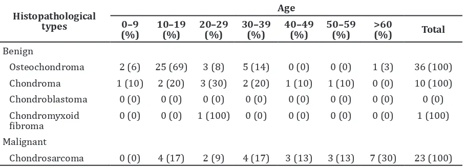
Table 2 Distribution of Benign and Malignant Cartilage-forming Neoplasm based on Histopathological Type and Age
Result
Seventy cases had been analyzed. Benign neoplasms were more common than malignant neoplasm (Table 1). The most common histopathological type of benign neoplasm was osteochondroma (51%) and chondroma (14%). Chondroblastoma was not found in this study. Osteochondroma was more prevalent among males but chondroma showed equal number between male and female. On the other hand, chondrosarcoma was more common in female than in male.
Osteochondroma as the most common benign neoplasm was more frequent among patients within the age of 10–19 years old, whereas chondroma was prevalent among patients within the age of 20–29 years old.
Therefore, both neoplasms were dominant among <30 years-old patients. However, chondrosarcoma as the malignant neoplasm was most frequent among >60 years-old patients (Table 2).
Overall, femur was the most common sites of osteochondroma and chondrosarcoma. It was followed by humerus and tibia in osteochondroma and humerus in chondrosarcoma. Meanwhile, chondroma was more frequent in humerus, digitorum manus, and sternum (Table 3).
There were some cases involvingmore than one bone. Three cases occurred with osteochondroma of tibia-fibula and metatarsal, humerus and femur, and radius and femur. There was also a case of chondrosarcoma involving femur and radius-ulna.
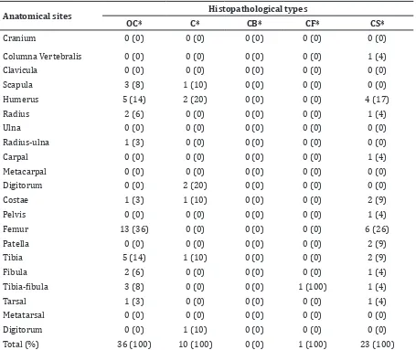
Table 2 Distribution of Benign and Malignant Cartilage-forming Neoplasm based on Histopathological Type and Anatomical Site
Anatomical sites Histopathological types
OC* C* CB* CF* CS*
Cranium 0 (0) 0 (0) 0 (0) 0 (0) 0 (0)
Columna Vertebralis 0 (0) 0 (0) 0 (0) 0 (0) 1 (4)
Clavicula 0 (0) 0 (0) 0 (0) 0 (0) 0 (0)
Scapula 3 (8) 1 (10) 0 (0) 0 (0) 0 (0)
Humerus 5 (14) 2 (20) 0 (0) 0 (0) 4 (17)
Radius 2 (6) 0 (0) 0 (0) 0 (0) 1 (4)
Ulna 0 (0) 0 (0) 0 (0) 0 (0) 0 (0)
Radius-ulna 1 (3) 0 (0) 0 (0) 0 (0) 0 (0)
Carpal 0 (0) 0 (0) 0 (0) 0 (0) 1 (4)
Metacarpal 0 (0) 0 (0) 0 (0) 0 (0) 0 (0)
Digitorum 0 (0) 2 (20) 0 (0) 0 (0) 0 (0)
Costae 1 (3) 1 (10) 0 (0) 0 (0) 2 (9)
Pelvis 0 (0) 0 (0) 0 (0) 0 (0) 1 (4)
Femur 13 (36) 0 (0) 0 (0) 0 (0) 6 (26)
Patella 0 (0) 0 (0) 0 (0) 0 (0) 2 (9)
Tibia 5 (14) 1 (10) 0 (0) 0 (0) 2 (9)
Fibula 2 (6) 0 (0) 0 (0) 0 (0) 1 (4)
Tibia-fibula 3 (8) 0 (0) 0 (0) 1 (100) 1 (4)
Tarsal 1 (3) 0 (0) 0 (0) 0 (0) 1 (4)
Metatarsal 0 (0) 0 (0) 0 (0) 0 (0) 0 (0)
Digitorum 0 (0) 1 (10) 0 (0) 0 (0) 0 (0)
Total (%) 36 (100) 10 (100) 0 (0) 1 (100) 23 (100)
Discussion
General distribution of cartilage neoplasm based on histopathological type on this study’s result showed that benign neoplasms (68%) were more frequent than malignant neoplasm (32%). Among all benign neoplasms, osteochondroma and chondroma were the most common types. This result was similar to those reported by Yulianti3 in 2011 in Dr
Hasan Sadikin General Hospital, Bandung reporting that osteochondroma as the most common benign cartilage neoplasm accounted for 23.3% among other benign bone lesion. Other study conducted by Norahmawati5 in 2009 in Malang, East Java, also reported that osteochondroma and chondroma as the most common benign cartilage neoplasms can be diagnosed by fine needle aspiration biopsy technique.
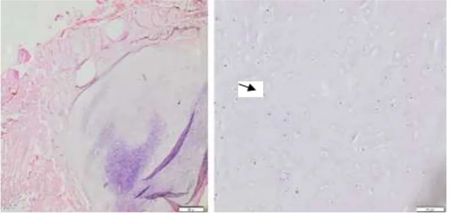
Figure 1 Microscopic Appearance of Osteochondroma from Left Distal Femur (Objective 10x)
Gender-specific incidence of cartilage neoplasm of this study showed that osteochondroma commonly affected male. This results similar to the study conducted by Solooki et al.4 in 2011 in Iran. They reported that 62.5% patients were men. The same number cases in male and female was shown by chondroma in this study and other study. WHO in 2002 stated that both gender are equally affected.6 Males were more frequent to be affected by cartilage neoplasm because chondrocyte growth was influenced naturally by testosterone hormone.7
While osteochondroma occurred more commonly in males, chondrosarcoma showed different results. It was more commonly in females than males. This result was not similar to other study’s results. Ocampo et al.8 in Mexico 2009 reported chondrosarcoma was dominant in male. Eyre et al.9 in northern England in 2010 also stated that chondrosarcoma was higher among males than females. Oemiati10 in 2011 stated that tumours or cancers in Indonesia were two times more frequent in females than males. These results are reffered to the data of Indonesian population proportion based on gender from Badan Pusat Statistik in 2007. The data showed that male and female proportion were almost equal in number.10 Indonesia Health Data released by Indonesia Health Ministry in 2011 reported that males was only accounted in 1.04 greater than females.11 Beside that, tumours or cancers were reported more frequent in females due to the probability that females had more attentions to her health
than males had.10
Age-specific incidence among benign and malignant cartilage neoplasms of this study showed that osteochondroma was prevalent among the patient within the age of 10–19 years old (69%). This result is similar to the study conducted by Solooki et al.4 in Iran in 2011. They reported that osteochondroma was prevalent among patients within the age of 5–24 years old. Lopez et al.12 in Cuba in 2005 also reported that osteochondroma as the most commonly occurred cartilage neoplasm within the 11–15 years age group. Chondroma as the second most common benign cartilage neoplasm, in this study, showed the peak incidence in the patients age of 20–29 years old (30%). Despite its peak incidence, this neoplasm had stable incidence in the age of 10–39 years old. This result is similar to the reference reporting that chondroma commonly affects the patients within the age of 20–40 years old.2
While benign neoplasms commonly affected <30 years old patients, chondrosarcoma had a peak incidence in >60 years old patients. Ocampo et al.8 in Mexico in 2009 also reported that chondrosarcoma was more prevalent in patients older than 60 years old. WHO in 2002 also stated that the incidence of chondrosarcoma would increase as the age of the patients increase.6
These age-specific distribution differences between benign and malignant neoplasms could be explained by their pathogenesis. Both osteochondroma and chondroma were
formed by abnormal growth of highly active bone structure in children. The patients may not become aware of the lesion until reaching their adolescence or early adult life.13 Chondrosarcoma as the malignant one had older age as one of the risk factors because older people had undergone some somatic mutation and decreased immune response to prevent malignancy development.2
Distribution of cartilage neoplasm based on anatomical sites in this study showed that most common sites of osteochondroma were femur, humerus, and tibia. This result was similar to the study conducted by Solooki et al.4 in Iran in 2011 statingthat femur, tibia, and humerus were the most common sites. Osteochondroma was formed by abnormality of growth direction and remodeling in metaphyseal region, the most actively growing end of long bones.13
In this study, the second most frequent benign neoplasm was chondroma, and more commonly was formed in humerus, digitorum manus, and sternum. WHO in 2002 reported that chondroma most commonly affected digitorum manus and pedis, then long bones such as humerus and femur, but rarely in flat bones like pelvis, vertebrae, and sternum.7 Chondroma is developed by growth abnormality of cartilaginous cells in epiphyseal plate in short bones during childhood. Its pathogenesis explained that the tendency of chondroma to be developed was on small bones.13
In this study, chondrosarcoma most commonly affected femur and humerus. According to WHO, primary chondrosarcoma affected ileum, humerus, femur, and costae as the most common sites.6 Qureshi et al.14 In Pakistan in 2010 also reported that pelvis, femur, costae, and skull as the most common sites.
The patient’s characteristic distributions shown by this study could not represent the real population because this study was done only in a general hospital. There were also the probabilitiesunder the diagnosed patients because not all patients in Dr. Hasan Sadikin General Hospital Bandung underwent histopathological test.
The conclusion of this study was both benign and malignant cartilage-forming neoplasms had different characteristical distributions. Study showed that benign neoplasms were two times more than malignant neoplasm. Among all the benign neoplasms, osteochondroma and chondroma were the most common types. Patient’s characteristics showed that all benign
neoplasms except chondroma commonly affected males, while chondroma showed the same ratio between males and females. Chondrosarcoma commonly affected females in this study. Benign neoplasms were more prevalent among <30 years old patients, while chondrosarcoma was prevalent among >60 years old patients. All neoplasms had femur as the most common anatomical site, except for chondroma that was dominant in humerus, digitorum manus and sternum.
From this study, it can be suggested to study larger population and another point of study such as revealing the risk factor of cartilage neoplasm to be done in the further study. Larger population was needed to obtain more accurate data.
References
1. Higuchi T, Taki J, Sumiya H, Kinuya S, Nakajima K, Namura M, et al. Characterization of cartilaginous tumors with 201Tl scintigraphy. Ann Nucl Med. 2005;19(2):95–9.
2. Robbins SL, Kumar V, Abbas AK, Cotran RS. Robbins and Cotran pathologicbasis of disease. 8th ed. Philadelphia: Saunders/ Elseiver; 2010. p. 273, 1227–30.
3. Yulianti LS. Epidemiologi tumor muskuloskeletal di Rumah Sakit Hasan Sadikin Bandung periode Januari 2010– Maret 2011. Proceedings of the 58th Continuing Orthopaedic Education Conference; 2011 June 16–18; Pekanbaru. Bandung: Universitas Padjadjaran; 2011. 4. Solooki S, Vosoughi AR, Masoomi V.
Epidemiology of musculoskeletal tumors in Shiraz, south of Iran. Indian J Med Paediatr Oncol. 2011;32(4):187–91.
5. Norahmawati E. Fine-needle aspiration biopsy has important role and high accuracy as preoperative diagnostic method for bone tumors. Jurnal Kedokteran Brawijaya. 2009; 25(2):77–82.
6. Fletcher CD, Unni KK, Mertens F, editors. World Health Organization classification of tumours, pathology and genetics of tumours of soft tissue and bone. Lyon, France: IARC Press, International Agency for Research on Cancer (IARC); 2002. p.233–58.
7. Mescher AL. Basic histology text & atlas. 12th ed. Singapore: McGraw-Hill Companies, Inc; 2010. p. 116.
City: retrospective clinicopathologic study of 566 patients at a referral institution. Ann Diagn Pathol. 2009;13(1):16–21. 9. Eyre R, Feltbower RG, James PW, Blakey K,
Mubwandarikwa E, Forman D, et al. The epidemiology of bone cancer in 0–39 year olds in northern England, 1981–2002. BMC Cancer. 2010;10:357.
10. Oemiati R, Rahajeng E, Kristanto AY. Prevalensi tumor dan beberapa faktor yang mempengaruhinya di Indonesia. Bul Penelit Kesehat. 2011;39(4):190–204.
11. Kementerian Kesehatan Republik Indonesia. Profil data kesehatan Indonesia tahun 2011. Jakarta: Kementerian Kesehatan Republik Indonesia; 2012
[cited 2013 February18]. Available from: http://[email protected].
12. Lopez AA, Lorenzo YG. Cartilaginous bone tumors in children: a twenty-year epidemiological report from our hospital. Acta Ortopedica Mexicana. 2005;19(Suppl 1):S47–S50.
13. Salter RB. Textbook of disorders and injuries of the musculoskeletal system. 3rd ed. Philadelphia: Lippincott Williams & Wilkins; 1999. p. 392–95.