A Guide to Clinical Practice
Humana Press Edited by
Jeannette M. Potts, MD
Essential
Urology
C URRENT C LINICAL U ROLOGY
Eric A. Klein,
MD,
SERIES EDITOREssential Urology: A Guide to Clinical Practice, edited by Jeannette M. Potts, 2004 Management of Prostate Cancer, Second Edition, edited by Eric A. Klein, 2004 Management of Benign Prostatic Hypertrophy, edited by Kevin T. McVary, 2004 Laparoscopic Urologic Oncology, edited by Jeffrey A. Cadeddu, 2004
Essential Urologic Laparoscopy: The Complete Clinical Guide, edited by Stephen Y. Nakada, 2003
Pediatric Urology, edited by John P. Gearhart, 2003
Urologic Prostheses: The Complete Practical Guide to Devices, Their Implantation, and Patient Follow-Up, edited by Culley C. Carson, III, 2002
Male Sexual Function: A Guide to Clinical Management, edited by John J. Mulcahy, 2001
Prostate Cancer Screening, edited by Ian M. Thompson, Martin I. Resnick, and Eric A. Klein, 2001
Bladder Cancer: Current Diagnosis and Treatment, edited by Michael J. Droller, 2001
Office Urology: The Clinician’s Guide, edited by Elroy D. Kursh and James C. Ulchaker, 2001
Voiding Dysfunction: Diagnosis and Treatment, edited by Rodney A. Appell, 2000
Management of Prostate Cancer, edited by Eric A. Klein, 2000
E SSENTIAL U ROLOGY
A G UIDE TO C LINICAL P RACTICE
Edited by
J EANNETTE M. P OTTS , MD
Glickman Urological Institute, Cleveland Clinic Foundation, Cleveland, OH
H UMANA P RESS
T OTOWA , N EW J ERSEY
Totowa, New Jersey 07512 www.humanapress.com
For additional copies, pricing for bulk purchases, and/or information about other Humana titles, contact Humana at the above address or at any of the following numbers: Tel.: 973-256-1699; Fax: 973-256-8341, E-mail: [email protected];
or visit our Website: http://humanapress.com All rights reserved.
No part of this book may be reproduced, stored in a retrieval system, or transmitted in any form or by any means, electronic, mechanical, photocopying, microfilming, recording, or otherwise without written permission from the Publisher.
All articles, comments, opinions, conclusions, or recommendations are those of the author(s), and do not necessarily reflect the views of the publisher.
Due diligence has been taken by the publishers, editors, and authors of this book to assure the accuracy of the information published and to describe generally accepted practices. The contributors herein have carefully checked to ensure that the drug selections and dosages set forth in this text are accurate and in accord with the standards accepted at the time of publication. Notwithstanding, as new research, changes in government regulations, and knowledge from clinical experi- ence relating to drug therapy and drug reactions constantly occurs, the reader is advised to check the product information provided by the manufacturer of each drug for any change in dosages or for additional warnings and contraindications.
This is of utmost importance when the recommended drug herein is a new or infrequently used drug. It is the responsibility of the treating physician to determine dosages and treatment strategies for individual patients. Further it is the responsi- bility of the health care provider to ascertain the Food and Drug Administration status of each drug or device used in their clinical practice. The publisher, editors, and authors are not responsible for errors or omissions or for any consequences from the application of the information presented in this book and make no warranty, express or implied, with respect to the contents in this publication.
Production Editor: Robin B. Weisberg
Cover illustration layout by Jeannette M. Potts. Cover illustration by Michelle Wolf. Cover design by Patricia F. Cleary.
This publication is printed on acid-free paper.
ANSI Z39.48-1984 (American National Standards Institute) Permanence of Paper for Printed Library Materials.
Photocopy Authorization Policy:
Authorization to photocopy items for internal or personal use, or the internal or personal use of specific clients, is granted by Humana Press Inc., provided that the base fee of US $25.00 per copy is paid directly to the Copyright Clearance Center at 222 Rosewood Drive, Danvers, MA 01923. For those organizations that have been granted a photocopy license from the CCC, a separate system of payment has been arranged and is acceptable to Humana Press Inc. The fee code for users of the Transactional Reporting Service is: [1-58829-109-X/04 $25.00].
Printed in the United States of America. 10 9 8 7 6 5 4 3 2 1 E-ISBN 1-59259-737-8
Library of Congress Cataloging-in-Publication Data
Essential urology : a guide to clinical practice / edited by Jeanette M.
Potts.
p. ; cm. -- (Current clinical urology) Includes bibliographical references and index.
ISBN 1-58829-109-X (alk. paper) 1. Genitourinary organs--Diseases.
[DNLM: 1. Urologic Diseases. 2. Urogenital Diseases. WJ 140 E78 2004] I. Potts, Jeanette M. II. Series.
RC871.E883 2004 616.6--dc22
2003016746
To my children, Bradley and Ellen,
and to my mentors, Jonathan Ross,
MDand Elroy Kursh,
MDDedication
Preface
vii
As a medical urologist with a background in family medicine, I have enjoyed the overlapping aspects of urology and primary care. Urological diseases are often brought to the attention of primary care providers who must then diagnose and manage these disorders. Essential Urology: A Guide to Clinical Practice is intended to provide support to primary care physicians through its review of common genitourinary problems. It is meant to enhance the recognition of urological disease as well as outline current manage- ment strategies.
Disorders of the urinary tract may be encountered during pregnancy, either as a maternal diagnosis or as a fetal anomaly detected in utero. Children as well as adults require screening and monitoring of genitourinary disorders, some of which are gender-specific. Urinary tract infections may be manifestations of risk factors, anatomical or functional abnormalities, specific to age and/or gender. These issues are presented in this text. Hematuria, frequently encountered in the primary care setting as an incidental finding, is discussed in a comprehen- sive chapter, followed by related chapters detailing urological imaging studies and the evalu- ation and management of nephrolithiasis, respectively. Urinary function is addressed in the chapters reviewing female incontinence, interstitial cystitis, and bladder outlet obstruction secondary to benign prostatic hyperplasia. Screening for urological cancers, particularly prostate and bladder cancers, is reviewed. We have included a chapter summarizing comple- mentary therapies in urology and a chapter that introduces alternative approaches to frequently diagnosed abacterial prostatitis/pelvic pain syndrome. Finally, we address the quality-of-life impact and medical significance of erectile dysfunction and its treatment.
Essential Urology: A Guide to Clinical Practice addresses various life stages and respective urological conditions and should be a valuable resource to family practitio- ners, internists, pediatricians, obstetricians, physician’s assistants, and nurse clinicians.
Jeannette M. Potts, MD
Preface ... vii List of Contributors ... xi Value-Added eBook/PDA ... xii 1 Management of Urologic Problems During Pregnancy:
A Rationale and Strategy ...1 Kevin R. Loughlin
2 Pediatric Potpourri ... 17 Jonathan H. Ross
3 Urinary Tract Infections in Children ... 33 Richard W. Grady
4 Screening and Early Detection for Genitourinary Cancer ... 47 Ian M. Thompson and Joseph Basler
5 Basic Imaging in Urology ... 61 Martin B. Richman and Martin I. Resnick
6 Hematuria ... 91 Mark J. Noble
7 Evaluation and Medical Management of Kidney Stones ... 117 John C. Lieske and Joseph W. Segura
8 Management of Female Urinary Incontinence ... 153 Raymond R. Rackley and Joseph B. Abdelmalak
9 Interstitial Cystitis ... 169 Kenneth M. Peters
10 Urinary Tract Infections in Adults ... 183 Joseph B. Abdelmalak, Sandip P. Vasavada,
and Raymond R. Rackley
11 Evaluation and Treatment of Benign Prostatic Hyperplasia ... 191 Elroy D. Kursh
12 Prostatitis/Chronic Pelvic Pain Syndrome ... 203 Jeannette M. Potts
13 Erectile Dysfunction ... 213 Drogo K. Montague and Milton M. Lakin
14 Complementary Medications in Urology ... 225 Elliot Fagelman, Bridgit Mennite, and Franklin C. Lowe
Index ... 235
Contents
ix
Contributors
xi
JOSEPH B. ABDELMALAK, MD • Glickman Urological Institute, Cleveland Clinic Foundation, Cleveland, OH
JOSEPH BASLER, MD • Division of Urology, University of Texas Health Science Center at San Antonio, San Antonio, TX
ELLIOT FAGELMAN, MD • Department of Urology, St. Luke’s-Roosevelt Hospital Center, College of Physicians and Surgeons, Columbia University, New York, NY
RICHARD W. GRADY, MD • Department of Urology, Children’s Hospital and Regional Medical Center, University of Washington School of Medicine, Seattle, WA ELROY D. KURSH, MD • Glickman Urological Institute, Cleveland Clinic Foundation,
Cleveland, OH
MILTON M. LAKIN, MD • Section of Medical Urology, Glickman Urological Institute, Cleveland Clinic Foundation, Cleveland, OH
JOHN C. LIESKE, MD • Division of Nephrology, Department of Medicine, Mayo Clinic and Medical School, Rochester, MN
KEVIN R. LOUGHLIN, MD • Division of Urology, Department of Surgery, Brigham and Women’s Hospital, Harvard Medical School, Boston, MA
FRANKLIN C. LOWE, MD, MPH • Department of Urology, St. Luke’s-Roosevelt Hospital Center, College of Physicians and Surgeons, Columbia University, New York, NY BRIDGIT MENNITE, BS • University of Albany, Albany, NY
DROGO K. MONTAGUE, MD • Center for Sexual Function, Glickman Urological Institute, Cleveland Clinic Foundation, Cleveland, OH
MARK J. NOBLE, MD • Glickman Urological Institute, Cleveland Clinic Foundation, Cleveland, OH
KENNETH M. PETERS, MD • Department of Urology, William Beaumont Hospital, Royal Oak, MI
JEANNETTE M. POTTS, MD • Section of Medical Urology, Glickman Urological Institute, Cleveland Clinic Foundation, Cleveland, OH
RAYMOND R. RACKLEY, MD • Glickman Urological Institute, Cleveland Clinic Foundation, Cleveland, OH
MARTIN I. RESNICK, MD • Department of Urology, University Hospitals of Cleveland, Case Western Reserve University, Cleveland, OH
MARTIN B. RICHMAN, MD • Department of Urology, University Hospitals of Cleveland, Case Western Reserve University, Cleveland, OH
JONATHAN H. ROSS, MD • Section of Pediatric Urology, Glickman Urological Institute, The Children’s Hospital at the Cleveland Clinic, Cleveland Clinic Foundation, Cleveland, OH
JOSEPH W. SEGURA, MD • Departments of Urology and Surgery, Mayo Clinic and Medical School, Rochester, MN
IAN M. THOMPSON, MD • Division of Urology, University of Texas Health Science Center at San Antonio, San Antonio, TX
SANDIP P. VASAVADA, MD • Glickman Urological Institute, Cleveland Clinic Foundation, Cleveland, OH
Value-Added eBook/PDA
This book is accompanied by a value-added CD-ROM that contains
an Adobe eBook version of the volume you have just purchased. This eBook can be viewed on your computer, and you can synchronize it to your PDA for viewing on your handheld device. The eBook enables you to view this volume on only one computer and PDA. Once the eBook is installed on your computer, you cannot download, install, or e-mail it to another computer; it resides solely with the computer to which it is installed. The license provided is for only one computer. The eBook can only be read using Adobe® Reader® 6.0 software, which is available free from Adobe Systems Incorporated at www.Adobe.com. You may also view the eBook on your PDA using the Adobe® PDA Reader® software that is also available free from Adobe.com.
You must follow a simple procedure when you install the eBook/PDA that will require you to connect to the Humana Press website in order to receive your license.
Please read and follow the instructions below:
1. Download and install Adobe® Reader® 6.0 software
You can obtain a free copy of Adobe® Reader® 6.0 software at www.adobe.com
*Note: If you already have Adobe® Reader® 6.0 software, you do not need to reinstall it.
2. Launch Adobe® Reader® 6.0 software
3. Install eBook: Insert your eBook CD into your CD-ROM drive PC: Click on the “Start” button, then click on “Run”
At the prompt, type “d:\ebookinstall.pdf” and click “OK”
*Note: If your CD-ROM drive letter is something other than d:
change the above command accordingly.
MAC: Double click on the “eBook CD” that you will see mounted on your desktop.
Double click “ebookinstall.pdf”
4. Adobe® Reader® 6.0 software will open and you will receive the message
“This document is protected by Adobe DRM” Click “OK”
*Note: If you have not already activated Adobe® Reader® 6.0 software, you will be prompted to do so. Simply follow the directions to activate and continue installation.
Your web browser will open and you will be taken to the Humana Press eBook registration page. Follow the instructions on that page to complete installation. You will need the serial number located on the sticker sealing the envelope containing the CD-ROM.
If you require assistance during the installation, or you would like more information regarding your eBook and PDA installation, please refer to the eBookManual.pdf located on your CD. If you need further assistance, contact Humana Press eBook Support by e-mail at [email protected] or by phone at 973-256-1699.
*Adobe and Reader are either registered trademarks or trademarks of Adobe Systems Incorporated in the United States and/or other countries.
xii
1
From: Essential Urology: A Guide to Clinical Practice Edited by: J. M. Potts © Humana Press Inc., Totowa, NJ
1 Management of Urological Problems During Pregnancy
A Rationale and Strategy Kevin R. Loughlin,
MDCONTENTS
INTRODUCTION
PHYSIOLOGICAL ALTERATIONS DURING PREGNANCY
ACUTE ABDOMENAND SURGICAL CONSIDERATIONS DURING
PREGNANCY
ANESTHETIC CONSIDERATIONS DURING PREGNANCY
URINARY TRACT INFECTIONSAND ANTIBIOTIC USE DURING
PREGNANCY
HYDRONEPHROSISAND RENAL RUPTUREIN PREGNANCY
RENAL CALCULI DURING PREGNANCY
PLACENTA PERCRETA INVADINGTHE URINARY BLADDER
UROLOGICAL CANCER DURING PREGNANCY
PREGNANCY AFTER URINARY DIVERSION
PREGNANCY AFTER RENAL TRANSPLANTATION
PREGNANCYAND URINARY INCONTINENCE
THE UROLOGISTINTHE DELIVERY ROOM
CONCLUSION
REFERENCES
INTRODUCTION
The pregnant patient presents unique management problems to the urologist. Atten- tion must be given to the unique physiological changes that occur throughout pregnancy and the impact that diagnostic, and therapeutic maneuvers may have on the fetus must also be considered. In this review, I will discuss the common urological problems that occur during pregnancy and outline an approach to their management.
PHYSIOLOGICAL ALTERATIONS DURING PREGNANCY
Changes occur in the cardiovascular, respiratory, hematological, gastrointestinal, and renal systems during pregnancy. Total blood volume increases by 25 to 40% by the end of pregnancy (1). This is predominantly a consequence of a 50% rise in plasma volume that begins in the first trimester and reaches a peak between 24 and 28 wk gestation (2).
A smaller increase of approx 15% occurs in red blood cell volume, and the consequence of this hemodilution is a fall in hematocrit. This hemodilution results in an increase of the free fraction of protein-bound drugs that can alter their effects and toxicity (3–5).
The cardiovascular system during pregnancy becomes hyperdynamic to meet in- creased metabolic demands. Cardiac output is increased by 30 to 50% by the third trimester (1) with a redistribution of cardiac output that effects increased blood flow to the placenta, uterus, skin, kidneys, and mammary glands. Simultaneously, systemic vascular resistance is reduced as a result of vascular relaxation caused by increased progesterone and prostacyclin (6,7).
The gravid uterus may cause compression of the great vessels during the second half of pregnancy. This can result in reduced aortic blood flow below the level of obstruction as well as decreased cardiac output when venous return is impaired (5).
The respiratory system is also dramatically affected during pregnancy. One of the most crucial changes is a 20% reduction in functional residual capacity by the fifth month of pregnancy (8). This phenomenon, coupled with a 15% increase in oxygen consump- tion, causes the pregnant mother to be at increased risk of becoming hypoxemic during periods of hypoventilation. Pregnant women have a more rapid rate of decline in PaO2 than nonpregnant women (9). Aside from the proportionally greater increase in plasma volume as compared with red cell volume, which results in the so-called physiological anemia of pregnancy, other critical hematologic changes occur. Most importantly, the blood of the pregnant patient becomes hypercoagulable. This is to the result of several activities, the first being an increase in factors VII, VIII, X, and fibrinogen during pregnancy (10). In addition, the fibrinolytic activity of the plasma is depressed (11,12), as well as both a reduction of the velocity of venous blood flow in the lower extremities and a rise in venous pressure (12,13). All of these factors contribute to a significantly increased risk of venous thromboembolism in the pregnant woman. This risk appears to be greatest in the third trimester or immediately postpartum and has been estimated to be five to six times greater than for nongravid, nonpuerperal women (11).
There are no prospective series of the use of prophylactic anticoagulation in patients undergoing surgery during pregnancy. However, some investigators (11,14) have advo- cated the use of low-dose heparin (which does not cross the placenta) in pregnant women who have a history of thromboembolism.
The gastrointestinal tract also undergoes alterations during pregnancy. Progesterone inhibits gastric and intestinal motility and relaxes the gastroesophageal sphincter (10).
In addition, the gravid uterus displaces the abdominal contents upward toward the dia- phragm, which may compromise the competence of the gastroesophageal sphincter further. It has also been shown that a delay in gastric emptying begins as early as 8 to 11 wk gestation (15) and that placental secretion of gastrin, which starts in the first trimes- ter, lowers the pH of gastric secretions (10). All of these factors contribute to an increased risk of perioperative aspiration in the pregnant patient.
Important physiological changes are also known to occur throughout the urinary tract during pregnancy. The renal calyces, pelves, and ureters dilate significantly beginning
in the first trimester (16). The cause of the dilation is probably both humoral and mechanical. Hsia and Shortliffe (17) have also demonstrated in an animal model that hydronephrosis in pregnancy may be the result of increased urinary tract compliance.
Schulman and Herlinger (18) reviewed 220 excretory urograms performed during preg- nancy and found the right side to be the more dilated side in 86% of the cases. The relative urinary stasis that occurs may explain why pregnant women have a higher incidence of pyelonephritis associated with bacteriuria than nonpregnant females.
Other renal changes that occur include a 30 to 50% increase in glomerular filtration rate and renal plasma flow during pregnancy (10). Therefore, normal ranges for serum creatinine and blood urea nitrogen are about 25% lower during gestation. Because of the increase in renal hemodynamics, medications administered in the perioperative setting may undergo rapid urinary excretion and dosage adjustment may, therefore, become necessary. In addition to upper tract changes, pregnancy causes changes in the bladder and urethra. Pregnancy induces a significant (greater than 50%) decrease in the contrac- tile response of the rabbit bladder to phenylephrine (19). The increased compliance and decreased responsiveness to α-adrenoceptor stimulation of both bladder neck and ure- thra that occurs during pregnancy may explain the stress urinary incontinence associated with pregnancy (20–22). The physiological alterations observed in pregnancy and their impact in the surgical patient are outlined in Table 1 (23,24).
ACUTE ABDOMEN AND SURGICAL CONSIDERATIONS DURING PREGNANCY
Making the correct diagnosis of the acute abdomen in the pregnant patient can be a daunting challenge. The incidence of nonobstetrical surgery during pregnancy is approx 1 in 500 deliveries (25). Appendicitis is the most common cause of nonobstetrical
Table 1
Physiological Changes of Pregnancy
System Change Clinical implications
Cardiovascular Uterine compression of vena cava Decreased cardiac output
Increase in venous stasis in lower extremities.
Respiratory Decrease in FRC; Increase in Increased risk of perioperative
oxygen consumption hypoxemia
Hematologic Increase in clotting factors and Increased rate of thromboembolism hypercoagulability
Gastrointestinal Decreased gastric motility and Increased risk of aspiration.
reduced competency of gastroesophageal sphincter.
Renal Dilation of collecting system; Increased risk of pyelonephritis Increase in glomerular filtration Changes in renal clearance of
rate some drugs
Modified from Barron (10) and Loughlin (24).
FRC, functional residual capacity.
surgery (1 in 1500 to 6600 deliveries; refs. 26,27) followed by intestinal obstruction (1 in 2500 to 3500 deliveries; ref. 28) and cholecystitis (1 in 1000 to 10,000 deliveries;
refs. 25,29). Drago et al. (30) reviewed their own experience and others in the literature and found the incidence of urolithiasis in pregnancy to be 1 in 1500 deliveries, which is the same as in the nonpregnant female.
However, pregnancy-specific causes of abdominal pain during pregnancy are much more common than the aforementioned problems. For example, ectopic pregnancy occurs in every 1 in 300 pregnancies; placental disruption occurs in every 1 in 100 pregnancies (31).
Smoleniec and James (32) have emphasized that two important clinical effects of increased uterine growth during pregnancy are the displacement of the appendix upward above the iliac crest and the separation of the viscera from the anterior abdominal wall, which may result in a decrease of somatic pain. These factors can make the evaluation of the acute abdomen during pregnancy extremely treacherous. Silen (33) pointed out that as a consequence of the cephalad displacement of the appendix, cholecystitis, right pyelonephritis, and appendicitis may be extremely difficult to differentiate during preg- nancy. The optimal time to perform nonobstetrical surgery during pregnancy appears to be the second trimester. There is an increased risk of miscarriage during the first trimes- ter (25,34) and an increased risk of premature labor during the last trimester (34).
ANESTHETIC CONSIDERATIONS DURING PREGNANCY
The goal of anesthetic management of the pregnant patient is maternal safety and fetal well-being. Gestation is associated with a decrease in drug requirements for both general and regional anesthesia. Palahniuk and associates (35) have reported a 25 to 40% reduc- tion in minimal alveolar concentrations by using halothane and isoflurane during ovine pregnancy. This is most likely because of the sedative effects of progesterone and the increased levels of endogenous opiates (36,37).
The requirement for local anesthetics is also reduced early in the first trimester of pregnancy (38). Progesterone induced enhancement of membrane sensitivity to local anesthetics has been proposed as the most likely mechanism for this observation (39).
Further decreases in drug requirements for spinal or epidural anesthesia occur with advancing gestation because epidural venous engorgement reduces the volume of cere- brospinal fluid and the epidural space. This vascular engorgement also increases the risk of unintended intravascular injection of local anesthetic.
Diazepam should not be given during pregnancy because two retrospective studies have demonstrated an association between maternal diazepam use and occurrence of cleft lip and/or palate in their progeny (40,41).
URINARY TRACT INFECTIONS
AND ANTIBIOTIC USE DURING PREGNANCY
The prevalence of bacteriuria among pregnant women has been published as ranging from 2.5 to 11% (42) with most investigators reporting a prevalence between 4 to 7%
(43). This is similar to the prevalence of bacteriuria among other sexually active women of childbearing age. Recurrent episodes of bacteriuria are more common among preg- nant women who have bacteriuria documented at their initial prenatal evaluation (44).
Unfortunately, the presence of symptoms traditionally associated with urinary tract infections has a low predictive value for identifying pregnant women with bacteriuria
(45). Therefore, it is generally accepted that all pregnant women should be screened with quantitative urine cultures. The purpose of screening programs is to prevent the compli- cations associated with bacteriuria in pregnancy. There is an association between the presence of bacteriuria in the first trimester and the subsequent development of acute pyelonephritis. Kass and associates reported that 20 to 40% of women with untreated bacteriuria will subsequently develop pyelonephritis (45–47). Zinner and Kass (43) have stated that 1 in 3000 pregnant women with pyelonephritis will eventually develop end- stage renal disease.
Maternal bacteriuria also is associated with adverse consequences for the fetus.
There is an increased risk of prematurity in pregnant women with pyelonephritis (48).
However, it is not clear whether treatment of asymptomatic urinary tract infections during pregnancy reduces the incidence of prematurity. It appears that increased risk of prematurity occurs mainly in women who have underlying renal involvement (49).
The real rationale for aggressive antibiotic treatment of bacterial cystitis during preg- nancy is to prevent upper-tract infection and its potential sequelae.
Antibiotic use during pregnancy must be based on the knowledge of potential increased drug clearance caused by increased maternal glomerular filtration rate as well as the potential drug-specific toxicities to the fetus. Table 2 is modified after Loughlin (24) and Krieger (43) and lists the potential toxicity of antibiotics to the mother and fetus.
Table 2
Potential Toxicity of Antimicrobial Drugs During Pregnancy Potential toxicity
Drug Fetal Maternal
Penicillin No known toxicity Allergy
Cephalosporins No known toxicity Allergy
Erythromycin base No known toxicity Allergy Sulfonamides Kernicterus hemolysis (G6PD) Allergy
Nitrofurantoin Hemolysis (G6PD) Interstitial pneumonia neuropathy Aminoglycosides Central nervous system toxicity Ototoxicity; nephrotoxicity
ototoxicity
Metronidazole — Blood dyscrasia
Clindamycin — Pseudomembranous colitis;
allergy
Isoniazid Neuropathy; seizures Hepatotoxicity
Tetracycline Tooth dysplasia; inhibition Hepatotoxicity; renal failure of bone growth
Chloramphenicol Gray syndrome Marrow toxicity
Trimethoprim- Folate antagonism Vasculitis
Sulfamethoxazole
Quinolones Abnormality of bone growth Allergy
Modified after Loughlin (24).
Kunin (50) has recommended that all pregnant women should be screened by quan- titative urine cultures at their first prenatal visit, preferably in the first trimester. Most investigators recommend an antibiotic course of 7 to 10 d for urinary tract infections in pregnancy. If there is demonstration of persistent bacteriuria during pregnancy, there is an increased risk of structural urinary tract abnormalities, and a thorough urological evaluation should be performed postpartum. Quinolones have been shown to cause cartilage erosion in animal studies and are contraindicated during pregnancy (51).
HYDRONEPHROSIS AND RENAL RUPTURE IN PREGNANCY Dilation of the upper urinary tract occurs commonly in pregnancy. Dilation starts at 6 to 10 wk of gestation and is present in approx 90% of women by the third trimester.
Both mechanical as well as hormonal factors likely contribute to the “physiological”
hydronephrosis of pregnancy, although the mechanical factors appear to be far more important. Although dilation of the upper urinary tract is common, spontaneous rup- ture of the kidney or collecting system is rare. These have been 17 cases of spontaneous rupture of the collecting system or renal parenchyma (52,53). Of these, five occurred in the second trimester and 12 occurred during the third trimester or immediately postpartum.
In six of the seven reported cases of spontaneous rupture of the renal parenchyma, a nephrectomy was performed. The seventh patient died before surgery could be per- formed. It would seem prudent to individualize the care of these patients based upon the degree of extravasation and presence of stable vital signs. In most patients, an attempt at conservative treatment with ureteral stent drainage appears reasonable. Rupture of the renal parenchyma with significant perinephric hemorrhage presents a cogent case for immediate flank exploration. If catheter drainage is unsuccessful, then open exploration, including drainage and repair, should be performed. Nephrectomy should only be per- formed if more conservative measures are unsuccessful.
RENAL CALCULI DURING PREGNANCY
As has been mentioned previously, it has been estimated that renal calculi occur in 1 in 1500 pregnancies (30). Most stones are diagnosed during the second and third trimes- ters of pregnancy (54–56).The initial management should be conservative because 50 to 80% of stones diagnosed during pregnancy will pass spontaneously (57,58). Urinary stones, however, can jeopardize the pregnancy by causing significant fever or pain, and stones have also been reported as causing initiation of premature labor (59,60).
The incidence of renal calculi is the same in pregnant women as it is in nonpregnant females (61). Because of the increase in glomerular filtration rate, the filtered trace of sodium, calcium, and uric acid increases during pregnancy. Sodium excretion is unchanged because of increased tubular reabsorption, but there is an increase in uric acid excretion (61). Calcium excretion can double during pregnancy (62). This is because there is increased gastrointestinal absorption of calcium as the result of an increase in plasma 1,25 dihydroxy-D3. More calcium is absorbed than is required for the fetal skeleton (61).
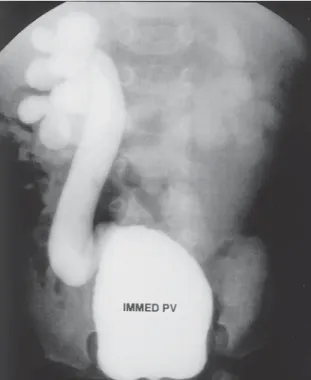
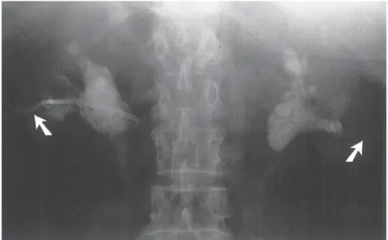
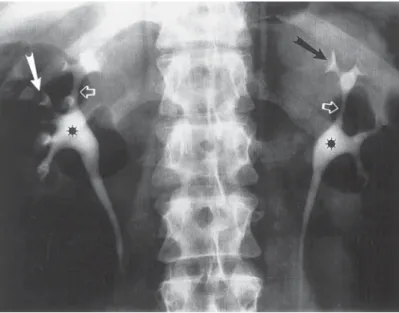
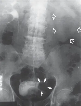
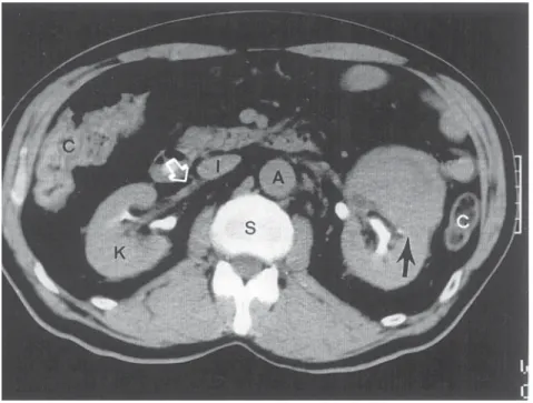
If initial conservative therapy of urolithiasis fails, then intervention of some type is indicated. However, because of the presence of hydronephrosis of pregnancy, the docu- mentation of urolithiasis during pregnancy may not always be straightforward. Hendricks et al. (57) have reported that ultrasound alone confirmed the diagnosis of urinary stones
in 47% of their patients. In symptomatic patients, where ultrasound was not diagnostic, they advocated a limited excretory urogram, which would normally expose the fetus to only 0.4 to 1.0 rad. Stothers and Lee (63) reported that ultrasound had a 34% sensitivity rate and an 86% specificity rate for stone detection in a symptomatic patient.
The use of radiation for diagnostic studies during pregnancy has been and remains controversial. Swartz and Reichling (64) noted the first trimester as the most signifi- cant risk period for limited ionizing radiation exposure during pregnancy. After that time, birth defects and spontaneous abortion were felt to be unlikely. However, sober- ing data are available that would suggest that, as urologists, we should make every effort to avoid fetal exposure to even low doses of radiation. Harvey and associates (65) have reported a case-control study investigating the relationship between prenatal x-ray exposure and subsequent childhood cancer. This was a retrospective study of twins born in Connecticut during a 40-yr period. Twins were chosen because a limited abdominal plain film was used to diagnose the presence of a twin gestation. It was estimated that the radiation dose to the fetus ranged from 0.16 to 4 rads, with an average dose of 1 rad, which is similar to the exposure of a limited intravenous pyelo- gram. Statistical analysis revealed a 1.6 relative risk of leukemia, a 3.2 relative risk of solid childhood cancers, and an overall risk of 2.4 for all childhood malignancies.
Other studies (66,67) also have suggested a relationship between fetal irradiation and subsequent childhood malignancy. It would therefore seem prudent for the urologist to avoid any radiation to the fetus during gestation, unless the radiographic study will leave a major impact on the care of the mother.
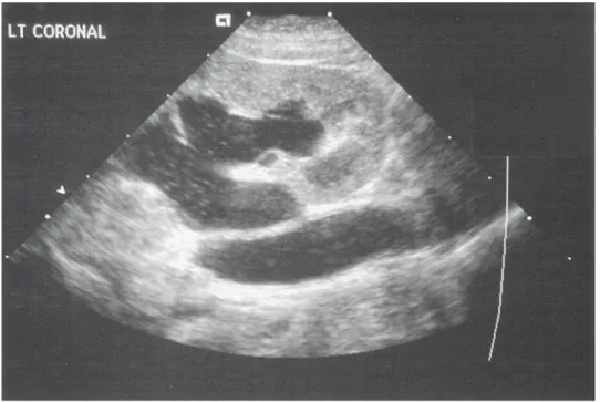
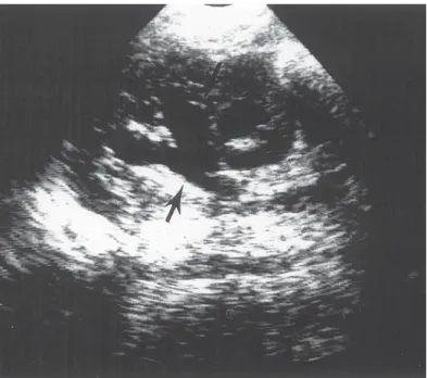
Although radiographic studies may continue to be required in some cases of renal colic during pregnancy, technological advances are making reliance on radiography less compelling. The limitations of transabdominal ultrasound in the diagnosis of ureteral stones in pregnancy have been well described (58,62) However, the use of Doppler ultrasound has been reported as increasing the accuracy of diagnosing ureteral stones (68,69). Doppler ultrasound has been applied to assess the mean intrarenal resistive index as a means of differentiating upper tract dilation from functional obstruction (70).
Hertzberg et al. (71) have demonstrated that the mean intrarenal resistive indices in pregnant women without urinary obstruction (physiologic hydronephrosis) was the same as in nonpregnant women without obstruction.
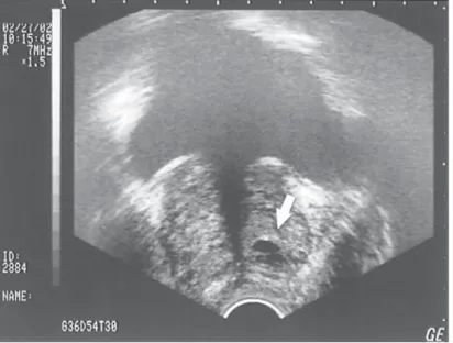
Another technological advance that may enhance the management of ureteral stones in pregnancy without the use of x-ray is the use of the vaginal ultrasound probe. Prelimi- nary experience at our institution suggests that the vaginal ultrasound probe enhances the diagnosis of distal ureteral calculi that may be missed by transabdominal ultrasound alone in both pregnant and nonpregnant patients (72).
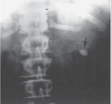
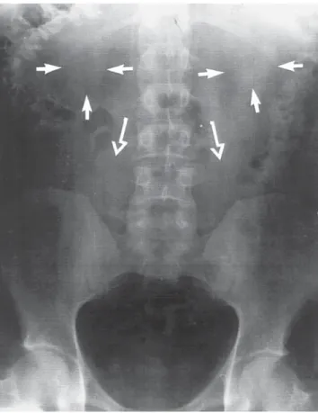
If initial conservative therapy of urolithiasis fails, then intervention of some type is indicated. Our own experience with placement of internal urinary stents has been quite favorable (73). Denstedt and Razvi (74) have stated that a potential drawback of stent placement in pregnancy is the need for x-ray confirmation of stent placement. However, we now use ultrasound alone to guide and confirm ureteral stent placement during pregnancy (75). Jarrard et al. (76) have reported a similar experience.
If a ureteral stent cannot be passed successfully from below using ultrasound guid- ance, then either ureteroscopic stone removal or placement of a percutaneous nephrostomy tube should be considered. Several investigators have reported on the use of a percutaneous nephrostomy for treatment of renal colic during pregnancy (74,75,77–
79). Placement of the nephrostomy tube can be achieved with local anesthesia under
ultrasound guidance. The experience with nephrostomy tubes in pregnancy has gener- ally been satisfactory, although Kroovand (55) cautions that nephrostomy tubes may result in a higher risk of infection and more patient discomfort than internal stents.
Denstedt and Razvi (74) advocate the placement of a percutaneous nephrostomy tube rather than an internal stent in the pregnant patient with urosepsis to ensure adequate drainage. Although there have been reports of successful percutaneous nephrolithotomy in pregnancy (79,80), we agree with Kroovand (55) that because of prolonged anesthesia requirements and the potential harmful effects of ionizing radiation that such procedures are rarely indicated during pregnancy.
A common complication of both ureteral stents and nephrostomy tubes has been stone encrustation (73,78). This may be exacerbated by hyperuricosuria and hypercalciuria that may occur during pregnancy (81,82). The optimal interval for stent or nephrostomy tube changes in pregnancy has not been determined. However, it would appear reason- able to encourage good hydration in all pregnant patients with stents or nephrostomy tubes and to consider tube changes at least every 8 wk during pregnancy.
However, both ureteral stents and nephrostomy tubes may cause the patient discom- fort. Some investigators have advocated ureteroscopic stone removal during pregnancy (83,84). If uteroscopy is used, it should be used without fluoroscopy (85). Extracorporeal shock wave lithotripsy has not been approved for use during pregnancy, and a recent report demonstrates intrauterine growth retardation after exposure to extracorporeal shock wave lithotripsy (ESWL) in an animal model (86). However, a recent report showed no birth defects in the children of six women who had inadvertently undergone ESWL during the first month of pregnancy (87). Nonetheless, the current recommenda- tion is that no pregnant women should undergo ESWL. In a small percentage of patients, open surgical removal of stones may be necessary.
PLACENTA PERCRETA INVADING THE URINARY BLADDER The placenta, normally confined to the decidual lining of the uterine cavity, can in some instances invade the muscular wall of the uterus, a condition known as placenta accreta. Less common is placenta increta, in which placental cotyledons become inter- twined with the muscular stroma of the uterus. Placenta percreta, in which the tropho- blastic tissues penetrate the serosa of the uterus and may extend directly to adjacent structures, including the bladder, is even more rare and potentially life threatening. We have reported our own experience with three cases of placenta percreta invading the urinary bladder and 10 other cases previously reported in the literature (88). All of these cases had a previous history of cesarean section and all presented with hematuria.
Any case of placenta percreta with invasion into the bladder presents the potential for massive blood loss, which the surgical team needs to anticipate. In any woman with a previous history of cesarean section, who presents with hematuria during pregnancy, the diagnosis of placenta percreta should be considered. Ultrasound evaluation is extremely valuable. If cystoscopy is performed, the bladder lesion should not be biopsied, as severe bleeding may occur.
UROLOGICAL CANCER DURING PREGNANCY
It has been estimated that the incidence of malignancy in pregnancy is approx 1 in 1000 (89). Although Gleicher et al. (90) have stated that pregnancy and cancer are the only two biological conditions in which antigenic tissue is tolerated by a seemingly intact
immune system, there does not seem to be any increased incidence of cancer in preg- nancy as compared with nonpregnant women of reproductive age (91).
Walker and Knight (92) have reviewed the subject of renal cell carcinoma in preg- nancy. They emphasize in their literature review that renal adenocarcinoma is the most common renal neoplasm in pregnancy, accounting for almost 50% of renal malignan- cies, with angiomyolipoma being second. The most common symptom of renal cell carcinoma in pregnancy is a palpable mass, which occurred in 88% of patients; hema- turia was noted in 47% of the patients. The diagnostic workup is tailored to limit radiation exposure to mother and child. Abdominal ultrasound and magnetic resonance imaging can adequately identify and stage a solid renal mass in most cases without exposing the mother or fetus to radiation (93). Management of the solid renal mass during pregnancy should be based on the premise that the urologist’s primary responsibility is to the mother. Surgery in the first trimester will result in fetal loss, whereas surgery in the third trimester usually will result in survival of both mother and fetus. When planning man- agement in an individual case, two important facts should be given consideration. First, the doubling time of renal carcinoma has been estimated to be 300 d (94). Second, with improved perinatal care, fetal survival continues to improve. At 28 wk gestation, neo- natal survival rates of over 90% can be expected (95,96). With these caveats, it would seem advisable to operate on all women with a solid renal mass diagnosed in the first trimester. If the mass is diagnosed in the second trimester, it would seem reasonable to continue the pregnancy to 28 wk gestation, test for fetal lung maturity, and then perform a radical nephrectomy with delivery of the infant if labor ensues. Successful cases of simultaneous cesarean section and radical nephrectomy have been reported at 28 wk gestation (97). If a solid renal mass is diagnosed in the third trimester, it is reasonable, after documenting fetal lung maturity, to proceed with radical nephrectomy. Obviously, the cases will be individualized to some extent, based on the wishes and concerns of the mother and other family members.
Ten cases of transitional cell carcinomas of the bladder have been reported during pregnancy (98–104) All pregnant patients with hematuria should have their upper tracts evaluated with renal ultrasound. It should be noted that four patients with blad- der cancer during pregnancy reported in the literature were initially thought to have vaginal bleeding rather than gross hematuria. Our own experience and others suggest that a well-performed bladder ultrasound may obviate the need for cystoscopy (105).
Successful transurethral resection of bladder tumors can be accomplished during preg- nancy and should be performed if a bladder tumor is found. Pheochromocytoma during pregnancy presents an interesting challenge to the obstetrical and surgical team. Pheo- chromocytoma in pregnancy is most dangerous if it remains undiagnosed (106–111).
In these circumstances, maternal mortality exceeds 50% (112–114). Deaths have been caused primarily by malignant hypertensive crisis and/or shock, which is most often precipitated by delivery. The signs and symptoms of pheochromocytoma during preg- nancy are essentially no different than in the nonpregnant patient. The patients most often present with severe preeclampsia or typical attacks of paroxysmal hypertension, sweating, and tachycardia. McCullough (115) has emphasized that supine hyperten- sion is an important clue to pheochromocytoma in the pregnant patient. Plasma cat- echolamines and/or urinary metanephrines and vanillylmandelic acid are measured to document the presence of pheochromocytoma. Radiographic localizing studies can be modified during pregnancy. Greenberg et al. (116) have reported the use of magnetic resonance imaging as an alternative to computed tomography scanning for localiza-
tion of pheochromocytoma during pregnancy. We have successfully used abdominal ultrasound to document the presence of pheochromocytoma during pregnancy. At the present time, there are no reports on the use of meta-(I-131) iodobenzylguanidine during pregnancy.
After the diagnosis of pheochromocytoma has been established, the α-adrenergic blockade should be instituted. Because of the potential mutagenic effects of pheno- xybenzamine, prazosin is considered the α-blocker of choice during pregnancy. Beta blockade may also be necessary, but may slow fetal heart rate and increase myometrial contractility (113,114). Usually, it is safest to proceed with surgical resection regardless of the gestational age of the fetus. With accurate preoperative diagnosis and proper pharmacologic management, mother and child have a good prognosis for survival is surgery if performed in the third trimester.
Janetschek et al. (111) have recently reported two successful cases of the laparoscopic removal of pheochromocytomas during pregnancy. The patients underwent surgery at 16 and 20 wk of gestation. Another case by Aishima et al. (115) has reported the laparoscopic removal of an adrenal during pregnancy because of Cushing’s syndrome.
However, further reports and wider experience will be necessary before the laparoscopic approach to the adrenal during pregnancy becomes the standard of care.
PREGNANCY AFTER URINARY DIVERSION
Successful pregnancies and deliveries have been reported after both continent and standard urinary diversions (115–120). It has been recommended that the mode of de- livery should be guided by obstetric indications (116), although vaginal delivery has been accomplished in the majority of these patients. If a cesarean section is necessary, a urologist should be available to the obstetrical team for consultation. Patients with urinary diversions do not appear to be at greater risk for urinary tract infections during pregnancy than the average patient (117). The physiological changes that occur during pregnancy usually do not compromise the continence mechanisms in patients with either urinary or fecal continent diversions (119–121).
Hill and Kramer (122) reported the management of pregnancy after augmentation cystoplasty in 15 patients. They found urinary tract infection or pyelonephritis in 9 of the 15 patients and premature labor occurred in 4 patients. Their recommendation was that a vaginal delivery could be performed in patients without a history of bladder neck reconstruction. However, if the patient has a history of placement of an artificial sphinc- ter or bladder neck reconstruction, cesarean section should be performed.
PREGNANCY AFTER RENAL TRANSPLANTATION
Murray and associates (123) reported the first successful pregnancy following renal transplantation. It has been estimated that pregnancy occurs in 1 of 200 women of reproductive age on dialysis and 1 in 50 women of childbearing age after successful renal transplantation. Complications during pregnancy are more common in renal transplant patients. Complications that are more likely to occur include urinary tract infections, preeclampsia, premature delivery, premature rupture of membranes, premature onset of labor, and babies that are small for their gestational age (124). Approximately 9% of patients will experience an episode of acute rejection during pregnancy, which is com- parable with that of the nonpregnant population (124). There does appear to be an increased risk of premature delivery in renal transplant patients, which may be the result
of pregnancy-induced hypertension, worsening renal function, or fetal distress (122).
The commonly used immunosuppressive drugs, such as prednisone, azathioprine, and cyclosporine A, present potential teratogenic risks (125,126). However, fetal malforma- tion caused by immunosuppressive drugs appears to be rare and does not contraindicate maintenance of maternal immunosuppression.
PREGNANCY AND URINARY INCONTINENCE
There are three areas of interest regarding pregnancy and urinary incontinence. First, if the patient has had anti-incontinence surgery before surgery does it alter the obstetrical management? Second, what influence, if any, does pregnancy have on urinary inconti- nence during gestation? Third, how does pregnancy impact postpartum continence rates?
The answer to the first question is that cesarean section appears to be the preferred method of delivery in women who have undergone previous anti-incontinence surgery.
A report by Dainer et al. (127) of a cohort of pregnant women who had undergone previous anti-incontinence surgery demonstrated a 73% postpartum continence status in women who had vaginal deliveries compared with a 95% continence rate in those man- aged by cesarean section (p = 0.0344)
The second issue is what effect does the state of pregnancy itself have on continence mechanisms? The answer is that urinary incontinence during pregnancy appears to be quite common. Mason et al. (128) reviewed the literature and found the prevalence of stress incontinence reported to be between 20 and 67%. They followed this with a questionnaire to over 1000 of their own patients and corroborated that 59% of the women responding confirmed stress incontinence during pregnancy, and in 31%, it persisted after delivery.
The third issue of postpartum incontinence is complex and is probably affected by multiple factors. As mentioned above, vaginal deliveries are associated with a higher rate of stress incontinence than cesarean sections. However, at least one study suggests (129) that birth weight per se is not predictive of postpartum incontinence.
THE UROLOGIST IN THE DELIVERY ROOM
Bladder injury has been reported to occur in 4 to 14% of women undergoing cesarean section (130–133). Injuries to the bladder dome are easily recognized and repaired.
However, injuries to the bladder base, which are more often associated with repeat cesarean sections, can transect the entire trigone and present a more difficult operative challenge. In managing such injuries, the ureteral orifices should be identified and cath- eterized before repair of the bladder.
Ureteral injuries occur during 1 in 1000 cesarean deliveries (131–133). Most often, ureteral injuries occur during efforts to control bleeding from the lateral extension of the uterine incision. When repairing ureteral injuries in these circumstances, the urologist should control the bleeding, obtain adequate exposure, and check for other unrecognized injuries to the urinary tract (134). The use of a ureteral catheter and suprapubic tube is preferable in most cases. The preferred operation in most circumstances is a ureteroneo- cystostomy with a psoas hitch (135).
CONCLUSION
In summary, the entire range of urological problems from urinary tract infections to malignancy can be encountered during pregnancy. Following the principles outlined
above, it should be possible to manage all of these problems in a rational manner while minimizing morbidity and mortality to mother and fetus.
REFERENCES
1. Lee MM, Taylor SH, Scott DB, et al. A study of cardiac output at rest during pregnancy. J Obstet Gynaecol Br Commonwealth 1967; 74:319–328.
2. Lund CJ, Donovan JC. Blood volume during pregnancy. Am J Obstet Gynecol 1967; 98: 393–408.
3. Steinberg ES, Santos AC. Surgical anesthesia during pregnancy. Int Anesthesiol Clin 1990; 28:
58–66.
4. Dean M, Stock B, Patterson RJ. Serum protein binding of drugs during and after pregnancy in humans. Clin Pharmacol Ther 1980; 28: 257–261.
5. Santos AC, Pederson H, Harmon TW, et al. Does pregnancy alter the systemic toxicity of local anesthetics? Anesthesiology 1989; 70: 991–995.
6. Veland K, Parer JT. Effects of estrogens on the cardiovascular system of the ewe. Am J Obstet Gynecol 1966; 96: 400–406.
7. Goodman RP, Killam AP, Brash AR, Branch RA. Prostacyclin production during pregnancy:
Comparison of production during normal pregnancy and pregnancy complicated by hypertension.
Am J Obstet Gynecol 1982; 142: 817–822.
8. Prowse CM, Gaensler EA. Respiratory and acid-base changes during pregnancy. Anesthesiology 1965; 26: 381–392.
9. Archer GW, Marx GF. Arterial oxygenation during apnea in parturient women. Br J Anaesth 1974;
46: 358–360.
10. Barron WM. Medical evaluation of the pregnant patient requiring nonobstetric surgery. Clin Peri- natol 1985; 12(3):481–496.
11. Hathaway WE, Bonnar J. Perinatal Coagulation. Grune and Stratton, New York, NY, 1978, pp.
27–51.
12. Letsky E. The hematological system. In: Hytten F, Chamberlain G, eds. Clinical Physiology in Obstetrics. Blackwell Scientific Publications, Oxford, England, 1980, pp. 43–78.
13. de Swiet M. The cardiovascular system. In: Hytten F, Chamberlain G, eds. Clinical Physiology in Obstetrics. Blackwell Scientific Publications, Oxford, England, 1980, pp. 3–42.
14. Kakkar VV. The current status of low-dose heparin in the prophylaxis of thrombophlebitis and pulmonary embolism. World J Surg 1978; 2: 3–13.
15. Simpson KH, Stakes AF, Miler M. Pregnancy delays Paracetamol absorption and gastric emptying in patients undergoing surgery. Br J Anaesth 1988; 60: 24–27.
16. Lindheimer MD, Katz AI. The renal response to pregnancy. In: Brenner BM, Rector RC, eds. The Kidney, 2nd Ed. W.B. Saunders Co., Philadelphia, PA, 1981, pp. 1762–1819.
17. Hsia TY, Shortliffe LM.: The effect of pregnancy on rat urinary tract dynamics. J Urol 1995; 154 (2pt2) 684–689.
18. Schulman A, Herlinger H. Urinary tract dilatation in pregnancy. Br J Radiol 1975; 48: 638–645.
19. Tong YC, Wein AC, Levin RM. Effects of pregnancy on adrenergic function in the rabbit urinary bladder. Urodyn 1992; 11: 525–533.
20. Foldspang A, Mommsen A, Law GW, Elving L. Parity as a correlate of adult female urinary incontinence prevalence. J. Epidemiol Common Health 1992; 46: 595–600.
21. Viktrup L, Lose G, Rolff M, Barfoed K. The symptom of stress incontinence caused by pregnancy or delivery in primiparas. Obstet Gynecol 1992; 79: 945–949.
22. Lee JG, Wein AJ, Levin RM. Effects of pregnancy on urethral and bladder neck function. Urology 1993; 42: 747–752.
23. Barron WM. The pregnant surgical patient: Medical evaluations and management. Ann Intern Med 1984; 101: 683–691.
24. Loughlin KR. Caring for your pregnant patient. Contemp Urol 1992; 4: 22–38.
25. Kammerer WS. Non-obstetric surgery during pregnancy. Med Clin North Am 1979; 6: 1157–
1164.
26. Babaknia A, Hossein P, Woodruff JD. Appendicitis during pregnancy. Obstet Gynaecol 1977; 50:
40–44.
27. Horowitz MD, Gomez GA, Santiesteban R, Burkett G. Acute appendicitis during pregnancy. Arch Surg 1985; 120: 1362–1367.
28. Welch JP. Miscellaneous causes of small bowel obstruction. In: Wekh J, ed. Bowel Obstruction:
Differential Diagnosis and Clinical Management. W.B. Saunders Co., London, England, 1990, pp.
454–456.
29. Woodhouse DR, Haylen B. Gallbladder disease complicating pregnancy. Aust NZ J Obstet Gynaecol 1985; 25: 223–237.
30. Drago JR, Rohner TJ Jr, Chez RA. Management of urinary calculi in pregnancy. Urology 1982;
20: 578–581.
31. Setchell M. Abdominal pain in pregnancy. In Studd J, ed. Progress in Obstetrics and Gynaecology, Vol. 6. Churchill Livingstone, London, England, 1987, pp. 87–99.
32. Smoleniec J, James D: General surgical problems in pregnancy. Br J Surg 1990; 77: 1203–1204.
33. Silen W. Cope’s Early Diagnosis of the Acute Abdomen, 17th Ed. Oxford University Press, New York, NY, 1987, pp. 210–213.
34. Hull LM, Johnson CE, Lee RA. Cholecystectomy in pregnancy. Obstet Gynecol 1975; 9: 291–293.
35. Palahniuk RJ, Schneider SM, Eger EI. Pregnancy decreases the requirements for inhaled anes- thetic agents. Anesthesiology 1974; 41: 82–83.
36. Merryman W: “Progesterone” anesthesia in human subjects. J Clin Endocrinol Metab 1954; 14:
1567–1568,.
37. Lyreras S, Nyberg F, Lindberg B, Terenius L. Cerebrospinal fluid activity of dynorphin-convert- ing enzyme at term pregnancy. Obstet Gynecol 1988; 72: 54–58.
38. Fagraeus L, Urban BJ, Bromage PR. Spread of epidural analgesia in early pregnancy. Anesthesi- ology 1983; 58: 184–187.
39. Datta S, Lambert DH, Gregus J. Differential sensitivities of mammalian nerve fibers during preg- nancy. Anesth-Analg 1983; 62: 1070–1072.
40. Safra M, Oakley GP. Association between cleft lip with or without cleft palate and prenatal exposure to diazepam. Lancet 1975; 2: 478–480.
41. Saxen I, Saxen L. Association between maternal intake of diazepam and oral clefts. Lancet 1975;
2: 498.
42. Krieger JN. Complications and treatment of urinary tract infections during pregnancy. Urol Clin North Am 1986; 13(4):685–693.
43. Sweet RL. Bacteriuria and pyelonephritis during pregnancy. Semin Perinatol 1977; 1: 25–40.
44. Kass EH. A symptomatic infection of the urinary tract. Trans Assoc Am Phys 1956; 69: 56–63.
45. Kass EH. The role of unsuspected infection in the etiology of prematurity. Clin Obstet Gynecol 1973; 16: 134–152.
46. Norden CW, Kass EH. Bacteriuria of pregnancy: a critical appraisal. Ann Rev Med 1968; 19:
431–470.
47. Zinner SH, Kass EH. Long term (10–14 years) follow-up of bacteriuria of pregnancy. N Engl J Med 1971; 285: 820–824.
48. McFadyen IR, Eykyn SJ, Gardner NH. Bacteriuria in pregnancy. J Obstet Gynaecol Br Common- wealth 1973; 80: 385–405.
49. Zinner SH. Bacteriuria and babies revisited. N Engl J Med 1979; 300: 853–855.
50. Kunin, CM. The Concepts of “significant bacteria” and asymptomatic bacteria, clinical syndromes and the epidemiology of urinary tract infections. In: Detection, Prevention and Management of Urinary Tract Infections, 4th Ed. Lea and Febiger, Philadelphia, PA, 1987, pp. 57–124.
51. The Medical Letter. 1991; 33 (849): 71–73.
52. Oesterling JE, Besinger, RE, Brendler CB. Spontaneous rupture of the renal collecting system during pregnancy: successful management with a temporary ureteral catheter. J Urol 1988; 140:
588–590.
53. El Halabi DAR, Humayun MS, Sharhaan JM. Spontaneous rupture of hydronephrotic kidney during pregnancy. Br J Urol 1991; 67(2):219–220.
54. Mostwin J. Surgery of the kidney and ureter in pregnancy. In: Marshall F, ed. Operative Urology.
W.B. Saunders Co., Philadelphia, PA, 1991, pp. 108–113 .
55. Kroovand RL. Stones in pregnancy and in children. J Urol 1992; 148: 1076–1078.
56. Lattanzi DR, Cook WA. Urinary calculi in pregnancy. Obstet Gynecol 1980; 56: 462–466.
57. Hendricks SK, Russ SO, Krieger JN. An algorithm for diagnosis and therapy of management and complications of urolithiasis during pregnancy. Surg Gynecol Obstet 1991; 172: 49–54.
58. Rodriguez PN, Klein AS. Management of urolithiasis during pregnancy. Surg Gynecol Obstet 1988; 166: 103–106.
59. Colombo PA, Pitino R, Pascalino MC, Quoronta S. Control of uterine contraction with tocolytic agents. Ann Obstet Gynecol Med Perinatal 1981; 102: 431–440.
60. Broaddus SB, Catalano PM, Leadbetter GW, Mann LI. Cessation of premature labor following removal of distal ureteral calculus. Am J Obstet Gynecol 1982; 143: 846–848.
61. Gorton E, Whitfield HN. Renal calculi in pregnancy. Br J Urol 1997; 56(1):4–9.
62. Maikranz P, Lindheimer M, Coe F. Nephrolithiasis in pregnancy. Balliere’s Clin Obstet Gynecol 1994; 8: 375–380.
63. Stothers L, Lee LM. Renal colic in pregnancy. J Urol 1992; 148: 1383–1387.
64. Swartz HM, Reichling BA. Hazards of radiation exposure for pregnant women JAMA 1978; 239:
1907–1908.
65. Harvey EB, Boice JD, Honeyman M, Flannery JT. Prenatal x-ray exposure and childhood cancer in twins. N Engl J Med 1985; 312: 541–545.
66. MacMahon B. Prenatal x-ray exposure and childhood cancer. J Natl Cancer Inst 1962; 28: 1173–1191.
67. Mole RH. Antenatal irradiation and childhood cancer: Causation or coincidence? Br J Cancer 1974;
30: 199–208.
68. Burge HJ, Middleton WD, McClennan BL, Dildebolt CF. Ureteral jets in healthy subjects and in patients with unilateral ureteral calculi, comparison with color Doppler US. Radiology 1991; 180:
437–442.
69. Platt JF, Rubin JM, Ellis JH. Acute renal obstruction: Evaluation with intrarenal duplex Doppler and conventional US. Radiology 1993; 186: 685–688.
70. Platt JF, Rubin JM, Ellis JH, DiPietro MA. Duplex Doppler US of the kidney: differentiation of obstructive from non-obstructive dilation. Radiology 1989; 171: 515–517.
71. Hertzberg BS, Carroll BA, Bowie JD, et al. Doppler US assessment of maternal kidneys: Analysis of intrarenal resistivity indexes in normal pregnancy and physiologic pelvicaliectasis. Radiology 1993; 186: 689–692.
72. Laing FC, Benson CB, DiSalvo DN, Brown DL, Frates MC, Loughlin KR. Detection of distal ureteral calculi by vaginal ultrasound. Radiology 1994; 192(2): 545–548.
73. Loughlin KR, Bailey RB Jr. Internal ureteral stents for conservative management of ureteral calculi during pregnancy. N Engl J Med 1986; 315: 1647–1649.
74. Denstedt JD, Razvi H. Management of urinary calculi during pregnancy. J Urol 1992; 148:
1072–1075.
75. Gluck CD, Benson, Bundy AL, Doyle CJ, Loughlin KR. Renal sonography for placement and monitoring of ureteral stents during pregnancy. J Endourol 1991; 5: 241–243.
76. Jarrard DJ, Gerber GS, Lyon ES. Management of acute ureteral obstruction in pregnancy utilizing ultrasound-guided placement of ureteral stents. Urology 1993; 42: 263–268.
77. Horowitz E, Schmidt JD. Renal calculi in pregnancy. Clin Obstet Gynecol 1985; 28: 324–338.
78. Rodriguez PN, Klein AS. Management of urolithiasis during pregnancy. Surg Gynecol Obstet 1988; 166: 103–106.
79. Kavoussi LR, Albala DM, Basler JW, Apte S, Clayman RV. Percutaneous management of uroli- thiasis during pregnancy. J Urol 1992; 148: 1069–1071.
80. Holman E, Toth C, Khan MA. Percutaneous nephrolithotomy in late pregnancy. J Endourol 1992;
6: 421–424.
81. Boyle JA, Campbell S, Duncan AM, Greig WR, Buchanan WW. Serum uric acid levels in normal pregnancy with observations on the renal excretion of urate in pregnancy. J Clin Pathol 1966; 19:
501–503.
82. Gertner JM, Coustan DR, Kliger AS, Mallette LE, Ravin N, Broaddus AE. Pregnancy as state of physiologic absorptive hypercalciuria. Am J Med 1986; 81: 451–456.
83. Vest JM. Ureteroscopic stone manipulation during pregnancy. Urology 1990; 35: 250–252.
84. Rittenberg MH, Bagley DH. Ureteroscopic diagnosis and treatment of urinary calculi during pregnancy. Urology 1988; 32: 427–428.
85. Shokei AA, Mutabagani H. Rigid ureterscopy in pregnant women. Br J Urol 1998; 81: 678–681.