저작자표시-비영리-변경금지 2.0 대한민국 이용자는 아래의 조건을 따르는 경우에 한하여 자유롭게
l 이 저작물을 복제, 배포, 전송, 전시, 공연 및 방송할 수 있습니다. 다음과 같은 조건을 따라야 합니다:
l 귀하는, 이 저작물의 재이용이나 배포의 경우, 이 저작물에 적용된 이용허락조건 을 명확하게 나타내어야 합니다.
l 저작권자로부터 별도의 허가를 받으면 이러한 조건들은 적용되지 않습니다.
저작권법에 따른 이용자의 권리는 위의 내용에 의하여 영향을 받지 않습니다. 이것은 이용허락규약(Legal Code)을 이해하기 쉽게 요약한 것입니다.
Disclaimer
저작자표시. 귀하는 원저작자를 표시하여야 합니다.
비영리. 귀하는 이 저작물을 영리 목적으로 이용할 수 없습니다.
변경금지. 귀하는 이 저작물을 개작, 변형 또는 가공할 수 없습니다.
Master’s Thesis of Science in Agriculture
Biofunctional Human Cystatin-C Expressed in Transgenic Chickens
형질 전환 닭에서 생산하는 생기능성 인간 시스타틴
CFebruary 2020
Seo Woo Kim
Department of International Agricultural Technology Graduate School of International Agricultural Technology
Seoul National University
Biofunctional Human Cystatin-C Expressed in Transgenic Chickens
A thesis
submitted in partial fulfillment of the requirements to the faculty of Graduate School of International Agricultural Technology
for the Degree of Master of Science in Agriculture
By Seo Woo Kim
Supervised by Prof. Tae Sub Park
Major of International Agricultural Technology Department of International Agricultural Technology Graduate School of International Agricultural Technology
Seoul National University
December 2019
Approved as a qualified thesis
for the Degree of Master of Science in Agriculture by the committee members
Chairman Byung-Chul Park, Ph.D.
Member Tae Min Kim, Ph.D.
Member Tae Sub Park, Ph.D.
Abstract
Bioreactor is the massive production system for bioactive substances as well as therapeutic proteins. Although the various methods such as microorganisms and animal cells, the efficient strategy should be necessary for the efficient production of biofunctional proteins. In the production of therapeutic proteins, post-translational modification is not properly performed in microorganisms. In addition, animal cell systems require more cost and time. To overcome these problems, a new method has been considered with transgenic animals such as the genetically- modified hen’s eggs. Human cystatin-C is a member of cystatin superfamily. It is produced in the body at a constant rate found in body fluids and secretions. Moreover, this protein acts as an inhibitor of cysteine protease and protects against microbial and parasitic infiltration.
We aim to construct transgenic chickens that produce human cystatin-C using a non-viral piggyBac transposon system. We generated transgenic chicken expressing human cystatin-C protein by using non-viral piggyBac transposon system. We analyzed the expression of human cystatin-C in DF1 cells and PGCs, and we produced transgenic chickens using the human cystatin-C expression vector. Expression of human cystatin-C protein in transgenic chickens were verified through Western blot and ELISA. Biofunctional activity of purified human cystatin-C in transgenic
chickens was confirmed through in vitro assay. Thus, these data suggests that transgenic chickens could be efficiently used as a bioreactor for production of various bioactive materials.
Key words: transgenic chicken, bioreactor, human cystatin-C Student Number: 2018-26749
Contents
Abstract ………..…………ⅰ Contents ……….ⅲ List of Tables ………..ⅵ List of Figures ……….ⅶ List of Abbreviation ………ⅸ
Introduction ……….. 1
Review of Literature ………... 4
1. Bioreactor ……… 4
1.1 Definition of bioreactor ……….. 4
1.2 Bacterial expression system ………. 5
1.3 Animal cell expression system ……….. 7
1.4 Transgenic animal expression system ……… 12
1.4.1 Transgenic chicken expression system
……… 14
2. Cystatin-C ……… 15
2.1 cystatin-C ………. 16
2.2 Kidney function biomarker ………. 18
2.3 function of cystatin-C ……… 21
3. Production of transgenic chicken using non-viral piggyBac transposon system ……….. 24
3.1 piggyBac transposon system ………. 24
3.2 PGCs ……… 25
Materials and Methods ………. 28
1. Construction of human cystatin-C expression vector system using piggyBac transposon .………. 28
2. Transfection of human cystatin-C expression vector to DF1 cell & PGCs ………..………. 28
3. Microinjection of cultured PGCs into recipient embryos ……… 29
4. Production of transgenic chickens ………. 29
5. Genomic PCR ……… 30
6. Purification of human cystatin-C in egg white and
muscle ………. 30
7. ELISA ……….. 31
8. Western blotting ………. 31
9. Antimicrobial activity test ……….. 32
Results ……….. 33
1. Construction of human cystatin-C expression vector ………. 33
2. Identification of vector expression in DF1 cells and PGCs ………. 35
3. Production of germ-line chimera and transgenic offspring ………. 37
4. Identification of human cystatin-C expression in transgenic chickens ……… 39
5. Purification of human cystatin-C in transgenic chickens’ egg white and muscle ……… 41
6. Antimicrobial activity of human cystatin-C in transgenic chickens’ egg white ……….. 43
Discussion ……….. 46
Conclusion ……….. 51
References ………. 52
Abstract in Korean ……….. 63
List of Tables
Table 1. Comparison of each expression systems producing
recombinant pharmaceutical proteins ……….. 11
Table 2. Total estimated costs-of-goods (COGS)
comparison between animal cell expression system and
transgenic animal expression system ……… 11
List of Figures
Figure 1. Comparison of production systems for approved biopharmaceuticals ……….. 10 Figure 2. Comparison of creatinine and cystatin-C as kidney markers ……….. 20
Figure 3. Antimicrobial activity of cystatin-C ……… 22 Figure 4. Antiviral activity of cystatin-C ……… 23
Figure 5. Construction of human cystatin-C expression
vector system using piggyBac transposon ……… 34
Figure 6. Identification of human cystatin-C in DF1 cells
and PGCs ………. 36
Figure 7. Production of germ-line chimera and transgenic
offspring ……….. 38
Figure 8. Expression of human cystatin-C in transgenic
chickens ……… 40
Figure 9. Purification of human cystatin-C in transgenic
chickens’ egg white and muscle ……… 42
Figure 10. Antimicrobial activity of human cystatin-C in
transgenic chickens’ egg white against E. coli ………….. 44
List of Abbreviation
CMV : Cytomegalovirus EF1 : Elongation factor 1
PBS : Phosphate-buffered saline PGC : Primordial germ cell
PCR : Polymerase chain reaction
ELISA : Enzyme-linked immunosorbent assay RIPA : Radioimmunoprecipitation
ECL : Enhanced chemiluminescence WT : Wild-type
TG : Transgenic EW : Egg white
H cystatin : Human cystatin
Introduction
Bioreactor is any manufactured device or system that supports a biologically active environment. It includes prokaryotic, eukaryotic cell system and transgenic animal using aerobic or anaerobic condition.
Through this system, it is possible from sewage treatment, fermentation to producing antibody, vaccine and recombination protein.
In 1980s, after recombinant DNA (rDNA) technology developed, recombinant human insulin was approved to FDA for the treatment and enter the market as a first recombinant pharmaceutical protein. Since then, recombinant protein production using prokaryotic cell like E.coli and yeast was developed (Ferrer-Miralles, Domingo-Espin et al. 2009).
However, bacteria and yeast have inaccurate post-translational modification (Jenkins 2007). Because of this disadvantage, recombinant pharmaceutical proteins can reduce efficacy, limit their shelf life, occur unwanted side-effects like immune reaction. Thus, in some cases, animal cell system is used to produce recombinant proteins. But compared to other production systems, production level is relatively low (Houdebine 2009). If production level is similar with others, the production cost will be increasing. Therefore, therapeutic protein production system using transgenic animal is getting attention.
In 2009, The FDA in USA first approved recombinant human anti-
thrombin-α in transgenic goat(Kling 2009). recombinant human anti- thrombin-α called ATryn produced in transgenic goat’s milk. Compared with human plasma version of ThrombateⅢ and ATrym, both efficacy is similar but production cost of ATryn is cheaper. Moreover, human plasma product can cause blood-borne diseases but ATryn is safe about these side-effect. However, there is a view that needs more research on protein maturation or secretion.
There are various transgenic animals for bioreactor. Among them, chickens have short generation and continue to produce eggs. So, it can be possible to get recombinant pharmaceutical proteins continuously. Also, it is easier to purification protein in egg white because it has no fat unlike milk. Moreover, most mammals except human produce α1,3- galactose but chicken has not. So there is no side effect like immune response when we use recombinant protein in chickens (Lillico, McGrew et al. 2005, Houdebine 2009).
Cystatin-C is single polypeptide chain structure containing 120 amino acid, molecular weight is 13kDa. Also, it is member of cystatin super family. In human, all cells with a nucleus produce it. So it can found in all tissues and body fluids like blood, sperm, saliva (Zi and Xu 2018). In addition, cystatin-C is as known as kidney function biomarker. The most commonly used kidney marker is creatinine, but some studies have shown that cystatin-C is a better indicator of glomerular filtration than creatinine (Dharnidharka, Kwon et al. 2002, Stevens, Coresh et al. 2008, Bagshaw
and Bellomo 2010). Acute kidney injury (AKI) in children and newborns is defined as a sudden decline in glomerular filtration rate and is most important to detect and treat early because AKI is asymptomatic, and the symptoms become apparent after renal function has declined significantly. Cystatin-C is a biomarker that has been studied a lot as one of AKI diagnostic methods (Bagshaw and Bellomo 2010, Nakhjavan- Shahraki, Yousefifard et al. 2017, Khosravi, Zadkarami et al. 2018).
Cystatin-C plays as cysteine protease (cathepsin B, H, L, S, elastase and papain) competitive inhibitor that blocks the cutting of peptide bonds (Shamsi and Bano 2017). The interaction of Cathepsin with cystatin-C inhibits angiogenesis and tumor growth (Wang, Sun et al. 2006). In addition, because cystatin-C inhibits cathepsin, serum cystatin-C can be used as an important marker for inflammation in COPD patients (Zhang, Li et al.
2016). Cystatin-C can protect body from virus and bacteria’s invasion, toxicity, reproduction by inhibiting cysteine protease of the microorganism (Sun, Liu et al. 2013, Hernandez, Marcet et al. 2014, Luthra 2015).
In this study, we produce transgenic chicken for bioreactor with human cystatin-C expression vector system using non-viral piggyBac transposon.
We designed the cystatin-C to appear throughout the chicken’s whole body. Therefore, our goal is to purify and use cystatin-C in all part of chicken not only egg white but also muscle.
Review of Literature
1. Bioreactor
1.1 Definition of bioreactor
A bioreactor is defined that any manufactured device or system that supports a biologically active environment. In addition, if one focuses only on the aspect of cell culture, a device or system for culturing cells or tissues is also called a bioreactor. This system can be in aerobic or anaerobic state. Depending on how it works, bioreactors are divided three types; batch cultures, fed-batch cultures, continuous cultures. Biocatalysts used in bioreactors include enzymes, microorganisms, animal cells, and plant cells, which are used in suspension or immobilized. In a bioreactor, specific biochemical reactions occur by enzymes, microorganisms, animal or plant-derived cells, or intracellular structures such as chloroplasts or mitochondria to obtain the desired materials.
Examples of bioreactors include sewage treatment, fermentation, and the manufacture of recombinant pharmaceuticals called pharming or molecular farming. In the case of recombinant drug production, animal cells are usually used. However, agitated environments such as microbial
bioreactor devices are disruptive to these cell types and tissues, and require highly specialized growth media for animal cells. Thus, quite different designs are required compared to general industrial bioreactors used to grow protein expression systems such as yeast and bacteria.
1.2 Bacterial expression system
In 1968, Swiss microbiologist Werner Arber discovered restriction enzymes (Piekarowicz 1979) and the following year US microbiologist Hamilton O. Smith purified type II restriction enzymes (Kelly and Smith 1970). The enzyme has been shown to be essential for genetic engineering due to its ability to cleave at specific sites in DNA as opposed to type I restriction enzymes. This discovery led to the emergence of recombinant DNA technology. Based on Smith's work, American molecular biologist Daniel Nathans showed that type II enzymes could be useful for genetic research in 1970-71 (Danna and Nathans 1972, Danna, Sack et al. 1973, Danna and Nathans 1999). At the same time, American biochemist Paul Berg developed a way to isolate DNA molecules from selected sites and attach molecular segments to DNA in viruses or plasmids to enter bacterial or animal cells (Jackson, Symons et al. 1972). In 1973, American biochemists Stanley N. Cohen and Herbert W. Boyer first cloned recombinant genes into bacterial cells and cloned them (Cohen, Chang et al. 1973). With the development of such recombinant DNA (rDNA)
technology in the 1970s, attempts have been made to create new gene combinations that are of value to science, medicine, agriculture and industry using recombinant DNA technology.
In October 1982, E. coli-produced recombinant human insulin(called Humulin) first became FDA approved and was produced (Quianzon and Cheikh 2012). Previously, insulin was isolated from pigs and bovines’
pancreas to treat diabetes. It was developed by Genentech which is the first US biotechnology company. Humulin licensed Eli Lilly and became the first marketable product through recombinant DNA technology. Also, it was the first recombinant drug approved by the FDA for use in the United States. Humulin is slower than normal insulin and has a longer activity period, making it suitable for use as a drug. Since Humulin received FDA approval, the production of useful proteins in E. coli or yeast has been increased.
In comparison with the expression system using microorganisms such as E. coli and yeast and the reaction process of the previous chemical industry, In the chemical industry, metal-based catalysts require a lot of energy and resources, but in the case of microorganisms, necessary materials are produced by enzymes and microbial biological functions.
Moreover, unlike other catalysts, enzymes act only on specific reactions.
Therefore, the bacterial expression systems do not produce unnecessary substances. It is possible to save resources, save energy, and to reduce the cost of construction and equipment since it can be a small factory.
In addition, it does not occur smoke, harmful substances, noise pollution, etc. The rapid growth of microorganisms allows the production of large quantities of material in a short time. Disadvantages include that the expressed protein is often mis-folded and can be biologically inactive, cannot be produced if the protein produced is toxic to microorganisms, and lacks the enzymes responsible for post-translational modifications compared to other species. There is no device for attaching sugar residues of glycoproteins in bacteria. If post-translational modifications are not carried out correctly, the useful protein may be reduced, the rate of protein degradation may be shortened, and the shelf life may be shortened, and when used as a drug, it is likely to cause side effects such as an immune response.
1.3 Animal cell expression system
Animal cell expression systems are widely used in recombinant protein production because of post-translational modifications that are not achieved in bacterial expression systems. Cultured mammalian cell lines such as Chinese hamster ovary (CHO), COS and human cell lines such as HEK and HeLa can be used to produce proteins. The vector is transfected into the cell and, in the case of stable transfection, DNA can be integrated into the genome or transfected temporarily by homologous recombination. Examples of mammalian expression vectors include the
adenoviral vectors (Berkner 1992), the pSV and the pCMV series of plasmid vectors, vaccinia and retroviral vectors (Hruby 1990), as well as baculovirus (Kost and Condreay 2002). The promoters for cytomegalovirus (CMV) and SV40 are commonly used in mammalian expression vectors to drive gene expression. Non-viral promoter, such as the elongation factor (EF)-1 promoter, is also known (Kim, Uetsuki et al. 1990).
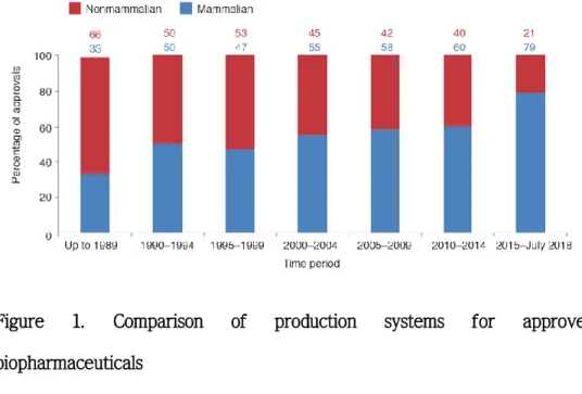
Examination of recombinant antibodies approved from 1989 to 2018 shows a gradual increase in products produced through mammalian cells (Figure 1). This suggests that the post-translational modification process is very important in the manufacture of recombinant pharmaceuticals.
However, the drawback of this animal cell expression system, which is mainly used for protein production, is its low output for the price.
Compared to bacterial expression systems, post-translational modifications and glycosylation are superior to mammalian cells. However, the yield is significantly lower compared to bacterial or transgenic animal systems (Table 1). Accordingly, there have been attempts to develop relatively high productivity recombinant cell lines through improvements in vector construction, selection of selectable markers and advances in gene- targeting and high throughput screening strategies (Gurtu, Yan et al.
1996, Pu, Cashion et al. 1998). Another disadvantage is the high cost of maintaining an animal cell culture system and the difficulty of scaling up the system for mass translation purposes. It would take three to five years to build a large manufacturing facility for mammalian cells and
cost $ 250 million (Dyck, Lacroix et al. 2003). Production costs are also more expensive for animal cell systems when compared to transgenic animal systems (Table 2)
Figure 1. Comparison of production systems for approved biopharmaceuticals
Relative use of mammalian- versus nonmammalian-based production cell lines in the manufacture of biopharmaceuticals approved over the indicated periods. Each dataset is expressed as a percentage of total biopharmaceutical product approvals for the period in question. (Figure adapted from (Walsh 2018))
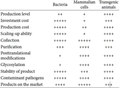
Table 1. Comparison of each expression systems producing recombinant pharmaceutical proteins
(Table adapted from (Wang, Zhao et al. 2013))
Table 2. Total estimated costs-of-goods (COGS) comparison between animal cell expression system and transgenic animal expression system
Presented by H.L. Levine, 2002 BIO International Biotechnology Convention and Exhibition, 9–12 June, Toronto, Ontario, Canada. (Table adapted from (Dyck, Lacroix et al. 2003))
1.4 Transgenic animal expression system
In February 2009, recombinant human anti-thrombin produced by transgenic goats became the first transgenic animal-producing drug under FDA approval and began to be produced under the name ATryn (Kling 2009). Anti-thrombin has the properties of an anticoagulant. Expression of ATryn in milk is achieved by linking antithrombin-α transgenes to the breast-specific promoter of milk proteins. The company that developed ATryn, rEVO Biologics (formerly GTC Biotherapeutics), says one transgenic goat can produce the same amount of anti-thrombin as 90,000 blood donations per year. The reason why they chose goat among many animals was that it was able to produce more rapidly than cows and produce more protein than rabbits and mice (Stix 2005). When comparing THROMBATE III, an anti-thrombin product extracted from human plasma, and ATryn, which is produced by transgenic goats, the efficacy of both products is the same, but anti-thrombin can be produced at a lower cost with transgenic goats. Human plasma products may also be a cause of blood-borne diseases, but they are safe from this risk when produced with transgenic goats.
The ability of transgenic animals to produce complex biologically active proteins in an efficient and economical way is superior to bacteria, mammalian cells, transgenic plants and insects. It takes three to five years to build a large (10,000 liter bioreactor) manufacturing facility for
mammalian cells and costs between $ 250 and $ 500 million, while transgenic farms with a single purification facility should not exceed $ 80 million (Dyck, Lacroix et al. 2003). Establishing a commercial production herd of transgenic goats in a company can be accomplished at a cost of about one tenth of the cost of building a commercial cell culture facility (Thiel 2004).
In general, because milk is easily collected in large quantities, it has been considered an organ of choice for expressing recombinant proteins in transgenic animal bioreactors. Milk is currently the best bioreactor.
Foreign proteins are generally reported to be produced in the milk of transgenic animals at the rate of several grams per liter. However, the production of protein in milk is limited by the relatively long intervals from birth to the first feeding experienced by domestic livestock, the discontinuous nature of the feeding cycle, and the considerable time and material investment required to produce genetically modified dairy animals.
In addition, certain bioactive proteins produced in milk can adversely affect animal health. This is especially true when high concentrations are produced and proteins are resorbed. These shortcomings have led to the search for other types of collectable fluids. Human α-AT was successfully obtained at high levels from the serum of transgenic rabbits (Massoud, Bischoff et al. 1991) and human hemoglobin was produced in the transgenic pig circulation (Swanson, Martin et al. 1992). In addition, recombinant antibodies were produced in the blood of transgenic goats,
pigs, and rabbits. However, blood is not an ideal body fluid for obtaining recombinant protein because it is invasive and can affect the health of the animal when producing bioactive proteins.
1.4.1 Transgenic chicken expression system
In 2015, the recombinant lysosomal acid lipase produced by transgenic chickens was approved by the FDA and began producing under the name Kanuma. To produce this drug, transgenic chickens were constructed using genetic modification techniques to produce recombinant LAL (rhLAL) in chicken egg whites (Sheridan 2016). It can then be extracted and purified from egg whites and used as a drug. At the time of FDA approval, Kunama was the first drug manufactured from chicken eggs and designed for human use. The first approved treatment for LAL-D patients is administered via intravenous infusion. LAL-D is a very rare disease and is defined as a disease that affects fewer than 20 patients per million people in the general population. It is almost always fatal in infants and children and adult patients are at high risk of fatal organ damage and premature death (Reiner, Guardamagna et al. 2014, Jones, Valayannopoulos et al. 2016). Kanuma (sebelipase alfa) is an innovative enzyme replacement therapy that addresses the root cause of lysosomal acid lipase deficiency (LAL-D) by reducing substrate accumulation in cellular lysosomes throughout the body. In clinical studies, Kanuma
treatment improved the survival rate of infants with LAL-D, and markedly improved lipid parameters as well as a significant reduction in ALT and liver fat content in children and adults with LAL-D.
The advantage of the transgenic chicken system is that it is easy to purify because the generation is shorter than other animals, and about 300 eggs are produced annually, which allows for continuous recombinant protein production. Egg white is easy to purify because it does not contain fat and consists only of water and protein. Also, unlike other mammals, there is α1,3-galactose, a glycoprotein not found in humans and apes. In humans, the gene for α1,3-galactosyl transferase is inactive and 1% of B lymphocytes respond to intestinal bacteria. It produces anti α1,3-galactose antibodies and contributes to the rejection of tissue
transplants in other mammals. Because of this, the presence of α1,3- galactose epitopes in recombinant drugs produced in mammals can cause side effects such as immune responses. However, in chickens, the recombinant protein produced through transgenic chickens is safe from this risk since it does not produce α1,3-galactose (Lillico, McGrew et al.
2005).
2. Cystatin-C
2.1 cystatin-C
Cystatin-C was first mentioned in 1961 when Jorgen Clausen described the name "γ-CSF" in the human cerebrospinal fluid (CSF) of the
"cerebrospinal fluid specific" protein (Clausen 1961). At the same time, Butler and Flynn described a new protein in human urine that moves in the γ- region of the electrophoretic pattern. It is named post-γ protein (Butler and Flynn 1961). In 1962, Hochwald and Thorbecke called γ-trace to the ascitic and pleural fluids of human plasma, cerebrospinal fluid, urine and proteins with γ-electrophoretic mobility (Hochwald and Thorbecke 1962). The complete amino acid sequence of human cystatin- C was finally determined by Anders Grubb and Helge Löfberg in 1981 (Grubb and Lofberg 1982).
Cystatin-C is a member of the cystatin superfamily. Cystatin superfamily can be divided into four types (Mussap and Plebani 2004).
Type 1 cystatin is also called stefins. It is mainly located intracellularly and typically human cystatin-A (stefin A), human cystatin-B (stefin B), and rat cystatin α and β belong to this type. Type 1 cystatin is generally considered to be a cytoplasmic protein but appears to be secreted or at least released in certain situations. Type 2 cystatins include cystatin-C, N, SN, SA, D, E / M, among which all the staphylostats identified as
ortholog have cystatin-C like proteins. Cystatin-D is considered an additional component of the oral non-immune protective system. Cystatin- F, also called leukocystatin, is primarily expressed in immune system cells.
Cystatin E / M is expressed in most human tissues, but expression levels vary from tissue to tissue. In particular, abundant expression in fetal skin and amniotic cells plays an important role in fetal development. Type 3 cystatin is a kininogen, primarily intravascular proteins, present in plasma and secretions. Kininogens inhibit cysteine peptidase. Type 4 cystatin is also called fetuins and is mainly synthesized in the liver. During fetal life it has been identified as a major protein mainly in the blood and brain and is considered a major protein in several metabolic pathways, including the regulation of bone formation and bone resorption. Furthermore, fetuins opsonize cationic macrophage-deactivating molecules and appear to be a key partner in the recovery step of an acute phase response to systemic inflammation.
The cystatin-C gene is located at the cystatin locus and comprises 3 exons, spanning 4.3 kilo-base pairs. It is found in high concentrations in fluids and is a housekeeping gene that expresses any nucleated cell.
Cystatin-C is a non-glycosylated, basic protein (isoelectric point at pH 9.3). Cystatin-C protein has 120 amino acids and is about 13 kDa in size.
The structural feature is a single polypeptide chain structure with short alpha helices and long alpha helices across a large antiparallel, five- stranded beta sheet. It has two disulfide bonds and about 50% of the
molecule carries a hydroxylated proline.
2.2 Kidney function biomarker
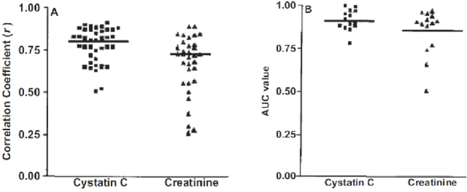
Cystatin-C has a low molecular weight and is removed from the bloodstream by glomerular filtration in the kidney. Reduced kidney function and glomerular filtration rate increase blood levels of cystatin- C. several studies demonstrate that serum levels of cystatin-C are more accurate tests of kidney function than serum creatinine levels (Figure 2).
Serum creatinine is the most widely used method of indirectly measuring glomerular filtration rate in clinical practice, but it can be influenced by various factors such as sex, age, muscle mass, diet, physical activity, and there is a drawback that does not reflect rapid changes in glomerular filtration rate or abnormal initial renal function. In addition, creatinine clearance will reflect the actual glomerular filtration rate excessively as creatinine is secreted from the proximal tubules in addition to glomerular filtration. Moreover, in chronic renal failure, the excretion of creatinine through the tubules is further increased, and it is difficult to accurately collect urine 24 hours. In comparison, the clinical utility of serum cystatin- C has recently been known. Several studies have shown that cystatin-C levels are more sensitive markers for measuring mortality in the elderly, cardiovascular disease, and acute kidney injury (AKI) in children (Salminen, Laine et al. 2016, Garcia-Carretero, Vigil-Medina et al. 2017, Nakhjavan- Shahraki, Yousefifard et al. 2017). However, it is difficult to commercialize
cystatin-C assays because they are three times less expensive and 20 times more expensive than creatinine assays.
Figure 2. Comparison of creatinine and cystatin-C as kidney markers Cystatin-C (Cys C) versus creatinine (Cr) as markers of glomerular filtration rate (GFR). (A) Scatterplot of correlation coefficients (r) for 1/Cys C and 1/Cr with measured GFR. Horizontal line represents the cumulative mean r of all studies analyzed. (B) Scatterplot of receiver operating characteristic plot area under the curve (AUC) values for Cys C and Cr. Horizontal line represents the cumulative mean of all 14 data sets. Adapted and reproduced from Dharnidharka et al7 with permission of the National Kidney Foundation. (Figure adapted from (Shlipak, Mattes et al. 2013))
2.3 function of cystatin-C
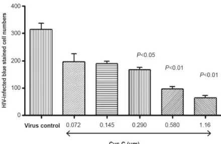
Despite the diversity of structure and distribution in the body, all members of the cystatin superfamily have in common cysteine proteinase inhibitory properties, suggesting the preservation of physiological functions through evolution. Their physiological role is to regulate the activity of endogenous proteolytic enzymes that are often secreted or leaked from lysosomes of dead or diseased cells. In addition, cystatin-Cnot only has antimicrobial activity against microorganisms (Figure 3), but also enters cells whose cell membranes have been damaged by viral attacks, which can inhibit intracellular cysteine proteinases as an important function (Korant, Brzin et al. 1985). It may interfere with viral replication requiring a host or viral cysteine proteinase, such as capsid maturation (Figure 4).
Especially, human cystatin-C totally blocks replication of Herpes simplex virus (Bjorck, Grubb et al. 1990), with an activity comparable with that of acyclovir (Bjorck 1990). It is also an effective inhibitor of the replication of Coronavirus (Collins and Grubb 1991), which can cause acute gastroenteritis. In order to increase the blocking efficacy of cysteine C's cysteine protease inhibitory activity, several peptidomimetic compounds that mimic their structure have been synthesized (Grubb, Abrahamson et al. 1990, Kasprzykowski, Schalen et al. 2000, Pikula, Smuzynska et al. 2017).
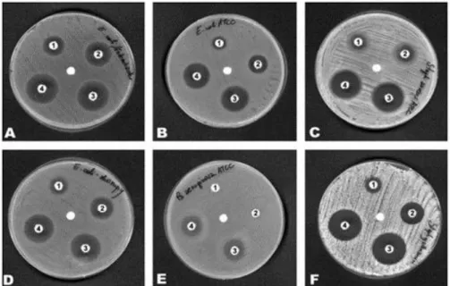
Figure 3. Antimicrobial activity of cystatin-C
Inhibition zones of six strains of (A) Escherichia coli [02], (B) Escherichia coli ATCC 25922, (C) Staphylococcus aureus ATCC 25923, (D) Escherichia coli S, (E) Pseudomonas aeruginosa ATCC 27853, (F) Staphylococcus gallinarum were spread on Muller-Hinton agar plates on which which paper disks impregnated with 80 μg (1), 100 μg (2), 120 μg (3), and 150 μg/disk (4) of chicken cystatin were placed. In the centre of the plate, the disk paper without cystatin added. Incubation period was 24 h at 37℃. (Figure adapted from Ref. World Journal of Microbiology &
Biotechnology 2005 21: 59-64)
Figure 4. Antiviral activity of cystatin-C
Cystatin-C exhibited a dose-dependant decrease of blue-stained HIV- infected cells as compared to virus only control. (Figure adapted from (Vernekar, Velhal et al. 2015))
3. Production of transgenic chicken using non-viral piggyBac transposon system
3.1 piggyBac transposon system
In general, when producing transgenic chickens, retroviral vectors are used to efficiently integrate and express genes in the host genome.
However, the size of transgenes that can be introduced into retroviral vectors is limited (Wu, Yang et al. 2010), and the vector is less efficient at integrating the transgenes into the host chromosome (McGrew, Sherman et al. 2004). In addition, safety issues due to the use of viral vectors cannot be ignored. Therefore, there is a need to find ways to make transgenic chickens safe and efficient.
DNA transposons are DNA sequences, also known as "jumping genes"
that can be moved and integrated into other locations within the genome.
DNA transposons can move in the DNA of an organism through single or double stranded DNA intermediates (Feschotte and Pritham 2007). DNA transposons were first discovered in maize (Mc 1950). DNA transposons have been found in both prokaryotes and eukaryotes. In prokaryotes, transposable elements can promote horizontal movement of other genes related to antibiotic resistance or toxicity. Among the DNA transposons, piggyBac transposon isolated from cabbage looper is a TTAA-specific, short repeating element, a transposon group that shares similarity in
structure and properties of movement (Fraser, Smith et al. 1983). PiggyBac has a fairly high potential efficiency and accommodates relatively large transgenes without compromising potential efficiency (Wu, Ying et al.
2007). Previous studies have shown that the piggyBac transposon system is efficient for the production of transgenic chickens (Park and Han 2012).
3.2 PGCs
PGCs can be identified based on the morphological features of the large globular nuclei, the developed Golgi and endoplasmic reticulum, and the presence of refractive lipids in the cytoplasm. PGCs, which temporarily circulate through the bloodstream and travel to the genital ridges, are easily distinguished from red blood cells because of their characteristic shape and larger size. These morphological properties of PGCs are widely preserved among bird species (for example, quails, ducks, pigeons, pheasants, etc.). In addition, PGC can be identified through staining (Meyer 1964) and immunological markers (Urven, Erickson et al.
1988, Maeda, Ohsako et al. 1994, Jung, Kim et al. 2005) for the cell surface glycoproteins present in the PGC such as SSEA-1 or the chicken vasa homolog (CVH) protein located in the cytoplasm. The main biological characteristics of algal PGCs are as follows. First, the unique route of passage to the genital ridges through the blood circulation, and second, the ability to propagate long-term in vitro, is limited to the gonads. These have led to the establishment of unique developmental techniques based
on PGC manipulation in algae to generate genetically modified poultry and to conserve poultry genetic resources (Tagami, Miyahara et al. 2017).
Unlike mammals, avian PGCs use the embryo's circulatory system to move from the embryonic crescent to the future gonads. The unique migration characteristics of PGCs provide opportunities for identification and isolation of cells.
In stage X chicken embryos consisting of 20,000–60,000 cells, approximately 30-130 VASA positive PGCs are scattered in the center of the pelvic area of the lobules. In this stage of development, most areas of pelvic cells remain on the surface to form the upper layer, the so- called epiblast, while other cells migrate to the diverticulum descent to form primary hypoblasts. Immediately after primary hypoblast formation, cell sheets derived from deep cells at the trailing edge of the brain germ layers move forward below the surface to form a lower layer called secondary hypoplasia. Then in the posterior marginal zone of step 2, the epiblast thickens (6-7 hours of culture). In step 3 (12 hours of incubation), VASA-positive PGCs are distributed in a horseshoe pattern in the anterior region of the primitive stripes. In stage 4 (18-19 h culture), putative amniotic cardiac vesicles of the genital crescent region develop. During steps 7-9 (23-33 hours of incubation), most PGCs are distributed in proamnion and conscientious vesicles. During primitive strike formation and somitogenesis, the total PGC count of chicken PGCs increases approximately three times as initially. At step 10 (33-38 hours of
incubation), the PGC begins to concentrate on the splanchnopleura towards the embryo. At step 11 (40-45 hours of incubation), PGC begins to migrate from the extra-embryonic zone to the intra-embryonic vascular system. Later in stage 11, PGC begins to be found in sinusoids and the number of PGCs decreases near the anterior empty vein. Most PGCs are axially concentrated at the end of the sinus and prefer to be transported axially to the embryonic circulation through the anterior tachycardia in stages 12-14. There is a limited number of PGCs available in an embryo during the blood circulation phase (steps 14-15). In order to effectively use a small number of PGCs, it is essential to increase the number by culturing these cells in vitro, and this culture technique for PGCs results in a wide range of applications for germline manipulation.
Materials and Methods
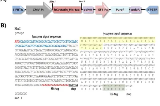
1. Construction of human cystatin-C expression vector system using piggyBac transposon
To insert the human cystatin-C gene into piggyBac transposon transgene expression system vector (System Biosciences, Palo Alto, CA, USA), NheⅠ and NotⅠ was used for digestion. Human cystatin-C was regulated by the cytomegalovirus (CMV) promoter. His-tag (His6) was added in front of stop codon of human cystatin-C. The promoter of elongation factor 1 (EF1) was used for expression of the puromycin- resistance gene in the transgene expression vector.
2. Transfection of human cystatin-C expression vector to DF1 cell & PGCs.
To establish the transgene expressing cell lines, transgene expression vector was co-transfected with piggyBac transposase using Lipofectamine 3000 (Invitrogen, USA) according to the manufacturer’s protocol. Once
the seeded cells reached 80% confluency on 6-well culture plates, they were washed with phosphate-buffered saline (PBS) and refreshed with 2ml of the culture media without antibiotic-antimycotic. 7.5μl Lipofectamine 3000 reagent in 250μl OPTI-MEM (Invitrogen, USA) and 10ul P3000 reagent with 2.5μg piggyBac transgene vector and piggyBac transposase plasmid in 250μl OPTI-MEM was added to each well. After 3 hour incubation in 37℃, each well were washed with PBS and refreshed with 3ml of the culture media with antibiotic-antimycotic. In next day, 10μg/ml puromycin was added to select the transfected cells.
3. Microinjection of cultured PGCs into recipient embryos.
For PGC transplantation, a small window was made on the pointed end of the recipient and the genetically-modified PGCs were microinjected with a micropipette into the dorsal aorta of the recipient embryo. The egg window of the recipient embryo was sealed with paraffin film and then the egg was incubated with the pointed end down until further screening and hatching.
4. Production of transgenic chickens
G0 chicken founders were mated through the testcross analysis and the donor PGC-derived offspring could be identified. Subsequently, transgenic chickens were identified by PCR analysis.
5. Genomic PCR
The DNA was extracted in feather pulp. The primers were hcyst his F&R and CMV cyst his F&R. The hcyst his F sequence was 5’- CACTGCTCCTCCTTGCGATC-3’; the hcyst his R sequence was 5’- GGTGATGGTGATGGGCATCC-3’. The CMV cyst his F sequence was 5’- GTGAACCGTCAGATCGCCTG-3’; the CMV cyst his R sequence was 5’- GTGAGGCTGGTCGTGGAAAG-3’. The annealing temperature was 65℃.
The cycle number of amplication for PCR reaction was 30.
6. Purification of human cystatin-C in egg white and muscle.
Egg white diluted with PBS and homogenized muscle were purified using MagListo™ His-tagged Protein Purification kit (Bioneer). The resin
removed supernatant were mixed with PBS and spread in the sample.
The sample with resin were shaken for 10min and centrifuged for 90sec at 4000 RPM, and removed supernatant. The sample with resin were washed with 3ml of Binding/washing buffer for 4 times and eluted with 500μl of elution buffer for 2 times.
7. ELISA
All samples were analyzed using human cystatin-C Quantikine® ELISA Kit (R&D Systems). Prepared samples (50μl) were added to each well contained 100μl of Assay Diluent buffer. After incubation for 3 hours at 2-8℃, each well was aspirated and washed by 400μl of Wash buffer 4 times. Each well was added 200μl of cold human cystatin-C conjugate and incubated for 1 hour at 2-8℃. The wells were washed like previous step and added 200μl of substrate solution and incubated for 30min at room temperature in dark place. After incubation, each well was added 50μl of stop solution and measured by ELISA reader.
8. Western blotting
Feather pulp and homogenized muscle proteins were extracted with 1×
radioimmunoprecipitation (RIPA) lysis buffer and purified muscle and egg white is in elution buffer (0.5M Imidazole). These samples separated on a 15% polyacrylamide gel followed by transfer to a nitrocellulose membrane. The primary antibodies used were mouse anti-β-actin (Santa Cruz Biotechnology) or anti-human cystatin-C (Abcam). HRP-conjugated anti-mouse IgG or anti-rabbit IgG (Bio-Rad) were used as secondary antibodies. The blots were treated with ECL substrate solutions and exposed in a ChemiDoc XRS System (Bio-Rad) to detect chemiluminescence.
9. Antimicrobial activity test
E. coli was subculture on LB agar plate. E. coli colony was streaked in four directions with a swab on LB agar plate. Make holes with 1000p tip. Each hole have 50μl test sample. The plate incubated 16 hours at 37℃ incubator. After 16 hours, check the clear zone size.
Results
1. Construction of human cystatin-C expression vector
We constructed piggyBac transposon vectors that contained the human cystatin-C gene (Figure 1). We use PB510B-1 Dual Promoter Vector Formats. CMV promoter controlled human cystatin-C gene to overexpress in whole chicken body. Because of CMV promoter, Human cystatin-C is expressed throughout the body of the chicken, thus there is no need to distinguish between male and female. Therefore, it is expected to help increase production by reducing the budget required for discriminating chick males and females and obtaining male cystatin from male tissue.
We insert the recombinant human cystatin-C gene using NheⅠ and Not
Ⅰ. His tag was attached to the end of the human cystatin-C gene to make selective purification possible. We used puromycin resistance gene for selection marker after transfection. The expected size of human cystatin-C protein is 13kDa.
Figure 5. Construction of human cystatin-C expression vector system using piggyBac transposon
(A) Expression vector of hCystatin_His-tag. The CMV promoter make over-expression of human cystatin-C_His-tag gene. The selection marker is puromycin resistance gene. (B) Sequences of human cystatin-C_His-tag gene and their amino acids. The bold ATG indicates the start codon and the blue sequences present the lysozyme signal sequences. The red sequences are the His-tag.
2. Identification of vector expression in DF1 cells and PGCs
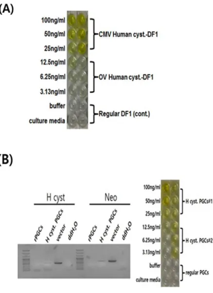
To check human cystatin-C expression vector, we identified expression of human cystatin-C for ELISA using transfected DF1 cells (Figure 2A).
Compared with control DF1 cells, transfected DF1 cells showed high expression of human cystatin-C over 50ng/ml. We proceed transfection in PGCs to use microinjection in recipient embryos. We identified human cystatin-C vector expression with genomic PCR analysis and ELISA (Figure 2B). The results showed that human cystatin-C vector entered the PGCs and human cystatin-C protein expression was over 50ng/ml. Both results suggested that human cystatin-C expression vector constructed by non- viral piggyBac transposon vector system was stably expressed the human cystatin-C proteins.
Figure 6. Identification of human cystatin-C in DF1 cells and PGCs (A) Identification of human cystatin-C expression in DF1 cells with ELISA.
DF1 with human cystatin-C expression vector successfully expressed human cystatin-C protein. (B) Identification of human cystatin-C expression in chicken primordial germ cells (PGCs) with genomic PCR analysis and ELISA.
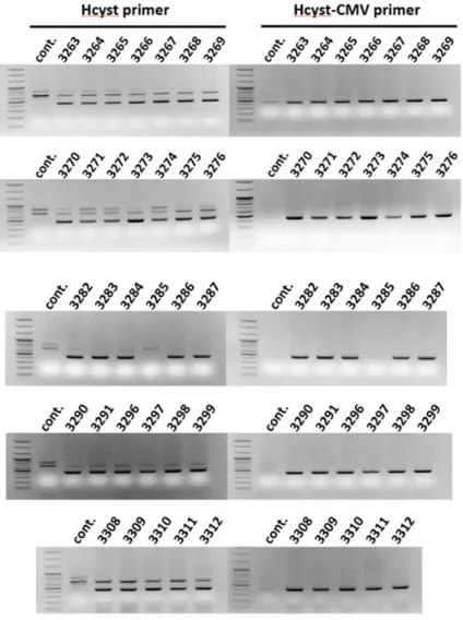
3. Production of germ-line chimera and transgenic offspring
In previous study, we confirmed that piggyBac transposition into primordial germ cells (PGCs) improved the efficiency of transgenic chicken production and led to high-level transgene expression through GFP expression (Park and Han 2012). The rate of germ-line transmission was 95.2% on average. In this study, we microinjected PGCs with human cystatin-C expression vector into recipient embryos to produce germ-line chimera. Subsequently, transgenic chickens were produced by testcross analysis using germ-line chimera. We extracted DNA in transgenic offsprings’ feather pulp for PCR screening using Hcyt primer and Hcyt- CMV primer to identify transgenic chickens. After testcross analysis, 30 of the 31 chicks, except 3285, were identified as human cystatin-C expression vector (Figure 3). The rate of transgenic chicken production is about 96%. This result is similar to the previous study. Therefore, there was a high probability that transgenic chickens were produced and human cystatin-C expression vector using non-viral piggyBac transposon system is efficient method of transgenic chicken production.
Figure 7. Production of germ-line chimera and transgenic offspring Production of transgenic chickens through the testcross analysis.
Transgenic chickens from the germline chimeras were identified by genomic PCR analysis.
4. Identification of human cystatin-C expression in transgenic chickens
After identifying the transgenic chickens to genomic PCR analysis, we investigated human cystatin-C protein in transgenic chicken to ELISA and western blot for identification of human cystatin-C protein expression in whole chicken body (Figure 4). We checked feather pulp, blood, egg white and muscle lysate. The human cystatin-C protein expression in feather pulp is less then 6.25ng/ml in ELISA, but in Western blot result, there was no abnormality in expression. All other tissues showed high expression of human cystatin-C. This result confirmed that the presence of sufficient human cystatin-C protein in other tissues. Therefore, it can be expected that human cystatin-C purification in other tissues as well as egg white is possible. Especially muscle has a lot of human cystatin- C protein. Since muscles make up most of the body, if human cystatin- C can be purified in muscles, the amount of human cystatin protein that can be obtained per chicken will increase dramatically.
Figure 8. Expression of human cystatin-C in transgenic chickens
In ELISA, human cystatin-C protein was expressed in transgenic chicken’s feather pulp (A), blood (C) and egg white (D). Additionally, Western blotting showed that human cystatin-C was expressed in transgenic chicken’s feather pulp (B) and muscle (E)
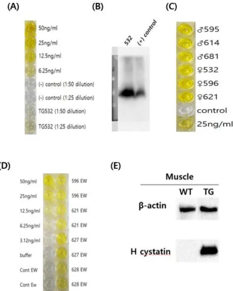
5. Purification of human cystatin-C in transgenic chickens’
egg white and muscle
Chickens can produce egg consistently. Thus, there have been attempts to purify recombinant proteins using eggs (Lillico, McGrew et al. 2005).
However, we also purified human cystatin-C in muscle because transgenic chickens we produced can express human cystatin-C in whole body.
Purified human cystatin-C using His tag was verified through western blot (Figure 5A). The level of the purified sample in egg white was lower than that in the muscle. However, in ELISA, it was found that both purified sample in egg white and muscle had more then 100ng/ml. In addition, when the dialysis sample for the antimicrobial activity test was confirmed by ELISA, the results show that the concentration was not greatly diluted. Compared TG egg white purification 1 and 2 in ELISA data, concentration of first elution sample is higher than second one.
Figure 9. Purification of human cystatin-C in transgenic chickens’ egg white and muscle
Muscle tissue was disrupted by homogenizer. Egg white was diluted 1:5 in PBS. We purified human cystatin-C with these samples and identified using western blot (A) and ELISA (B). In ELISA, TG muscle is purified human cystatin-C in TG muscle. TG EW is purified human cystatin-C in TG egg white. TG EW dialysis is purified and dialysis human cystatin-C in TG egg white.
6. Antimicrobial activity of human cystatin-C in transgenic chickens’ egg white
To check the biofuctional activity of purified human cystatin-C, we tried an antimicrobial activity experiment. First, we checked antimicrobial activity of transgenic chickens’ egg white against E. coli. (Figure 10A).
Compared with human cystatin-C 50ng/ml, the clear zone was small but accurate. It is assumed that the clear zone is small because the egg white is so large that the concentration of cystatin is diluted. Next, we examined the antimicrobial activity of the purified cystatin-C in TG egg white (Figure 10B). Compared with human cystatin-C 100ng/ml and 50ng/ml, there were no clear zone in purified cystatin-C. Since the antimicrobial activity experiment was conducted immediately after protein purification, It is thought that the modification of protein is unlikely to occur after purification. It is assumed that the cause is in the purification method. In other studies, The egg whites are pretreated before purification for remove ovomucin (Wang and Wu 2014). In future studies, we will add a new pretreatment method to purify human cystatin and then confirm the antimicrobial activity.
Figure 10. Antimicrobial activity of human cystatin-C in transgenic chickens’
egg white against E. coli
(A) Identification of TG egg white’s antimicrobial activity. 1 is human cystatin-C 50ng/ml, 2 is water, 3 is TG EW. (B) Identification of purified human cystatin-C’s antimicrobial activity. 1 is human cystatin-C 100ng/ml,
2 is human cystatin-C 50ng/ml, 3 is purified and dialysis human cystatin- C in TG egg white.
Discussion
We constructed a vector using non-viral piggyBac transposon system to produce transgenic chickens expressing human cystatin-C and confirmed that the vector works normally by transfection in DF1 cells and PGCs. PGCs transfected with the identified vectors were microinjected into embryos to produce germline chimeras. Transgenic chickens were selected by testcross analysis among the chimeras’ offspring. When we identified the expression of human cystatin-C in various parts of the transgenic chickens, it was detected in all parts. This indicates that transgenic chickens expressing human cystatin-C have been produced.
In general, chicken is livestock that produce eggs. Thus, the males are eliminated from the chick stage. In addition, even when used as a bioreactor, the male chickens were excepted because the recombinant protein produce in egg white. In this study, the CMV promoter was used to express human cystatin-C throughout the chicken body, rather than the egg white specific promoter. This allows human cystatin-C to be purified not only on egg white but also on muscles and other tissues. It has the advantage that the recombinant protein can be obtained even in the muscles of males without eliminating the males when producing transgenic chickens. Using CMV promoters will have a positive impact on the productivity of recombinant proteins, reducing the effort of screening
male and female.
Human cystatin-C purification must be accompanied for use as medicines or diagnostic kits. In a previous study, they introduced a method for purifying cystatin from chicken egg whites using affinity chromatography (Wang and Wu 2014). However, we attached His tag back of the human cystatin-C sequence when we constructed the vector, only human cystatin-C protein was purified selectively using Ni-NTA magnetic nanobead. The protein was purified from egg white and homogenized muscle. But the amount of protein purified from muscle was higher due to differences in quantity of egg white and muscle. But the easier form to purify was egg white. In fact, when the purification purity was identified through SDS PAGE, purified sample in muscle had not only human cystatin-C but also other proteins. This is considered to be a problem of the purification method, and since the purification kit that we use is using magnetic nanobead, it is more likely that unwashed proteins will elute together when receiving the purification solution. In the case of muscle, the tendency to lump together with the resin was observed. Egg white was also agglomerated together with the resin, but after freeze-drying, diluting in PBS and once or twice with a sieve, it was confirmed that the resin agglomeration greatly reduced.
Cystatin is a cysteine protease inhibitor that has been known to have antimicrobial and antiviral properties against microorganisms and viruses through several papers (Szpak, Trziszka et al. 2014, Luthra 2015, Pikula,
Smuzynska et al. 2017). Using this property, we are conducting antimicrobial activity experiment to verify the activity of purified human cystatin-C. Antibiotics are one of the most commonly used treatments for bacterial infections. Antibiotic resistant microorganisms continue to emerge as the abuse of antibiotics and the ability of microorganisms to adapt. Many studies have been conducted to prevent the production of antibiotic resistant bacteria or to effectively eliminate them (Li, Li et al.
2018, Luna, Ershova et al. 2019, Sorbara, Dubin et al. 2019). If this experiment shows that the purified human cystatin-C has the antimicrobial activity, we can suggest the possibility of using the human cystatin-C protein as a new antibiotic.
We were concerned whether cystatin’s role as cysteine protease inhibitor would affect the basic mechanism of the transgenic chicken, but our transgenic chickens were not particularly different from those of normal chickens. In other papers, cystatin-C protein was never fed or injected as a drug, but there were no cases of health abnormalities when cystatin-C overexpressed individuals were produced and confirmed. Rather, it has been found to be possible as a therapeutic agent for inhibiting cancer or for neurological diseases such as Alzheimer’s disease (Kaeser, Herzig et al. 2007, Yan, Zhou et al. 2015, Coronado, Barrios et al. 2017, Zou, Chen et al. 2017, Kaur, Gauthier et al. 2018, Li, Wang et al. 2018).
In addition, cystatin expressed by parasites can induce the production of IL-10, which can suppress allergy and intestinal inflammation (Schnoeller,
Rausch et al. 2008, Jang, Cho et al. 2011). It is also being studied as a potential treatment for collagen-induced arthritis (CIA) (Liu, Cheng et al.
2016). Therefore, it was found that cystatin-C can be purified and used as a therapeutic agent in human cystatin-C expressing transgenic chickens.
Cystatin-C is generally known to have a characteristic of being filtered by glomeruli so that the glomerular filtration rate (GFR) can be calculated from the serum concentration of cystatin-C. Cystatin-C can thus be used as a marker for kidney disease. Traditionally, kidney function was assessed by serum creatinine concentrations. However, various studies have concluded that serum cystatin-C shows higher sensitivity and specificity than serum creatinine (Onopiuk, Tokarzewicz et al. 2015) and that more accurate data can be obtained with both creatinine and cystatin-C than with creatinine alone (Stevens, Coresh et al. 2008). However, when compared to creatinine, the cost of cystatin analysis is three times higher than the enzyme creatinine test and 20 times more than the creatinine test using the jaffe reaction (Shlipak, Mattes et al. 2013). Since the main cost is reagents, cystatin analysis will be more common if the reagent price can be reduced. This study is expected to contribute to the reduction in cystatin assay reagent prices.
With the advancement of gene editing techniques into zinc-finger nuclease, TALEN and CRISPR-Cas9, it is easier than ever to modify, add or delete the genes we want. Methods of making transgenic animals have also evolved to make faster and cheaper than before, which is an
economic advantage when using transgenic animals as bioreactors. Not many transgenic animals will grow rapidly, be easy to manage and produce protein continuously. Through this study, we believe that chicken is the most suitable animal for these conditions. In addition, the transgenic chicken production technique used in this study could be of great commercial value in the recombinant protein market if it is incorporated into other useful proteins.
Conclusion
In this study, the results can be summarized in two ways. First, transgenic chicken production using a non-viral piggyBac transposon system. For existing commercialized transgenic chickens, transgenic chickens were produced using a lentiviral vector system. The way in which viral vectors are used can pose safety concerns. In this study, we successfully produced transgenic chickens using the non-viral piggyBac transposon system, which we believe will help in the production of other useful recombinant drugs.
Second, presenting a new method for the production of cystatin-C.
Cystatin-C is well known as a marker for kidney disease and is being actively researched as a therapeutic agent for neurological diseases.
Human cystatin-C, which is on sale, is produced from animal cells and costs about 970,000 won per 10μg. The use of transgenic chickens continues to cost less than traditional animal cell-based production systems for the production of recombinant pharmaceuticals. Therefore, the production of human cystatin-C from transgenic chickens could lower the production cost and contribute to the study of the potential of human cystatin-C as a therapeutic agent.
References
Bagshaw, S. M. and R. Bellomo (2010). "Cystatin-C in acute kidney injury."
Curr Opin Crit Care 16(6): 533-539.
Berkner, K. L. (1992). "Expression of heterologous sequences in adenoviral vectors." Curr Top Microbiol Immunol 158: 39-66.
Bjorck, L. (1990). "Proteinase inhibition, immunoglobulin-binding proteins and a novel antimicrobial principle." Mol Microbiol 4(9): 1439-1442.
Bjorck, L., A. Grubb and L. Kjellen (1990). "Cystatin-C, a human proteinase inhibitor, blocks replication of herpes simplex virus." J Virol 64(2): 941-943.
Butler, E. A. and F. V. Flynn (1961). "The occurrence of post-gamma protein in urine: a new protein abnormality." J Clin Pathol 14: 172-178.
Clausen, J. (1961). "Proteins in normal cerebrospinal fluid not found in serum." Proc Soc Exp Biol Med 107: 170-172.
Cohen, S. N., A. C. Chang, H. W. Boyer and R. B. Helling (1973).
"Construction of biologically functional bacterial plasmids in vitro." Proc Natl Acad Sci U S A 70(11): 3240-3244.
Collins, A. R. and A. Grubb (1991). "Inhibitory effects of recombinant human cystatin-C on human coronaviruses." Antimicrob Agents Chemother 35(11): 2444-2446.
Coronado, S., L. Barrios, J. Zakzuk, R. Regino, V. Ahumada, L. Franco, Y. Ocampo and L. Caraballo (2017). "A recombinant cystatin from Ascaris lumbricoides attenuates inflammation of DSS-induced colitis."
Parasite Immunol 39(4).
Danna, K. and D. Nathans (1999). "Specific cleavage of simian virus 40 DNA by restriction endonuclease of Hemophilus influenzae. 1971." Rev
Med Virol 9(2): 75-81.
Danna, K. J. and D. Nathans (1972). "Bidirectional replication of Simian Virus 40 DNA." Proc Natl Acad Sci U S A 69(11): 3097-3100.
Danna, K. J., G. H. Sack, Jr. and D. Nathans (1973). "Studies of simian virus 40 DNA. VII. A cleavage map of the SV40 genome." J Mol Biol 78(2): 363-376.
Dharnidharka, V. R., C. Kwon and G. Stevens (2002). "Serum cystatin-C is superior to serum creatinine as a marker of kidney function: a meta- analysis." Am J Kidney Dis 40(2): 221-226.
Dyck, M. K., D. Lacroix, F. Pothier and M. A. Sirard (2003). "Making recombinant proteins in animals--different systems, different applications." Trends Biotechnol 21(9): 394-399.
Ferrer-Miralles, N., J. Domingo-Espin, J. L. Corchero, E. Vazquez and A.
Villaverde (2009). "Microbial factories for recombinant pharmaceuticals."
Microb Cell Fact 8: 17.
Feschotte, C. and E. J. Pritham (2007). "DNA transposons and the evolution of eukaryotic genomes." Annu Rev Genet 41: 331-368.
Fraser, M. J., G. E. Smith and M. D. Summers (1983). "Acquisition of Host Cell DNA Sequences by Baculoviruses: Relationship Between Host DNA Insertions and FP Mutants of Autographa californica and Galleria mellonella Nuclear Polyhedrosis Viruses." J Virol 47(2): 287-300.
Garcia-Carretero, R., L. Vigil-Medina, O. Barquero-Perez, R. Goya- Esteban, I. Mora-Jimenez, C. Soguero-Ruiz and J. Ramos-Lopez (2017).
"Cystatin-C as a predictor of cardiovascular outcomes in a hypertensive population." J Hum Hypertens 31(12): 801-807.
Grubb, A., M. Abrahamson, I. Olafsson, J. Trojnar, R. Kasprzykowska, F.
Kasprzykowski and Z. Grzonka (1990). "Synthesis of cysteine proteinase
inhibitors structurally based on the proteinase interacting N-terminal region of human cystatin-C." Biol Chem Hoppe Seyler 371 Suppl: 137- 144.
Grubb, A. and H. Lofberg (1982). "Human gamma-trace, a basic microprotein: amino acid sequence and presence in the adenohypophysis." Proc Natl Acad Sci U S A 79(9): 3024-3027.
Gurtu, V., G. Yan and G. Zhang (1996). "IRES bicistronic expression vectors for efficient creation of stable mammalian cell lines." Biochem Biophys Res Commun 229(1): 295-298.
Hernandez, H. M., R. Marcet and J. Sarracent (2014). "Biological roles of cysteine proteinases in the pathogenesis of Trichomonas vaginalis."
Parasite 21: 54.
Hochwald, G. M. and G. J. Thorbecke (1962). "Use of an antiserum against cerebrospinal fluid in demonstration of trace proteins in biological fluids." Proc Soc Exp Biol Med 109: 91-95.
Houdebine, L. M. (2009). "Production of pharmaceutical proteins by transgenic animals." Comp Immunol Microbiol Infect Dis 32(2): 107-121.
Hruby, D. E. (1990). "Vaccinia virus vectors: new strategies for producing recombinant vaccines." Clin Microbiol Rev 3(2): 153-170.
Jackson, D. A., R. H. Symons and P. Berg (1972). "Biochemical method for inserting new genetic information into DNA of Simian Virus 40:
circular SV40 DNA molecules containing lambda phage genes and the galactose operon of Escherichia coli." Proc Natl Acad Sci U S A 69(10):
2904-2909.
Jang, S. W., M. K. Cho, M. K. Park, S. A. Kang, B. K. Na, S. C. Ahn, D.
H. Kim and H. S. Yu (2011). "Parasitic helminth cystatin inhibits DSS- induced intestinal inflammation via IL-10(+)F4/80(+) macrophage
recruitment." Korean J Parasitol 49(3): 245-254.
Jenkins, N. (2007). "Modifications of therapeutic proteins: challenges and prospects." Cytotechnology 53(1-3): 121-125.
Jones, S. A., V. Valayannopoulos, E. Schneider, S. Eckert, M. Banikazemi, M. Bialer, S. Cederbaum, A. Chan, A. Dhawan, M. Di Rocco, J. Domm, G. M. Enns, D. Finegold, J. J. Gargus, O. Guardamagna, C. Hendriksz, I. G. Mahmoud, J. Raiman, L. A. Selim, C. B. Whitley, O. Zaki and A.
G. Quinn (2016). "Rapid progression and mortality of lysosomal acid lipase deficiency presenting in infants." Genet Med 18(5): 452-458.
Jung, J. G., D. K. Kim, T. S. Park, S. D. Lee, J. M. Lim and J. Y. Han (2005). "Development of novel markers for the characterization of chicken primordial germ cells." Stem Cells 23(5): 689-698.
Kaeser, S. A., M. C. Herzig, J. Coomaraswamy, E. Kilger, M. L. Selenica, D. T. Winkler, M. Staufenbiel, E. Levy, A. Grubb and M. Jucker (2007).
"Cystatin-C modulates cerebral beta-amyloidosis." Nat Genet 39(12):
1437-1439.
Kasprzykowski, F., C. Schalen, R. Kasprzykowska, B. Jastrzebska and A.
Grubb (2000). "Synthesis and antibacterial properties of peptidyl derivatives and cyclopeptides structurally based upon the inhibitory centre of human cystatin-C. Dissociation of antiproteolytic and antibacterial effects." APMIS 108(7-8): 473-481.
Kaur, G., S. A. Gauthier, R. Perez-Gonzalez, M. Pawlik, A. B. Singh, B.
Cosby, P. S. Mohan, J. F. Smiley and E. Levy (2018). "Cystatin-C prevents neuronal loss and behavioral deficits via the endosomal pathway in a mouse model of down syndrome." Neurobiol Dis 120: 165- 173.
Kelly, T. J., Jr. and H. O. Smith (1970). "A restriction enzyme from
Hemophilus influenzae. II." J Mol Biol 51(2): 393-409.
Khosravi, N., M. Zadkarami, F. Chobdar, R. Hoseini, N. Khalesi, P. Panahi and A. Karimi (2018). "The Value of Urinary Cystatin-C Level to Predict Neonatal Kidney Injury." Curr Phar