저작자표시-비영리 2.0 대한민국 이용자는 아래의 조건을 따르는 경우에 한하여 자유롭게
l 이 저작물을 복제, 배포, 전송, 전시, 공연 및 방송할 수 있습니다. l 이차적 저작물을 작성할 수 있습니다.
다음과 같은 조건을 따라야 합니다:
l 귀하는, 이 저작물의 재이용이나 배포의 경우, 이 저작물에 적용된 이용허락조건 을 명확하게 나타내어야 합니다.
l 저작권자로부터 별도의 허가를 받으면 이러한 조건들은 적용되지 않습니다.
저작권법에 따른 이용자의 권리는 위의 내용에 의하여 영향을 받지 않습니다. 이것은 이용허락규약(Legal Code)을 이해하기 쉽게 요약한 것입니다.
Disclaimer
저작자표시. 귀하는 원저작자를 표시하여야 합니다.
비영리. 귀하는 이 저작물을 영리 목적으로 이용할 수 없습니다.
수의학 박사학위논문
Inhibition of porcine endogenous retrovirus by multi-targeting RNA
interference in porcine cells
다중 표적 RNA 간섭에 의한 돼지 내인성
레트로바이러스 억제
2020 년 2 월
서울대학교 대학원
수의학과 수의미생물학 전공
정 희 천
Inhibition of porcine endogenous retrovirus by multi-targeting RNA
interference in porcine cells
By
Chung, Hee Chun
February, 2020
Department of Veterinary Medicine The Graduate School of
Seoul National University
Inhibition of porcine endogenous retrovirus by multi-targeting RNA
interference in porcine cells
By
Chung, Hee Chun
Supervisor: Prof. Park, Yong Ho, D.V.M., M.Sc., Ph.D.
A dissertation submitted to the faculty of the Graduate School of Seoul National University in partial fulfillment of the requirements
for the degree of Doctor of Philosophy in Veterinary Microbiology
February, 2020
Department of Veterinary Medicine The Graduate School of
Seoul National University
i
Inhibition of porcine endogenous retrovirus by multi-targeting RNA
interference in porcine cells
Chung, Hee Chun
(Supervised by Prof. Park, Yong Ho)
Veterinary Microbiology, Department of Veterinary Medicine, the Graduate School of Seoul National University
Abstract
For xenotransplantation, pigs present numerous advantages as donor animals as compared with other non-human primates, and are recognized as recipient animals for organ xenotransplantation in preclinical studies. However, all pigs have the porcine endogenous retrovirus (PERV) genome inserted in their germ line, which is transmitted to offspring. Because of the potential risk of interspecies transmission, PERV is a major threat in xenotransplantation experiments. Ribonucleic acid interference (RNAi) technology is used to knockdown gene expression, which reduces risk and improves the overall safety of xenotransplantation. Herein, an RNAi strategy was employed to inhibit
ii
PERV expression in porcine cells by targeting multiple PERV genes.
As stated in chapter 1, polymerase (pol)-targeting RNAi prevented infection through the reduction of reverse transcription and replication. The quantity of group- specific antigen (gag) mRNA, which encodes capsid protein essential for budding, was reduced by RNAi by blocking the shedding of viral particles. Therefore, if gag and pol genes of PERV can be suppressed simultaneously, it is an important strategy for significant inhibition of PERV gene expression. Among the four siRNAs targeting the gag and pol genes of PERV that were designed, two were more effective siRNAs (gag2, pol2) in reduction of the expression of PERVs. Concurrent treatment of porcine cells with these two siRNAs (gag2 + pol2) showed knockdown efficiency up to 88% as compared to the negative control. Despite the high initial knockdown efficiency by siRNA 48 hours after transfection, that effect may be a mere transient effect for PERV suppression. A multi-targeting vector was designed containing both gag and pol genes, and making use of the POL II microRNA (miRNA) expression vector, this vector allows for simultaneous targeting of multiple genes. The sequence of miRNA for the combined genes (gag2 + pol2) was designed in the same region as siRNAs which target gag2 and pol2 separately. Through the antibiotic-resistance characteristics of this vector, miRNA- transfected porcine kidney (PK) 15 cells (gag2 + pol2) were selected over 2 weeks.
Reduction of mRNA expression for pol and gag gene targets was 88.1% and 72%, respectively. In addition, two assays were performed: 1) reverse transcriptase assay (RT) activity analysis and 2) fluorescence in situ hybridization (FISH) assay and it demonstrated the highest knockdown efficiency in the multi-target (gag2 + pol2) miRNA
iii
group. According to the results above, using gene knockdown systems (siRNA and shRNA) through a multi-targeting (gag2 + pol2) strategy are effective methods to inhibit PERVs.
In chapter 2, the objective was to target the long terminal repeats (LTR) region with a dual LTR1 + LTR2 miRNA. The miRNA expression vector was designed using PERV LTRs sequences from porcine organs eligible for xenotransplantation. The targets for the LTR region of miRNAs were automatically selected via the online program BLOCK-iT RNAi Designer. The inhibition efficiency among the miRNAs was compared based on their inhibition of different PERV genes, LTR, gag, and pol. Relative quantitative real-time polymerase chain reaction (qPCR) and C-type reverse transcriptase activity were performed. The miRNA targeting the LTR region degraded the target sequence, but simultaneously inhibited the mRNA expression of both gag and pol genes of PERV. The LTR1, LTR2, and dual LTR1+LTR2 miRNA inhibited 76.2%, 22%, and 76.8% of gag gene expression, respectively. Similarly, miRNA knock-downed pol gene expression by 69.8%, and 25.5% for the single targeting miRNA (LTR1 and LTR2), respectively and 77.7% for the multi-targeting miRNA (LTR1 + LTR2). A stable PK15 clone constitutively expressed dual LTR1 + LTR2 miRNA and exhibited higher inhibition up to 82.8% and 92.7% of the expression for the gag and pol genes, respectively. Also, co-cultivation of dual LTR1 + LTR2 miRNA-transfected PK15 cells with a human cell line (HeLa cells) showed that dual LTR1 + LTR2 miRNA inhibited expression of LTR, gag, and pol genes of PERV mRNAs so that PERV infectivity was reduced in a human cell line.
iv
Dual mRNA targeting of LTR produced a marked reduction in PERV expression.
However, the experimental design for the study in chapter 2 did not include primary porcine kidney cells and or examine inhibition of the env gene.Therefore, in chapter 3, this study was performed in primary porcine kidney cells in vitro to determine whether miRNAs selected that target specific regions of the LTR could exert an inhibitory effect on the expression of LTR (the U3 promoter region of PERV), gag, pol, and env genes.
Two miRNAs (LTR1 and LTR2), and dual LTR1 + LTR2, were selected to inhibit the expression of PERV in primary porcine kidney cells. The inhibition efficiency of the miRNAs was compared based on their inhibition of different PERV regions, specifically LTRs (U3 promoter region of PERV), gag, pol, and env. Gene expression was quantified using real-time polymerase chain reaction (RT-PCR) and the C-type reverse transcriptase (RT) activity was determined. The mRNA expression of the PERV LTR (U3 promoter region of PERV) and env region was determined in HeLa cells co-cultured with primary porcine kidney cells. The mRNA expression of the LTR (U3 promoter region of PERV), gag, pol, and env regions of PERV was dramatically inhibited by dual miRNA (LTR1 + LTR2) from 24 hours to 144 hours after transfection, with the highest inhibition observed for the LTR (U3 promoter region of PERV) and pol regions at 120 hours. Changing the co-culture incubation time from 48 hours to 120 hours resulted in the largest change in the PERV amount in the negative vector control group contrary to HeLa cells co-cultured with primary porcine kidney cells transfected with dual LTR1 +LTR2 miRNAs.
In conclusion, these three studies confirm that miRNA techniques targeting the regions of PERV can positively inhibit the expression of PERV in porcine kidney cells.
v
Multi-targeting miRNAs for LTR1 +LTR2 of PERV reduced gene expression for the LTR region as well as the expression of functionally important PERV genes such as gag, pol, and env. The methods on co-cultivation and gene expression profiling offer new approaches to generate data to help evaluate the risk/benefit balance for PERV inhibition for safer xenotransplantation, and ultimately for the future creation of transgenic pigs.
Keywords: Inhibition; miRNA vector; Porcine endogenous retrovirus;
Transfection; Human cell; Multi-targeting; RNA interference Student number: 2017-27680
vi
Contents
Abstract ... i
Contents ... vi
List of Figures... ix
List of Tables ... xv
Abbreviations ... xvi
General introduction ... 1
Literature review ... 4
1. PERV in xenotransplantation ... 5
1.1. PERV historical perspective ... 5
1.2. Xenotransplantation trials ... 6
1.3. PERVs and potential to cause xenozoonotic disease ... 8
1.4. PERV risk of xenotransplantation ... 9
1.5. The need to screen for PERVs in xenotransplantation ... 11
2. PERV structure and biology ... 13
2.1 PERV molecular structure ... 13
2.2 PERV biology ... 16
3. Control of PERV ... 18
3.1. RNA inference (RNAi) ... 18
vii
3.2. small interfering RNA (siRNA) ... 19
3.3. small hairpin RNA (shRNA) ... 20
3.4. microRNA (miRNA) ... 23
3.5. Clustered regularly interspaced short palindromic repeats (CRISPR) ... 25
3.6. Latest strategy………..27
Chapter I. Inhibition of porcine endogenous retrovirus in PK15 cell line by efficient multitargeting RNA interference...29
Abstract ... 30
1.1. Introduction ... 31
1.2. Materials and methods ... 33
1.3. Results ... 38
1.4. Discussion ... 42
Chapter II. Inhibition of porcine endogenous retrovirus by multi-targeting microRNA against long terminal regi on……….……….55
Abstract ... 56
2.1. Introduction ... 58
2.2. Materials and methods ... 61
viii
2.3. Results ... 66
2.4. Discussion ... 69
Chapter III. Regulation of porcine endogenous retrovirus by dual LTR1+2 (Long Terminal Region) miRNA in primary porcine kidney cells...80
Abstract ... 81
3.1. Introduction ... 82
3.2. Materials and methods ... 85
3.3. Results ... 91
3.4. Discussion ... 94
General conclusion ... 104
References ... 108
Abstract in Korean ... 124
ix
List of Figures
Figure 1 PERV. (A) Genomic RNA. (B) PERV structure. (C) Proviral DNA and nucleotide positions of the main elements.
15
Figure 2 Replication cycle of PERV and strategies of PERV detection in xenotransplantation.
17
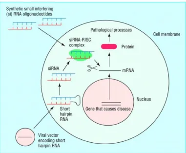
Figure 3 Natural mechanism of RNA interference. 19
Figure 4 Targeting disease by shRNA. 21
Figure 5 Overview of microRNA processing in animals, from transcription to the formation of the effector complex.
24
Figure 6 Diagram of the CRISPR prokaryotic antiviral defense mechanism.
26 Figure 7 The miRNAs transfection. Lipofectamine2000 (Invitrogen)
Transfection reagent was used to transfect the plasmid into PK15 cells (a) gag2 miRNA transfection (b) pol2 miRNA transfection (c) gag2-pol2 miRNA transfection (d) Neg-vector transfection.
45
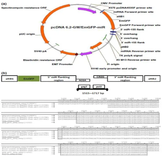
Figure 8 Constructs design. (a) Map and features: pcDNA 6.2- GW/EmGFP-miR Expression Vector for targeting of porcine endogenous retroviruses
(PERV). CMV promoter, green fluorescent protein coding sequence (GFP), PERV multitargeting genes inserted into the attB2 sites, EM7 promoter, and blasticidin resistance gene; (b) The strategy of designing the constructs: Both of gag2 (1515~1579 bp) and pol2 (1653~1717 bp) were inserted to pcDNA 6.2-GW/EmGFP-miR Expression Vector (Invitrogen).
46
Figure 9 Efficiency of siRNA in reduction of porcine endogenous retroviruses (PERV) mRNA expression in PK15 cells. The control did not affect any experiment results through Invitrogen Web because the sequence of siRNAs did not target any gene
47
x
product as standard control (RQ = 1), comparisons showed differences in suppression efficiency among siRNA targeting different sites of (a) gag and (b) pol genes. Porcine endogenous retrovirus expression was measured by a two-step quantitative real-time PCR. All experiments were repeated three times.
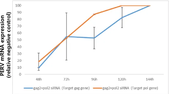
Figure 10 Knockdown efficiency of siRNAs (gag2+pol2) expression in transfected PK15 cells. Real-time PCR was used to detect the expression level of PERV mRNA in PK 15 cells relative to standard negative control (RQ = 100).
48
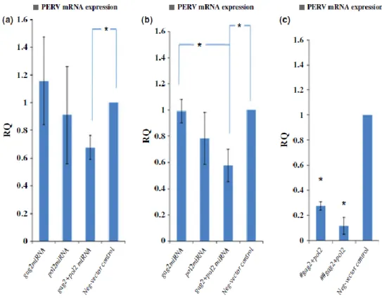
Figure 11 Suppression efficiency among miRNAs targeting different sites (a) gag, (b) pol, and (c) after transfection, selected blasticidin- resistant colonies; gag2-pol2 miRNAs (#) targeting gag region, (##) targeting pol region. The neg-vector-transfected control is regarded as standard control (RQ = 1), the multitargeting shRNA vector was then designed for expressing miRNAs targeting both gag and pol gene sites. It showed the most knockdown efficiency among RNAi groups. Porcine endogenous retroviruses mRNA expression was measured by a two-step quantitative real-time PCR.
49
Figure 12 Porcine endogenous retroviruses reverse transcriptase activity among miRNA targeting different sites. (a) Indicating a practical value of negative control as a standard, each of sample values was presented as above by % level. (b) Standard curve for C-type RT activity kit was obtained with the serial dilutions of MMuLV rRT against the concentration of MMuLV present (LOT number 11071). The equation for the curve is as follows: y = 2.671x + 0.092 (R2 = 0.997).
50
Figure 13 Fluorescence in situ hybridization assay for targeted pol gene mRNAs of porcine endogenous retroviruses in PK15 cells. (a)
51
xi
gag2-pol2 miRNAs vector transfected into PK15 cells. (b) neg- vector miRNAs transfected into PK15 cells. (c) negative control.
Figure 14 Expression of porcine endogenous retroviruses (PERV) in HEK293 cell line after cocultivation with inhibition of PERV in PK15 cells according to the change of incubation times (a) Genomic DNA, (b) mRNA was extracted from HEK293 cell and quantitative real-time RT-PCR assay was performed to measure the amount of PERV (copies/ ul).
52
Figure 15 Map of miRNAs. (A) Both of the LTR1 and LTR2 miRNAs are located at the U3 region of the LTR of PERV. The positions of miRNAs were based on Genbank accession number FJ357768.
(B) Alignment between miRNAs and the LTR sequences of PERV infected PK15 cells (shaded areas). Mismatches between LTR1 and LTR2 miRNAs and the LTR sequences of PERV were indicated by squares. Gaps in sequences of miRNA were introduced due to the algorithm of BLOCK-iT RNAi Designer.
72
Figure 16 miRNA-mediated degradation of LTR at RNA level. Column indicated level of transcribed RNA of LTR in PK15 cells transfected by single miRNA (LTR1, LTR2), dual miRNA (LTR1 + LTR2), and negative miRNA expression vector.
73
Figure 17 LTR miRNA induced suppression of the gag gene (A) and the pol gene (B). Column indicated level of gene inhibition by single miRNA (LTR1, LTR2), dual miRNA (LTR1 + LTR2), and negative miRNA expression vector.
74
Figure 18 (A) After transfection; LTR1+2 miRNA, selected blasticidin- resistant colonies during 14 days (original magnification 200X;
fluorescence microscope). (B) LTR1+2 miRNAs (#) targeting LTR region, (##) targeting gag region, and (###) targeting pol region. The neg vector-transfected control is regarded as standard
75
xii control (RQ = 100).
Figure 19 RT activity of PERV in PK15 cells transfected by single miRNA, multi-miRNA, and negative expression vector. (A) Standard curve for C-type RT activity kit was obtained with the serial dilutions of MMuLV rRT against the concentration of MMuLV present (lot number 11071). The equation for the curve is as follows: y = 4.384x + 0.057 (R2 = 0.999). (B) Indicating a practical value of negative control as a standard, each sample value was presented as above by percentage.
76
Figure 20 Gene expression of PERV in HeLa cells after co-culture in PK15 cells. Red line was HeLa cells co-cultured with PK15 cells transfected by off-target expression vector. Blue line was HeLa cells co-cultured with PK15 cells transfected by dual LTR1 + LTR2 miRNA.
77
Figure 21 Confirmation of miRNA targeting site in primary porcine kidney cells and HeLa cells (A) Both LTR1 and LTR2 miRNAs are located at the U3 region of the LTR of PERV. Alignment between miRNAs and the LTR sequences (GenBank accession numbers FJ357767 and FJ357768) was confirmed and primary porcine kidney cells were used for this study. Matches between the LTR1 and LTR2 miRNAs and the LTR sequences of PERV are indicated. (B) Genotyping by RT-PCR in primary porcine kidney cells and HeLa cells using envelop primer sets.
96
Figure 22 Determination of miRNA transfection efficiency. Lipofectamine 2000 (Invitrogen) reagent was used to transfect the plasmids (dual LTR 1+2 miRNA, and negative vector control miRNA) into primary porcine kidney cells. Mock-transfected cells were subjected to the transfection process without the addition of miRNA (cells were treated with transfection reagent only).
97
xiii
Figure 23 Knockdown efficiency of miRNA (dual LTR1+LTR2)- transfected primary porcine kidney cells. Real-time PCR was used to detect the expression level of PERV mRNA in primary porcine kidney cells relative to the standard negative vector control (RQ = 100).
98
Figure 24 Selected blasticidin-resistant colonies and analysis of PERV pol gene mRNA levels based on passage numbers. (A) Following transfection with dual LTR 1+2 miRNA, blasticidin-resistant colonies were selected after 20 days at passage 6. (B) Dual LTR 1+2 miRNA targeting the LTR, gag, and env regions. The negative vector-transfected control is regarded as the standard control (RQ = 100 value).
99
Figure 25 PERV pol gene analysis by 2 methods: reverse transcriptase (RT) activity and mRNA levels in blasticidin-resistant primary porcine kidney cells over 20 days. (A) Plots show the negative vector control as the standard and sample values are represented by percentage for passages 2, 4, and 6. (B) A standard curve for the C-type RT activity kit was obtained with serial dilutions of MMuLV rRT with respect to the concentration of MMuLV present. The equation for the curve is as follows: y = 1.4551x + 0.0611 (R2 = 0.99). (C) Real-time PCR was used to detect the expression level of the PERV pol gene mRNA in primary porcine kidney cells relative to the standard negative vector control (RQ
= 100).
100
Figure 26 Gene expression of PERV in HeLa cells after co-culture with primary porcine kidney cells stably expressing miRNA. Gray line indicates HeLa cells cocultured with primary porcine kidney cells transfected with off-target expression vector. Orange line denotes HeLa cells co-cultured with primary porcine kidney cells
101
xiv
transfected with dual LTR1 + LTR2 miRNA. Blue line represents naive HeLa cells.
xv
List of Tables
Table 1 The siRNAs targeted against porcine endogenous retroviruses (PERVs) in PK15 cells.
53 Table 2 The miRNAs select oligos designed to contain all of these
sequence elements for miR RNAi Vector.
54
Table 3 The sequence of miRNA. 78
Table 4 Primers for quantitative PCR measuring the level of gene inhibition.
79
Table 5 Primers used for the detection of PERV regions. 102
xvi
Abbreviations
APOBEC Apolipoprotein B mRNA editing enzyme catalytic AZT Azidothymidine
CA Capsid
Cas9 Associated protein 9
CRISPR Clustered regularly interspaced short palindromic repeats
env Envelope
ERV Endogenous retrovirus gag Group specific antigen
GAPDH Glyceraldehydes 3-phosphate dehydrogenase HEK-293 Embryonic kidney cell
IN Integrase
INDELs Insertions and deletions LTR Long terminal repeat
MA Matrix
miRNA microRNA
NC Nucleocapsid
NHDFs Normal dermal human fibroblasts NHEJ Nonhomologous end joining NHP non-human primate
PBMCs Peripheral blood mononuclear cells PBS Phosphate buffered saline
PCR Polymerase chain reaction
PERT Product enhanced reverse transcriptase PERV Porcine endogenous retrovirus
PK-15 Porcine kidney-15
Pol Polymerase
PPT Poly purine tract pri-miRNA primary microRNA
pro Protease
RNAi RNA interference
RISC RNA induced silencing complex RT Reverse transcription
SA Splice acceptor shRNA Small hairpin RNA
xvii siRNA Small RNA
sgRNA Single guide RNA
SU Surface
TM Trans membrane
1
General introduction
Pig organs and tissues are well suited for organ transplantation to humans (Petersen et al., 2009, Takeuchi et al., 1998) and organs are approximately the same size as human organs. However, all pigs have the porcine endogenous retrovirus genome inserted in the pig germ line and transmitted to offspring. Porcine endogenous retrovirus (PERV) is a member of the Retroviridae family. The virus is classified into an infectious group (PERV-A, -B, and -C) and a non-infectious group (γ2 to γ5, and β1 to β4) (Garcia- Etxebarria et al., 2014, Patience et al., 2001). The genomic structures of PERV are comprised of gag (group specific antigen), pol (polymerase), and env (envelope) genes which are flanked with 5’ and 3’ long terminal repeats (LTR) possessing regulatory elements (Lower et al., 1996). The expression of PERV genes and their adjacent genes is controlled by transcription regulatory elements in the LTR (Kowalski et al., 1999).
The majority of PERVs are transcriptionally inactive, because deletions and point mutations interrupt the coding potential of the gag, pol, and env genes (Lee et al., 2008c).
Until now, the fact that PERV-A and PERV-B can infect human cells in vitro is recognized but without direct corroborating evidence (Czauderna et al., 2000, Patience et al., 1997). Furthermore, PERVs have wide range of hosts including mouse, chimpanzee, dog, cat, horse, mink, and cow as well as pig (Wilson et al., 2000). The recombination rate of PERV-A/C is much higher than that of PERV-A alone for infection of HEK293 cells. Elimination of PERV from pigs is difficult (Kaulitz et al., 2011b, Takeuchi et al., 1998), and PERV increases risk during organ xenotransplantation.
Recently, RNA interference (RNAi) technology was developed to knockdown gene
2
expression, and to potentially increase the safety of xenotransplantation (Denner, 2011;
Li et al., 2006; Miyagawa et al., 2005). In one study using this technique, the expression of PERV was significantly inhibited by up to 94% in all organs tested in two transgenic piglets (Dieckhoff et al., 2008). In fact, short hairpin RNA (shRNA) vectors can be transfected into primary pig fibroblasts, allowing for production of PERV-controlled transgenic pigs, in which PERV expression is suppressed for prolonged periods. On that basis, primary porcine kidney cells, rather than immortalized cancer cell lines, are available for in vitro inhibition studies and for safer somatic nuclear transfer for the creation of transgenic pigs for xenotransplantation. Additionally, genome-wide inactivation of PERV was achieved using the CRISPR/Cas9 procedure (Yang et al., 2015), an essential technique for safe xenotransplantation. However, Joachim Denner (Denner et al., 2018) said current experiences with clinical xenotransplantation implies cell and tissue transplantation in the absence of immunosuppressed conditions (Güell et al., 2017). In support of the contention that complete PERV inhibition occurs and that there is no in vivo PERV delivery, clinical data are lacking, particularly information in immunocompromised patients with hypertension organ transplants (Denner, 2015).
Furthermore, co-culture models (HEK293 or HUVEC cells) showed types of PERV infection still occur, and further investigation is required to demonstrate complete suppression of PERV (Denner, 2015, Denner et al., 2018). For the complete inhibition of PERV, an essential technique for safe xenotransplantation, further research is still needed.
The overall objective of this thesis was to investigate inhibition of PERV by
3
RNAi techniques in porcine cells. In order to achieve this objective, three sub-objectives were accomplished by three chapters of studies. The objective of chapters I, II, and III are inhibition of porcine endogenous retrovirus in PK15 cell line and primary porcine kidney cells by efficient multitargeting RNA interference whether miRNAs that target specific regions of the gag and pol, LTR could simultaneously exert an inhibitory effect on the expression of LTR, gag, pol, and env genes.
Taken together, these experiments show significant inhibition of PERV expression using an RNAi strategy which exploits multi-targeting of PERV genes.
4
Literature review
5 1. PERV in xenotransplantation
1.1 PERV historical perspective
In 1971 a porcine kidney cell line (PK 15 cells), was reported to spontaneously release retrovirus particles type C (Armstrong et al., 1971).Over the next 10 years indicated that PERV replication was limited to porcine induced cells (Lieber et al., 1975), and attempts to confirm causative associations between PERV and cancerous were unsuccessful (Strandström et al., 1974). Nearly 20 years later, few field of clinical research in transplantation medicine emerged that involved the use of living non-human cells or organs to treat human disease, termed xenotransplantation (Wilson, 2008, Ibrahim et al., 2006). In the 1990s, the field of xenotransplantation began moving away from the utility of non-human primates, towards the use of pigs as the primary source for xenotransplantation organs, based on a number of considerations: i) easier animal husbandry, ii) relatively similar to anatomic size of organs. physiologic compatibility (Ibrahim et al., 2006), and iii) the assumption that pigs would be safer from a microbiological point of view (Allan, 1998). During the 1970s, PERVs had a narrow host range exclusive of human cells, the increased risk of porcine to human xenotransplantation certified further study of this question based on modern tools of micro virology (Wilson, 2008). Robin Weiss et al., used the same cell line that Armstrong used, PK 15cells, and demonstrated that PERV could be transmitted to human cells in vitro (Patience et al., 1997). One year later, it was shown that primary porcine peripheral blood mononuclear cells (PBMCs), upon mitogenic stimulation, also
6
released virus that could be transmitted to and replicate in human cell lines (Wilson, 2008). Thus, PERV research returned to the mainstream with new goals that included the development of the following: i) baseline knowledge of PERV biology, replication and potential for pathogenesis to increase our comprehension of the risks in porcine to human xenotransplantation; ii) methods with improved sensitivity and specificity for detecting evidence of PERV transmission in xenotransplantation product recipients; and iii) means to prevent transmission or to treat disease, should it develop in xenotransplantation product recipients (Wilson, 2008).
To date, research of clinical xenotransplantation studies using pig cells, tissues, and organs have failed to demonstrate transmission of PERV to humans including transplantation of porcine pancreatic islets and over 200 individuals exposed to pig cells or tissues or in vitro perfusion of pig organs or pig cell–based bioreactors (Morozov et al., 2017).
1.2. Xenotransplantation trials
Porcine materials such as livers, splenic or kidney perfusion ex vivo, fetal pig neural cells, porcine islets, corneas, and skin have been used to treat different human diseases (Sasaki et al., 2009, Fink et al., 2000, Deacon et al., 1997). In addition, porcine heart valves have been widely used for many years in replacement cardiac valve surgery (Gu et al., 2008).
However, nonliving animal biological materials are classified as medical devices, drugs,
7
or biological products, but not as xenotransplantation products. A few xenotransplantation clinical trials, such as the investigation of the safety and effectiveness of DIABECELL® (immunoprotected alginate-encapsulated porcine islets for xenotransplantation), have been conducted in patients with type 1 diabetes mellitus (O’Connell et al., 2013). Retrospective studies have also been conducted to assess possible PERV infection in human xenograft recipients (Heneine et al., 1998, Patience et al., 1998, Paradis et al., 1999). Paradis et al. (Paradis et al., 1999) collected peripheral blood mononuclear cells (PBMCs) and serum samples from 160 patients who underwent different xenotransplantation procedures, such as extracorporeal liver, splenic or kidney perfusion, pancreatic islet cell transplantation, and skin xenografts. While 81% of those samples were PERV DNA-negative, some samples were found to be positive for the presence of pig centromeric or mitochondrial DNA, indicating microchimerism. No PERV RNA was found in the serum or saliva. Similar results were obtained in other studies, suggesting a lack of PERV infection in patients exposed to various porcine materials, including pig islets, skin grafts, livers, kidney and splenic perfusions, and heart valves (Elliott et al., 2000, Cunningham et al., 2001, Patience et al., 1997). However, the ability of PERVs to infect human cells in vitro has led to the development of diagnostic tools to detect viral infection in patients exposed to pig cells and tissues (Specke et al., 2001, Li et al., 2006a).
8
1.3. PERVs and potential to cause xenozoonotic disease
The expression of PERVs may differ, depending on the breed of pig and the tissue (Clemenceau et al., 1999, Tacke et al., 2003, Sypniewski et al., 2005), but the PERV DNA copy number in the whole organism is about 50 copies per haploid genome (Patience et al., 2001, Klymiuk et al., 2006). Moreover, there are variations in PERV integration sites among breeds (Yu et al., 2011, Groenen et al., 2012). Groenen et al.
(Groenen et al., 2012) analyzed the genome sequence of a domestic Duroc pig and compared it with the genomes of wild and Europe and Asia domestic pigs. The authors identified 20 almost intact PERV γ1 loci and four β-retroviral PERVs, but with defects in the gag, pol, or env, indicating that these proviruses are not replicable. Moreover, these loci were different in the studied pigs, which might suggest considerable PERV polymorphisms. Endogenous retroviruses are proviruses integrated into the germ line of the host and inherited by the offspring. PERVs can be activated to emerge as potentially infectious virus particles; therefore, the existence of PERVs in exogenous form has been proposed, and a PERV-A/C recombinant, which appears to exist in vivo, has been isolated from PBMCs but has not been found in the germ line of the same individuals (Scobie et al., 2004b, Wood et al., 2004, Scobie et al., 2004a). Martin et al. (Martin et al., 2006) demonstrated the presence of the recombinant PERV-A/C provirus in the genome of some porcine cells in some organisms. Some endogenous retroviruses can induce diseases, but they are generally nonpathogenic in their original hosts. Moreover, many endogenous proviral elements are transcriptionally silent or defective, carrying
9
deletions or point mutations, and are thus incapable of producing an infectious virus (Herring et al., 2001b, Machnik et al., 2005). However, some gamma retroviridae, such as feline leukemia virus, murine leukemia virus, gibbon ape leukemia virus, and koala retrovirus induce leukemia and immunodeficiency in the infected host (Denner, 2007).
PERVs are not known to cause disease, although a recent work reported an increased incidence rate of PERV-A/C viraemia in pigs suffering from clinical conditions including diarrhea, wasting, and respiratory disease compared to healthy pigs (Pal et al., 2011).
1.4. PERV risk of xenotransplantation
The ability of PERVs to infect human cells in vitro raises afraid, especially in the context of the eventual use of porcine cells, tissues, and organs in xenotransplantation. The possibility of PERV transmission to various human cell lines such as PBMCs (Clemenceau et al., 2001, Cullen, 2004b), embryonic kidney cell (HEK-293) line, and normal dermal human fibroblasts (NHDFs) has been confirmed in vitro (Kimsa et al., 2013). However, PERV transmission in vivo has not been reported among patients with type 1 diabetes, after pancreatic islets xenografts (Heneine et al., 1998), recipients of pig’s nerve cells, patients with porcine liver cell based on bio artificial liver (Di Nicuolo et al., 2010) porcine skin graft recipients, and butchers exposed to contact with pig tissues. It is possible, that in the case of in vitro studies, it’s not able to reproduce the
10
involuted dependence networks that have a significant impact on the defense of cells against PERV infection in vivo (Garkavenko et al., 2004).
Currently, the risk of PERV transmission is considered to be low, assuming that the pigs are adequately and continuously monitored. To minimize the risk of PERV transmission during human xenotransplantation, donor pigs should be selected using the absence of PERV-C and the lowest expression of PERV-A and -B. Biological materials such as animal saliva or blood should be used for screening. However, if the number of PERV copies in the organ for xenotransplantation differs compared to the material used for screening, an investigation of the whole animal or of its sisters or brothers should be performed (Łopata et al., 2018).
Even a small change in the genetic code of the PERV may be dangerous (Gemeniano et al., 2006; Argaw et al., 2008). In addition, the barriers associated with the tropism of PERVs to human cells can be overcome with the use of other receptors or their corresponding domains from other viruses or by recombination with other PERV subtypes (Harrison et al., 2004).
Expression of the PERVs’ env proteins plays an important activity in the development of the placenta. It provide fusogenic activity for the syncytiotrophoblast formation and regulate its homeostasis (Denner, 2016a). Their immunosuppressive properties contribute to preventing the rejection of the semi allotransplant embryo (Denner, 2016a).
PERVs can influence the regulation of the innate immunity, and some may even protect their hosts against viral infection (Chuong et al., 2016).
It has been estimated that about 8% of the human genome consists of retroviral
11
sequences. Potential recombination of one of the three subtypes of PERVs with closely related human ERVs (HERVs), especially HERV-R or HERV-E, cannot be excluded, especially if both elements are located near to one another. Such a situation could theoretically give rise to a new virus with unknown pathogenic potential. Recombination could occur during packaging of PERV and HERV transcripts into a single retroviral particle. Changes in the expression of HERV genes have been observed in studies of the HEK 293 cell line exposed to PERVs. Both the mRNA and protein abundance of env were significantly higher than in the control cells (Machnik et al., 2005). Conversely, studies on infected HEK 293 cells indicated that the recombination potential of PERVs and HERVs was low (Suling et al., 2003). Moreover, the evolution gap of about 20 million years between the time of PERV and HERV incorporation into the host genome probably permitted primates to develop adequate defense mechanisms for inactivating foreign ERVs, for example, restriction factors such as apolipoprotein B mRNA editing enzyme catalytic subunit 3 (APOBEC3).
(Denner, 2016b). However, the final exclusion of PERV–HERV recombination would necessitate experiments in vivo studies (Suling et al., 2003).
1.5. The need to screen for PERVs in xenotransplantation
PERV DNA cannot be completely eliminated from materials used in xenotransplantation (Cyganek-Niemiec et al., 2012, Li et al., 2013). It is also difficult to eliminate PERV by
12
designated pathogen-free pig breeding, as their presence in the host genome means they are inherited by the offspring. Thus the need to monitor transplant recipients for PERV infection has long been recognized (Herring et al., 2001a). Moreover, there is a need to look for virus-human junction fragments to provide unambiguous evidence of infection of human cells. Moalic et al. (Moalic et al., 2006, Moalic et al., 2009), by cloning and mapping PERV integration sites in infected human embryonic kidney 293 cells (HEK293), revealed an integration preferences of the PERV DNA genome near the transcriptional start sites and CpG islands of transcriptional active genes in the chromosomes, similar to murine leukemia virus. These authors also revealed 224 hot spots in the human genome (Moalic et al., 2009). In fact, it is important that screening for PERVs be carried out in both donors and recipients using sensitive and specific methods, and that it be carried out at the genome, transcriptome, and proteome stages.
The methods described in these studies could be used to evaluate the risk of PERV transmission in human recipients, enhance the effectiveness and reliability of monitoring procedures, and stimulate discussion regarding the development of improved, more sensitive methods of detecting PERVs in the future.
13 2. PERV structure and biology
2.1. PERV molecular structure
The genomic RNA of PERV is composed of two identical single strands with positive polarity and includes both coding and noncoding genome sequences (Czauderna et al., 2000, Güell et al., 2017). The non-coding sequences are localized at both ends of the RNA, which includes the R and U5 regions at the 5’-end and the U3 and R regions at the 3’-end (Lower et al., 1996). Between noncoding sequences, there are sequences encoding the gag, pol, and env proteins, that is, the gag (group-specific antigen), pol (polymerase gene), and env (envelope gene) genes, respectively (Figure 1A). The gag gene encodes the structural proteins of the matrix (MA), the capsid (CA), and the nucleocapsid (NC) (Figure 1B).
MA is associated with the inner lipid bilayer that descends from a host cell during budding. CA is the main structural protein of PERV, with a molecular weight of about 27 kDa. NC is the third structural protein, with a molecular weight of about 10 kDa , and it is responsible for the efficient packaging of RNA in the virion (Akiyoshi et al., 1998).
The pol gene has the information to encode reverse transcriptase (RT), and intergrase (IN) enzymes. RT is responsible for the transcription of viral ssRNA into dsDNA, which is subsequently incorporated into the genome of the host with the help of IN (Denner and Tönjes, 2012). The viral envelope glycoprotein is encoded by env gene and this glycol protein is made up with surface (SU) and transmembrane (TM) domains (Denner
14
and Tönjes, 2012).Env glycoprotein has several glycosylation sites: about 10 in PERV- A, 6 in PERVB, and 8 in PERV-C. Glycosylation may influence the binding to the host receptor (Lee et al., 2008a, Lee et al., 2008b).The tropism of the retrovirus depends on the env proteins.The TM protein is buried in the lipid bilayer and anchors the SU protein to the surface of viral particles. The TM protein mediates the membrane fusion reaction (Watanabe et al., 2005).
The primer-binding site (PBS), the sequence responsible for starting the first RNA strand-reverse transcription (RT), is located between the U5 region and gag.
The splice acceptor (SA) site is located between the pol and env genes. The polypurine tract (PPT) is located between the env region and U3. PPT is required for RT as the primer for synthesis of the second strand of the DNA copy (Magre et al., 2003). The cap is situated on the 5’-side of the genomic RNA, while the 3’-end contains a poly A tail (Akiyoshi et al., 1998, Magre et al., 2003).
PERV, occurs mainly in the form of provirus integrated within the DNA of the host genome. The length of the provirus is about 9000 bp (Czauderna et al., 2000, Preuss et al., 2006, Tonjes et al., 2004). Just like the virus genome, the provirus contains coding sequences gag, pol, and env. These sequences are flanked by noncoding sequences, LTRs, with U3, R, and U5 regions at both the 5’- and 3’-ends. The length of these LTRs is about 600 to 800 bp (Akiyoshi et al., 1998, Magre et al., 2003) (Figure 1C).
15
16
Figure 1. PERV. (A) Genomic RNA. (B) PERV structure. (C) Proviral DNA and nucleotide positions of the main elements (Łopata et al., 2018). PBS, primer-binding site;
SD, splice donor site; %, packaging signal psi; SA, splice acceptor site; PPT, polypurine tract; MA, matrix; CA, capsid; NC, nucleocapsid; PR, protease; RT, reverse transcriptase;
IN, integrase; SU, surface envelope protein; TM, transmembrane envelope protein; LTR, long terminal repeat; gag, group-specific antigen; pol, polymerase; env, envelope.
2.2. PERV biology
LTRs play an important role in the integration of the provirus within the host genome and the replication cycle of the virus. In addition, they contain promoter, enhancer, and other regulator sequences important for the subsequent proviral transcription. U3 appears to be the most heterogeneous region, with many binding sites for numerous transcription factors (Akiyoshi et al., 1998, Magre et al., 2003, Wilson et al., 2003).
The replication cycle of PERVs is similar to that of other orthoretroviruses, especially gammaretroviruses such as MLV, and can be divided into early and late phases (Łopata et al., 2018). The early phase includes adsorption onto the cell surface, entry into the cell, RT, and integration within the genome of the host cell (Figure 2). The late phase includes the expression of retrovirus genes, the release, and maturation of descendant virions.
17
Figure 2. Replication cycle of PERV and strategies of PERV detection in xenotransplantation (Łopata et al., 2018). 1, detection of viral RNA; 2, detection of viral DNA; 3, evaluation of reverse transcriptase activity; 4, detection of PERVs proteins; 5, detection of PERVs antibodies. RT, reverse transcriptase; cross and question mark, PERV-C there is no body of evidence for the possibility of human cells infection in vivo.
18 3. Control of PERV
3.1. RNA inference (RNAi)
Small interfering RNA (siRNA), sometimes known as short interfering RNA or silencing RNA, is a class of double-stranded RNA molecules, 20-25 base pairs in length and operating within the RNAi pathway. It interferes with the expression of specific genes with complementary nucleotide sequences by degrading mRNA after transcription, preventing translation (Fabian et al., 2010).
Over the past decade “RNA interference” has emerged as a natural mechanism for silencing gene expression (Wang et al., 2011). This ancient cellular antiviral response can be utilities to allow specific inhibition of the function of any chosen target genes, including those involved in causing diseases such as AIDS, cancer, and hepatitis. RNAi is already proving to be an invaluable research tool, allowing much more rapid characterization of the function of known several genes. In addition, the technology considerably bolsters functional genomics to help in the identification of novel genes involved in disease processes (Downward, 2004). RNAi is also probably important in maintaining order in the genome by suppressing the movement of mobile genetic elements such as transposons and repetitive sequences. The RNA interference machinery may also have a role in fine tuning normal cellular gene expression (Downward, 2004).
19 3.2. Small interfering RNA (siRNA)
Not content with just degrading the viral double stranded RNA, the cell uses an enzyme complex called RISC (RNA induced silencing complex) to use the short pieces of RNA produced by dicer as a template to seek out and destroy single stranded RNA with the same sequence, such as mRNA copies used by the virus to direct synthesis of viral protein. Together, dicer and RISC make up the RNAi system whereby double stranded RNA is recognized and used as a guide to prevent expression of similar sequences by destroying mRNA transcripts, a process sometimes termed post-transcriptional gene silencing (Downward, 2004).
20
Figure 3. Natural mechanism of RNA interference (Downward, 2004) . The appearance of double stranded (ds) RNA within a cell—for example, as a result of viral infection—triggers an RNA interference response. The cellular enzyme dicer binds to the dsRNA and cuts it into short pieces of 20 or so nucleotide pairs in length known as small interfering RNAs or siRNAs. These bind to a cellular enzyme complex RISC (RNA induced silencing complex) that uses one strand of the siRNA to bind to single stranded RNA molecules such as mRNA of complementary sequence. RISC then degrades the mRNA, thus silencing expression of the viral gene. In mammals, other antiviral responses to dsRNA also exist.
3.3. Small hairpin RNA (shRNA)
A short hairpin RNA or small hairpin RNA (shRNA/ Hairpin Vector) is an artificial RNA molecule with a tight hairpin turn that can be used to silence target gene expression via RNAi (Brummelkamp et al., 2002, Paddison et al., 2002). Expression of shRNA in cells is typically accomplished by delivery of plasmids or through viral or bacterial vectors. shRNA is an advantageous mediator of RNAi in that it has a relatively low rate of degradation and turnover. However, it requires use of an expression vector, which has the potential to cause side effects in medicinal applications (Wang et al., 2011). The promoter choice is essential to achieve robust shRNA expression. At first, polymerase
21
III promoters such as U6 and H1 were used; however, these promoters lack spatial and temporal control (Wang et al., 2011). As such, there has been a shift to using polymerase II promoters to regulate shRNA expression.
Figure 4. Targeting disease by shRNA (Wang et al., 2011). Diseases caused by aberrant gene expression include viral diseases and cancer. A gene implicated in causing the disease state can be silenced by RNA interference. Two of the most commonly used methods for artificially inducing RNA interference are shown here. Synthetic small interfering RNA molecules can be introduced into cells by using reagents such as cationic lipids to promote uptake across the cell membrane. Alternatively, engineered viral vectors can be used to deliver an expression construct to the cell, which will direct the production of a short hairpin RNA. This is then processed within the cell to form an
22
siRNA. The siRNAs from either route then use the cellular RNA machinery to degrade mRNA with complementary sequence, in this case chosen to target the gene that causes the disease.
23 3.4. microRNA (miRNA)
A microRNA (miRNA) is a small non-coding RNA molecule (containing about 22 nucleotides) found in plants, animals and some viruses, that functions in RNA silencing and post-transcriptional regulation of gene expression(Ambros, 2004) miRNAs function via base-pairing with complementary sequences within mRNA molecules (Bartel, 2009).
As a result, these mRNA molecules are silenced, by one or more of the following processes: (i) cleavage of the mRNA strand into two pieces, (ii) destabilization of the mRNA through shortening of its poly (A) tail, and (iii) less efficient translation of the mRNA into proteins by ribosomes (Fabian et al., 2010). miRNAs resemble the siRNAs of the RNAi pathway, except miRNAs derive from regions of RNA transcripts that fold back on themselves to form short hairpins, whereas siRNAs derive from longer regions of double-stranded RNA (Fabian et al., 2010). miRNAs are abundant in many mammalian cell types (Lagos-Quintana et al., 2002) and appear to target about 60% of the genes of humans and other mammals (Lewis et al., 2005). Many miRNAs are evolutionarily conserved, which implies that they have important biological functions (Fabian et al., 2010). For example, 90 families of miRNAs have been conserved since at least the common ancestor of mammals and fish, and most of these conserved miRNAs have important functions, as shown by studies in which genes for one or more members of a family have been knocked out in mice (Fabian et al., 2010).
24
Figure 5. Overview of microRNA processing in animals, from transcription to the formation of the effector complex (Fabian et al., 2010). There are two pathways, one for microRNAs from independent genes and one for intronic microRNAs. Enzymes in the picture: Drosha, Pasha (pri-miRNA → pre-miRNA) Spliceosome (pre-mRNA → intron lariat) Debranching enzyme (intron lariat → RNA that can fold into pre-miRNA) RAN- GTP, Exportin-5 (export from nucleus) Dicer (pre-miRNA → miRNA)
25
3.5. Clustered regularly interspaced short palindromic repeats (CRISPR)
CRISPR (clustered regularly interspaced short palindromic repeats) is a family of DNA sequences found within the genomes of prokaryotic organisms such as bacteria and archaea (Barrangou, 2015). These sequences are derived from DNA fragments of viruses that have previously infected the prokaryote and are used to detect and destroy DNA from similar viruses during subsequent infections. These sequences play a important role in the antiviral defense system of prokaryotes (Barrangou, 2015).
Cas9 (or "CRISPR-associated protein 9") is an enzyme that uses CRISPR sequences as a guide to recognize and cleave specific strands of DNA that are complementary to the CRISPR sequence. Cas9 enzymes together with CRISPR sequences form the basis of a technology known as CRISPR-Cas9 that can be used to edit genes within organisms (Zhang et al., 2014). This editing process has a wide variety of applications including basic biological research, development of biotechnology products, and treatment of cancer or diseases (Zhang et al., 2014).
The CRISPR/Cas9 system has been harnessed to create a simple, RNA-programmable method to mediate genome editing in mammalian cells, and can be used to generate gene knockouts (via insertion/deletion) or knockins (Barrangou, 2015). To create gene disruptions, a single guide RNA (sgRNA) is generated to direct the Cas9 nuclease to a specific genomic location. Cas9-induced double strand breaks are repaired via the NHEJ DNA repair pathway. The repair is error-prone, and thus insertions and deletions (INDELs) may be introduced that can disrupt gene function (Zhang et al., 2014).
26
A single guide RNA (sgRNA), consisting of a crRNA sequence that is specific to the DNA target, and a tracrRNA sequence that interacts with the Cas9 protein, binds to a recombinant form of Cas9 protein that has DNA endonuclease activity. The resulting complex will cause target-specific double-stranded DNA cleavage. The cleavage site will be repaired by the nonhomologous end joining (NHEJ) DNA repair pathway, an error-prone process that may result in insertions/deletions (INDELs) that may disrupt gene function (Zhang et al., 2014).
Figure 6. Diagram of the CRISPR prokaryotic antiviral defense mechanism (Zhang et al., 2014).
27 3.6. Latest strategy
The strategies of removal the potential problems related with PERVs rely on the search of specific vaccines strategy (Kaulitz et al., 2011a), the use of antiretroviral drugs (mainly azidothymidine; AZT) (Denner, 2017), attempts to suppress the PERVs expression by RNAi (Semaan et al., 2012a, Semaan et al., 2012b), or inactivation of all PERV proviruses in the pig germline genome by the CRISPR/Cas technique (Niu et al., 2017, Yang et al., 2015).
CRISPR is a remarkable technology that enables accurate and efficient tools of several mammalian genomes. Since its first application in xenotransplantation, it has confirmed the means to simultaneously knock down multiple porcine several genes that contribute to the xenoimmune response (Martens et al., 2017)to knock protective transgenes into detrimental loci, and even to eradicate all copies of PERV from the porcine genome (Yang et al., 2015). With the application of the CRISPR-Cas9 technique, 62 copies of PERV pol gene were deactivated, leading to a 1000 times reduction in the virus ability to infect human cells (Yang et al., 2015). Long-term studies are underway to monitor the impact of PERV inactivation and gene editing on PERV-inactivated pigs (Niu et al., 2017). Previously, the lengthy period required to generate and breed multi-modified donor pigs was a considerable brake on progress. Now, with the acceleration provided by CRISPR, there is a plethora of new pigs available, and the bottleneck has become the strict testing of these candidate pigs in non-human primate (NHP) models (Cowan, 2018).
However, the question of whether such genetically engineered pigs could serve in the
28
future as a safe resource of tissues and organs for xenotransplantation remains open (Denner et al., 2018, Walters and Burlak, 2017).
Although the functionality has been shown in in vitro cell culture, with inherent low translation value to the pig-to- human clinical situation as outlined above, it needs to be shown in an in vivo situation that the inactivation of PERVs in the pig donor makes sense, also in relation to the off-target effects of the gene editing procedure (Denner et al., 2018).
This aside, the possibility of gene editing resulting in inactivated PERVs raised the question whether conventional pigs can still be used for xenotransplantation, or whether only CRISPR/Cas9-inactivated pigs have to be used as source animals for future xenotransplantations (Scobie et al., 2017). Furthermore, off-target effects by CRISPR/Cas9 may happen, but they will be detected when analyzing the health of the animals, and animals with defects will be eliminated (Scobie et al., 2017, Denner et al., 2018).
29
Chapter I
Inhibition of porcine endogenous retrovirus in PK15 cell line by efficient multitargeting
RNA interference
30 Abstract
To effectively suppress porcine endogenous retroviruses (PERVs), RNAi technique was utilized. RNAi is the up-to-date skill for gene knockdown which simultaneously multitargets both gag and pol genes critical for replication of PERVs.
Previously, two of the most effective siRNAs (gag2, pol2) were found to reduce the expression of PERVs. Concurrent treatment of these two siRNAs (gag2+pol2) showed knockdown efficiency of up to 88% compared to negative control. However, despite the high initial knockdown efficiency 48 h after transfection caused by siRNA, it may only be a transient effect of suppressing PERVs. The multitargeting vector was designed, containing both gag and pol genes and making use of POL II miR Expression Vector, which allowed for persistent and multiple targeting.
This is the latest shRNA system technique expressing and targeting like miRNA.
Through antibiotics resistance characteristics utilizing this vector, miRNA-transfected PK15 cells (gag2-pol2) were selected during 10 days. An 88.1% reduction in the level of mRNA expression was found. In addition, the performed RT-activity analysis and fluorescence in situ hybridization assay, and it demonstrated the highest knockdown efficiency in multitargeting (gag2+pol2) miRNA group. Therefore, according to the results above, gene knockdown system (siRNA and shRNA) through multitargeting strategy could effectively inhibit PERVs.
Key words: inhibition, multi-targeting, PERVs, PK15 cells, RNA interference
31 1.1. Introduction
Pig organs and tissues are well suited for transplantation of human (Petersen et al., 2009, Takeuchi et al., 1998) and are approximately the same size as human organs.
However, all pigs always have the porcine endogenous retrovirus (PERV) genome inserted in the pig germ line and transmitted to offsprings. Until now, the fact that PERV- A and PERV-B could infect human cells in vitro has been revealed without direct evidence (Czauderna et al., 2000, Patience et al., 1997). Furthermore, PERVs are known to have wide host range including mouse, chimpanzee, dog, cat, horse, mink, and cow as well as pig (Wilson et al., 2000). Recombination rate of PERV-A/C is much higher than PERV-A to infect HEK293 cells. It is very difficult to eliminate PERV from pigs (Kaulitz et al., 2011b, Takeuchi et al., 1998). Therefore, PERV is one of the major threats in xenotransplantation because of potential special risk. Recently, RNA interference technology was developed to knockdown gene expression, and it can be a good alternative to increase the safety of xenotransplantation (Denner, 2011, Li et al., 2006b, Miyagawa et al., 2005). In many RNAi technologies, small interfering RNAs (siRNA) could induce high knockdown efficiency. It prevents protein translation by disrupting the mRNA encoding the same sequence of RNAi (Kubo et al., 2012). Also, short hairpin RNAs (shRNA) could target multiple of specific genes (Dieckhoff et al., 2008). This vector system could be transfected into primary pig’s fibroblast and allow to produce PERV-controlled transgenic pigs (Abbas-Terki et al., 2002, Fishman et al., 2012) in
32
which PERV expression would be suppressed for a long time (Semaan et al., 2012b).
Therefore, shRNA vector-based system should be considered for long-term inhibition of PERV (Dieckhoff et al., 2008, Semaan et al., 2012b). Through these technologies, PK15 cell lines derived from pigs that have PERV-A and PERV-B (Kaulitz et al., 2011b) will be available on the several inhibition studies through in vitro experiments for safety xenotransplantation (Martineau and Pyrah, 2007). Indeed, three functional genes of PERV encode capsid protein (gag), reverse transcriptase (pol), and envelope glycoprotein (env) which are different in the copy number of gag, pol, and env from the organs of pig (Bittmann et al., 2012, Moon et al., 2010). The copy number of pol gene of PERV was higher than gag, envA, envB, and envC (Zhang et al., 2010). The pol- targeting RNAi could prevent infection through reduced reverse transcription and replication because of the most important role on PERV processing. Also, the mRNA level of gag, which encodes capsid protein essential for budding, was reduced by RNAi, blocking viral particles from shedding (Chan and Kim, 1998, Patience et al., 1997).
Therefore, if gag and pol genes of PERV can be suppressed simultaneously, it will be a great strategy for significant inhibition of PERV expression. This could significantly inhibit PERV expression for the first time using RNAi strategy by multi-targeting of PERV gag and pol genes in PK15 cells.
33 1.2. Materials and methods
1.2.1. Cell culture and transfection
Transfection reagent Lipofectamine 2000 (Invitrogen, Carlsbad, CA, USA) was used to transfect the siRNAs and miRNAs into PK15 cells (ATCC CCL-33) according to the manufacturer’s reverse transfection method. A preliminary experiment was conducted to determine the best transfection condition with highest efficiency, and it was 30 000 cells/well (6-well plate), and 5 μl lipofectamine for 100 pmol siRNAs. For miRNA (1.6 μg), it was 10 000 cells/well (12-well plates) and 4 μl lipofectamine. We followed the reverse transfection method as it was more efficient than forward transfection.
Using these conditions, these got 70% transfection efficiencies into PK15 cell for both siRNA and miRNA (Figure 7). Transfected PK15 cells were maintained in DMEM supplemented with 10% fetal bovine serum (FBS), and without antibiotic, in 5% CO2 incubator (Dieckhoff et al., 2008).
1.2.2. The siRNAs design
The siRNAs targeting gag, pol and the negative control siRNAs were purchased from Invitrogen (Table 1). Each of target gene and negative control could be designed automatically through Invitrogen Web (siRNAs design software) which did not affect experiment results.
34 1.2.3. The miRNAs design and delivered plasmids
Through the screen of siRNAs, two efficient siRNAs were recently selected to inhibit expression of PERV: gag2 miRNA and pol2 miRNA. A multitargeting vector of pol and gag gene of PERV was made through shRNA vector system (Table 2). The sequence of miRNA (gag2-pol2) was designed to be the same as siRNA targeting gag2 and pol2.
Both gag2 and pol2 targeting sequences were cloned through POL II miR RNAi Expression Vector Kits (Invitrogen) (Figure 8a).
1.2.4. Generating a stable inhibition of PERV in PK15 cells
Concentration of blasticidin used was sufficient to kill untransfected PK15 cells (5 μg/ml). Transfected PK15 cells were plated at 60% confluence in a set of 24-well plates, and cells were allowed to adhere to it overnight. The next day, the culture medium was replaced with DMEM containing 5 μg/ml concentrations of blasticidin. The DMEM containing blasticidin was replenished every 2 days, and the percentage of surviving cells was observed until blasticidin-resistant colonies could be identified (generally at 10–14 days after selection). At least 10 blasticidin-resistant colonies per construct were picked and identified each clone. Then, the assay was performed to find the targetgene knockdown, comparing uninduced cells with cells stably transfected with negative control plasmid (Cullen, 2004a).
35 1.2.5. Quantification of RT activity
C-type RT activity kit (Cavidi) was intended for quantifying RT activity of PERV pol gene according to the manufacturer’s protocol. Then, RT activity was determined for wells giving an A405 within the linear range of the reading. Also, standard curve for C- type RT activity kit was obtained through the serial dilutions of MMuLV rRT against the concentration of MMuLV present (LOT number 11071).
1.2.6. RNA extraction and quantitative real-time RT-PCR
Cellular RNA was extracted from PK15 cells (ATCC CCL-33) using the RNA Plus Kit (Qiagen) according to the manufacturer’s protocol. Then, RNA concentrations were measured with a UV spectrophotometer at 260 nm. Total RNA concentration was adjusted to be 500 ng/20 μl. Then, contaminated genomic DNA was removed by adding DNase I (Fermentas). Step one is 37 °C, 30 min after which was inactivated by incubating step two at 67 °C, 10 min. Oligo dTs primer (100 pmol) and 2 μl of RNA was mixed, heated at 95 °C for 5 min, and chilled on ice immediately (Moon et al., 2010).
The remaining reagents including 59 first strand buffer, 10 mM DTT, and 0.3 mM each dNTP were added in a final volume of 20 μl. To analyze the expression of PERV gag and pol mRNA, a SYBER Green qPCR was performed with the qPCR Kit (Fermentas) master mix. To enhance the sensitivity and accuracy, the specific primer was designed as follows: (GAPDH for: 5´- CACCCTGTTGCTGTAGCCAAA, GAPDH rev: 5´- CGACCACTTCGTCAAGCTCAT) the shRNA expression cassette (pol for:
36
5´- CATCCTCTTAC CTTCCACCACAT, pol rev: 5´- GACTGGAGAAGTGCTAA CCTGGTT) and (gag for: 5´-CCTACCTTCAGCCGTGTTG TAGT, gag rev: 5´- AGATTGACATGGGATTTCCCTTAA) under the following temperature conditions:
95 °C, 10 min; 40 cycles (95 °C, 30 s; 60 °C 30 s; 72 °C, 30 s). Each of the expression levels of target genes was calculated as relative quantity (RQ) values by comparing the PERV gag, pol, both gag and pol, cellular GAPDH RNA expression through real-time qRT-PCR (Stepone plus-applied Biosystems and Thermo) (Dalzell et al., 2010, Semaan et al., 2012b).
1.2.7. FISH (fluorescence in situ hybridization) assay
The slides were fixed by ethanol and 4% formaldehyde, using the QuantiGene View RNA FISH kit (Affymetrix), and View RNA Probe Set (Affymetrix) is designed to specifically hybridize to pol gene (accession AF038600_2) probe (Cy5 650-nm filter set) of PERV and Pig B2M gene (accession NM_213978) probe (FITC 488-nm filter set) of PK15 cells according to