Sooraj Baijnath conducted the animal experiments, consulted on the development of the LC-MS/MS method, assisted with data analysis and assisted in the drafting of the manuscript. Sooraj Baijnath contributed to the development and optimization of all molecular biology protocols and helped draft the manuscript.
Introduction
Importance of molecular imaging in drug development
Commonly used MI techniques include computed tomography (CT), positron emission tomography (PET), magnetic resonance imaging (MRI), single photon emission computed tomography (SPECT), ultrasound and optical imaging [1-4]. The average resolution of the most commonly used molecular imaging technologies, the resolution can vary from instrument to instrument.

Mass Spectrometry Imaging: the latest technology in molecular imaging
The specificity of the technique is unquestionable as tandem mass spectrometry (either MS/MS or TOF/TOF) can be used to obtain fragmentation patterns that allow positive identification of the analyte of interest. Therefore, DESI-MSI can be used to generate 2D images and to determine the relative abundance of analytes of interest.

LC-MS/MS: the gold standard in drug quantitation
In MS mode only a signal product ion is detected, while in MS/MS mode a precursor ion is selected, which is then subjected to physical breakdown (either electrical as in a collision cell or gaseous as in collision induced dissociation) , leading to product ions , known as molecular fragments. MS/MS mode is preferred for drug analysis as it is highly specific, the precursor ion is also known as the qualifier ion while the fragment ion is the quantifier ion [24].
Tuberculosis
The permeability of the blood brain barrier (BBB) is a major obstacle for potential drug candidates in the treatment of TBM. However, antibiotics do not exhibit homogeneous distribution in CNS compartments (eg CSF and brain compartments).
Clofazimine
The inclusion of bedaquiline in MDR treatment regimens shows that other promising anti-TB drugs have great potential in the treatment of TB and TBM [ 35 ]. Clofazimine (CFZ) and linzezolid (LIN) are two such drugs that remain as unknowns in the treatment of mycobacterial infections.
Linezolid
Some studies have shown that LIN can also be used in the treatment of CNS infections [55]. LIN has shown excellent efficacy in the treatment of drug-resistant tuberculosis both in vitro and in vivo [58, 59].

Rifampicin
Despite the drug concentration being four times the MIC for tuberculosis, RIF showed poor permeability compared to other first-line drugs. Given its proven efficacy against tuberculosis and its penetration into the central nervous system, it is important to investigate the distribution of RIF in the brain using MSI and correlate this with PET studies using radiolabeled forms of the drug .
Tetracyclines: Tigecycline and Doxycycline
- Tigecycline
- Doxycycline
These conflicting results call for investigations of TIG penetration into the CSF using direct tissue measurements rather than relying on CSF quantification. Therefore, direct visualization of DOX in the brain using MSI will provide great insight into why it is such a useful antibiotic in the treatment of CNS infections.

Gatifloxacin
Pretomanid
Drug Penetration in the Brain
This highlights the need for the pharmaceutical scientist to measure brain drug concentrations and investigate the tissue distribution of drug candidates to better guide clinical treatment regimens. At this point, it is also important to measure drug concentrations in the absence of infection, as these values would represent the minimal concentrations reached in the early stages of CNS infection [107].
Molecular Imaging of the Lung and its Associated Difficulty
Due to the nature of its function, the lung consists of a number of air-filled spaces to facilitate gas exchange. In order for this process to occur, the lung is maintained in an "open" state by the balance of pressures inside and outside the chest cavity [112].
![Figure 12. A collapsed lung vs. an inflated lung. This figure shows how the lung collapses when the thoracic cavity is pierced causing air to flow into the cavity and the subsequent loss of lung structure [113]](https://thumb-ap.123doks.com/thumbv2/pubpdfnet/10643970.0/34.918.319.592.333.545/figure-collapsed-inflated-collapses-thoracic-pierced-subsequent-structure.webp)
Cryopreservation and Cryoprotectants
Research Questions
Outline of thesis
Addie, R.D., et al., Current status and future challenges of mass spectrometry imaging for clinical research. Wiseman, J.M., et al., Atmospheric pressure tissue imaging using desorption electrospray ionization (DESI) mass spectrometry.
Paper 1
A and B show the axial view of the 25 mg/kg and 100 mg/kg dose, respectively, while C and D show the drug distribution in coronal view. This is the first study to confirm the penetration and distribution of CFZ in the brain of healthy mice.

Paper 2
This will help to understand the role of LIN in the treatment of neurological disorders. However, extremely high levels of the drug can be observed in the brainstem after week 1 (Figure 4). Baijnath, S., et al., Evidence for the presence of clofazimine and its distribution in healthy mouse brain.
Youssef, S., et al., Role of vitamin B6 in prevention of hematologic toxic effects of linezolid in cancer patients. Tukaram, S.A., et al., Systemic safety evaluation of central nervous system function in rats with oral administration of linezolid.

Paper 3
Hypothesis: The use of cryoprotectants as tissue inflation media will help preserve the structural integrity of the lung during MSI experiments involving small molecule delivery. This allows the lungs to be maintained in a state that more closely resembles their physiological appearance, while preserving all histological features. This provides further evidence that DMEM causes displacement of the drug from multiple cell layers and leads to its deposition in the lung walls in response to the distribution pattern.
This study demonstrated that DMSO is a suitable cryoprotectant as inflation medium for biomedical MSI of the lung. Shobo, A., et al., MALDI MSI and LC-MS/MS: Towards preclinical determination of the neurotoxic potential of fluoroquinolones.

Paper 4
Several studies have demonstrated the presence of RIF in the brain, five of which have reported quantification by measurements of this drug in cerebrospinal fluid (CSF) [10-14]. This study aims to investigate the penetration and distribution of RIF in the brain of healthy rats using MALDI MSI after intraperitoneal administration of the drug. Axial and coronal views (Figure 3) support this theory with the observed presence of RIF concentration in the brainstem on time-dependent images.
The detected RIF in the isocortex region of the brain supports the point that administration of RIF reduces brain injury after both permanent and transient focal cerebral ischemia in mice [27–29] . Gratzl, Rifampin concentrations in various compartments of the human brain: a new method for determining drug levels in the cerebral extracellular space.

Paper 5
Subsequent extraction procedure of the drug from the rat brain tissue samples was the same as described for the plasma. To evaluate the linearity of the method, seven calibration standards were examined over a calibration range of 150-1200 ng/ml for TIG in rat brain homogenate. After suitable optimization of the chromatographic separation conditions, a reproducible separation of TIG from plasma was achieved.
These results confirmed the suitability of the method for the analysis of rat brain samples (Table 2.). However, other studies suggest that inflamed meninges may facilitate drug penetration into the brain [12, 14].

Paper 6
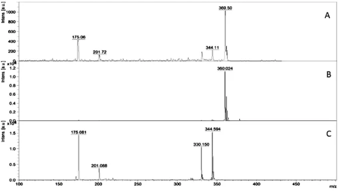
LC-MS/MS was used to quantify the drug in plasma and brain homogenates, and MALDI MSI was used to determine the distribution of the analyte. Matrix effects were determined using a calculated ratio of the peak area in the presence of matrix (measured by analyzing blank matrix added after extraction with analyte) to the peak area in the absence of matrix (pure solution of the analyte) expressed as a percentage. After sufficient optimization of the chromatographic separation conditions, a reproducible separation of DOX from plasma and brain homogenates was achieved.
Based on previous dose-response studies by Jantzie et al., DOX reached a therapeutic dose when 10 mg/kg of the drug was administered intraperitoneally [15]. The intensity increased with time, which showed the penetration and accumulation of the drug in the brain.

Paper 7
The suitability of this method was validated by its use for the quantification of the analytes in the biological samples. The H&E-stained coronal brain section is located in the center of the brain images (C= cortex; H= hippocampus). After that, the drug completely localized to the cortex of the brain as shown in the images for time points 120-240 min.
Tewson, T., et al., The synthesis of fluoro-18 lomefloxacin and its preliminary use in human studies. Babich, J.W., et al., 18 F labeling and biodistribution of the new fluoroquinolone antimicrobial, trovafloxacin (CP 99,219).

Paper 8
Our findings showed that the drug localizes in specific compartments of the rat brain, viz. This made it possible for the visualization and mapping of the drug in the brain sections. At this stage, the distribution of the drug appears to be higher in the CC over the C.
As is clear in the images, the distribution of the drug at this time is not very different. Shobo, A., et al., MALDI MSI and LCMS/MS: Toward preclinical assessment of the neurotoxic potential of fluoroquinolones.

Summary
In Chapters 5, 6, 7 and 8, we have shown how LC-MS and MSI can be used to evaluate a wide range of antibiotics, focusing on their utility in CNS investigations focusing on drug distribution in the brain. This highlights how the chemical properties of a drug strongly influence its interaction with the BBB and, consequently, its distribution to various structures in the brain. This technique has also been used to great effect in the evaluation of pretomanid, a drug currently in clinical trials, and is similarly used to evaluate other antibiotics of this type.
These findings further highlight how MSI can be used to streamline the drug development process and aid in the selection and identification of promising candidates while eliminating less promising candidates. Studies to be carried out will include investigating the distribution of the anti-mycobacterial drugs in the brains of TB-infected animals (such as mice and rabbits).


![Figure 11. The blood brain barrier. Endotheial tight junctions govern the movement of substances from blood into the brain, while astrocytes are responsible for selective permeability in the opposite direction [111]](https://thumb-ap.123doks.com/thumbv2/pubpdfnet/10643970.0/33.918.137.780.177.446/endotheial-junctions-substances-astrocytes-responsible-selective-permeability-direction.webp)

