http://mji.ui.ac.id
Delayed diagnosis of nasopharyngeal carcinoma in a patient
with early signs of unilateral ear disorder
Abstrak
Karsinoma nasofaring (KNF) merupakan keganasan kepala leher paling sering di Indonesia. Keterlambatan diagnosis sering terjadi pada KNF karena gejala tidak
spesiik seperti gangguan telinga unilateral. Kasus ini
dilaporkan untuk mengingatkan pasien, keluarganya, dan dokter tentang gejala awal KNF dan faktor-faktor penyebab keterlambatan diagnosis. Laki-laki, 44 tahun, gejala awal gangguan telinga unilateral sudah konsultasi ke dokter (THT-KL, mata, syaraf, dan gigi) tetapi baru didiagnosis KNF stadium IVA (T4N1M0) 1 tahun
kemudian. Gejala awal KNF tidak spesiik seperti keluhan
telinga unilateral. Pasien, keluarganya dan dokter harus mecurigai KNF terhadap pasien dengan keluhan telinga unilateral yang tidak sembuh dengan pengobatan yang diberikan.
Abstract
Nasopharyngeal carcinoma (NPC) is the most frequent head and neck malignancy in Indonesia. Misdiagnosis of NPC is common because of unspeciic symptoms as unilateral ear complaint. This case reminds doctors of the early symptoms of NPC and of other factors which lead to misdiagnosis and addresses also patients and their families. Reported is a 44 years old man with unilateral ear disorder that had been treated by otorhinolaryngologists, an ophthalmologist, a neurologist, and dentist irst, but diagnosed with nasopharyngeal carcinoma stage IVA (T4N1M0) one year later. NPC has unspeciic early symptoms such as unilateral ear disorder. Primarily doctors, but also patients and their families should be aware of unilateral ear complaint.
Keywords: misdiagnosis, nasopharyngeal carcinoma, unilateral ear disorders
pISSN: 0853-1773 • eISSN: 2252-8083 • http://dx.doi.org/10.13181/mji.v23i1.689 • Med J Indones. 2014;23:52-7 Correspondence author: Dadan Rohdiana, dadan_halsel@yahoo.com
C a s e R e p o r t
Copyright @ 2014 Authors. This is an open access article distributed under the terms of the Creative Commons Attribution-NonCommercial-ShareAlike 4.0 International License (http://creativecommons.org/licenses/by-nc-sa/4.0/), which permits unrestricted non-commercial use, distribution, and reproduction in any medium, provided the original author and source are properly cited.
Nasopharyngeal carcinoma (NPC) has a high incidence rate in Southern China, South East Asia, Japan, North Africa and the Middle East.1,2
The incidence rate of NPC in Southern China ranges from 15 to 50 per 100,000 inhabitants.3 The
nasopharyngeal malignancy is the most common malignancy of head and neck in Indonesia, the fourth
most common cancer after ovarium, breast and skin cancer. The incidence rate of NPC in Indonesia is 6.2 per 100,000 population or 12,000 new cases every
year.4
Early detection of NPC is dificult, but early diagnosis is important to achieve optimum results of treatment. The dificulty of early NPC diagnosis is caused by unspeciied early symptoms such as
ear and nose complaints. Ear complaints in this
malignancy have unilateral characteristic, such as
Eustachian tube dysfunction, otitis media effusion,
conductive hearing loss and ear pain.5-7
NPC patients in early stage (T1, T2, N0-1 without
metastasis) have a 5 years survival rate of 85%, whereas in advanced stage (T3, T4 dan N2, N3 without metastasis) the 5 years survival rate is 65%.5,8
The 10 years free disease rate in early stage is 67-71% and for advanced stage it is 29 - 54%. Therefore, it is
important to recognize factors related to the delay of
symptoms and misdiagnosis to improve identiication
and early management.9
CASE REPORT
We reported one case of a 44-year-old man, Sumatran
ethnicity from Lampung Province with chief
complaint about intermittent tinnitus in right ear since one year. The tinnitus disappeared in physical
activity. Patient also complained about loss of hearing
in the right ear, fullness in right ear, headache, and pain in the right retroorbital region. There was no history of otorrhea and head trauma. There was Marlinda Adham, Dadan Rohdiana, Ika D. Mayangsari, Zanil Musa
http://mji.ui.ac.id
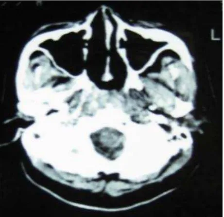
Figure 1. Computed tomography with normal result, while there is asymmetry of the right nasopharyngeal area
no complaint on the left ear. This patient did not complain about stuffed nose or bleeding from the
nose. He presented to the irst otorhinolaryngologist and was told that he did not have any disease and should consult to a dentist. He was given ear drops.
He consulted to a dentist and was told that there was a decay in left upper tooth. His tooth was
extracted but the complaints did not decrease. Three
months later, the patient complained of tinnitus and fullness in the right ear. He consulted to the second otorhinolaryngologist and was told that there was
no disorder. The patient received ear drops and 10
times ear thermal therapy, but there was still no
improvement.
Three months after he had presented to the second otorhinolaryngologist, the patient complained
about double vision in the right eye. There was no blurred vision and no complaint of the
nose. The patient presented again to the second otorhinolaryngologist and was consulted to a neurologist and an ophthalmologist. Patient was transferred to do computed tomography and
no abnormality was found. He was only given
analgetics until 4 months. The ophthalmologist stated that the eye complaint was only caused by
tiredness in the eye and recommended vitamins to improve his vision.
In February 2012, four months after he consulted with the neurologist the patient went to the third
otorhinolaryngologist with the same complaints and was told again that there was no disorder. The patient was suggested to go to another dentist for
further evaluation. Then, he underwent panoramic radiography. The radiographic examination showed sclerotic inding around the radices of the upper
right teeth number 4 through 8 with the possibility of chronic periodontitis. The upper left teeth number
7 and 8, right lower region number 6 and 7, and left lower region number 6 and 8 had already fallen
out. There was no radical cyst or granuloma. The
dentist extracted 4 teeth in the upper right region, but complaints did not improve.
Figure 2. Panoramic examination showed chronic periodontitis
of upper right teeth number 4 through 8
In April 2012, exactly one year after his irst complaints,
the patient presented to the fourth otorhinolaryngologist with the same complaints and was subjected to
audiologic examination with tune fork, audiometry, and timpanometry. From Weber examination there
was lateralization to the right ear. From the audiometry
result there was a mild conductive hearing loss with
threshold of 35 dB at the right ear and 10 dB at the left ear. The tympanometry results were type B on the right ear and type A at the left ear.
The patient was diagnosed with right otitis media
effusion and consulted to the ifth otorhinolaryngologist
for nasopharyngeal malignancy suspicion.
The ifth otorhinolaryngologist examined the right
ear with result of wide ear canal, no secrete, no cerumen, intact tympanic membrane and decreased
cone of light relection. The left ear examination
showed wide ear canal, no secrete, no cerumen, intact tympanic membrane and normal cone of
http://mji.ui.ac.id
Figure 4. Nasoendoscopic examination showed mass with
smooth surface on the right nasopharyngeal roof
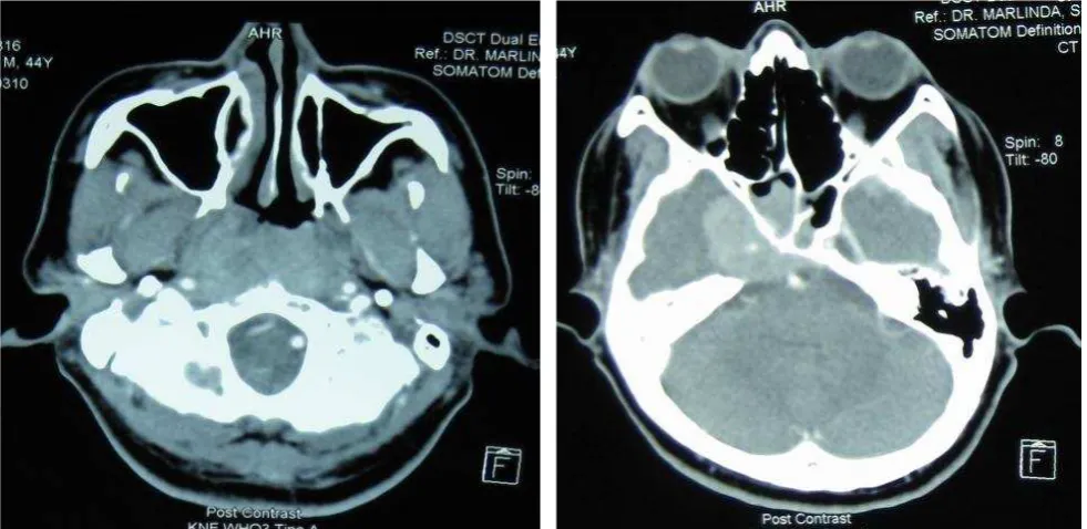
Figure 5. Computed tomography showed nasopharyngeal mass with extension to the surrounding structure iniltrating the intracra
-nium and right temporal lobe. Lymph nodes enlargement of left submandibular and colli bilateral with biggest size of 1.6 x 0.9 cm including sinusitis maxilaris sinistris
Figure 3. Audiometry examination of the right ear showed mild
conductive hearing loss at threshold 35 dB
inferior turbinate, no deviation of septum and mass. From throat examination, the pharyngeal arch was symmetrical, uvula in the middle, tonsil T1/T1, and normal posterior pharyngeal wall. The examination in the right neck region revealed lymph node enlargement level II sized 1.5 x 1 cm2.
Nasoendoscopic examination resulted in mass with
smooth surface on right nasopharyngeal roof which obliterates Rosenmuller fossa, torus tubarius, and narrowed Eustachian tube.
Computed tomography re-examination showed nasopharyngeal mass with extension to the surrounding structure and iniltrate intracranially and also iniltrate to the right temporal lobe. There
was lymph nodes enlargement in left submandibular region and colli bilateral region with biggest size of
1.6 x 0.9 cm2 and left maxillary sinusitis.
http://mji.ui.ac.id
malignancy. Thorax x-ray, abdominal ultra sonography,
and bone scan showed no further metastases.
The patient was also consulted to an ophtalmologist, a dentist, and neurologist. In accordance with
consultation reports, the eye movement showed no
abnormality in each direction, anterior and posterior segment showed no abnormality, funduscopy was within normal limit, impression of right and left eye was normal. The dentist found no focal infection
in the oral cavity, so that the dental treatment was inished and the patient could be radiated.
Neurologist consultation found that there were
lesions at the right trigeminal nerve; irst, second,
and third branch (sensoric), diplopia (right abduscen
nerve paresis), right facialis nerve paresis. Thus, the
conclusions were multiple cranial palsy with suspect
of iniltration into cranial base.
In conclusion, the patient had NPC WHO type 3 T4N1M0 (stage IV A) and treated with
chemoradiation: 33 times radiation with 66 Gy and chemotherapy weekly with cisplatin 60 mg/m2. Eight
weeks post-treatment, the result from clinical and
radiologic examination showed complete response.
DISCUSSION
This report is about 44 year old male patient with unilateral ear disorder and late diagnosis of nasopharyngeal carcinoma. The ear disorder is the most common initial complaint of NPC, reaching
89% as reported by Abdullah, et al10 or 60% based
on the report of Adham, et al.4
Ear disorder is not a speciic for nasopharyngeal symptom even though it is a common complaint in
early stage of NPC and therefore, diagnosis for NPC is often late. Ear disorder in NPC is usually unilateral, with tinnitus as a form of Eustachian tube disorder,
otitis media effusion, conductive hearing loss, and
otalgia. The ear symptom may be diagnosed as another common ear disease or to be caused by upper tract respiratory infection. This happens because “ear symptom” is not pathognomonic for NPC, especially in low rate incidence area.4,7,11
Our patient had no enlargement of cervical lymph
nodes as further complaint and nasal symptom.
The enlargement of cervical lymph nodes and nasal
symptoms are the most common reasons that couse patients come to health care professionals. Muthanna and Alaryani12 reported that among 100 patients with
NPC, 60% had enlarged cervical lymph nodes and 50% had nasal symptom, while Alabi, et al13 reported
that 96.7% of 30 patients had enlarged cervical lymph nodes and 66.7% of them had nose bleeding.
The desire of patients to overcome their ear complaint
is high. This can be seen from the high number of
patients who visit an otorhinolaryngologist directly after suffering ear symptoms and in this case from the
history of previous multiple visits to other specialists who did not manage to improve the patient’s symptoms.
The doctors did not consider ear symptoms as an early indication of NPC, but considered dental disorder as
the cause. The medical history of double vision is a symptom of advanced NPC which caused paralysis of cranial nerve 3, 4, and 6.
Some studies state that there are two factors as the cause of misdiagnosis of NPC, which are patient factors (patient delay) and health system factors (professional delay). Improper diagnosis, lack of suspicion of NPC by professional health practitioners
and consideration of dental abnormalities play a very
important role in late diagnosis.14-16
Delay in diagnosis may be up to 12 months counted
from the irst consultation to an otorhinolaryngologist. This is similar to the report by Prassad and Pua15 that
health professionals diagnose NPC within 127 days
on the average, which means, more than 4 months.
Patients with nasal complaint as an early symptom
will be diagnosed of NPC within 26 days, with intracranial symptoms within 51 days, with cervical
lymph nodes enlargement in 3 months, and with ear
symptoms in nearly 9 months.15
Patients’ unilateral otitis media efusion may be
caused by Eustachian tube dysfunction as the result of tumor depression.17 This symptom can be examined
by otoscopy, audiometry, and tympanometry, while nasoendoscopy and nasopharyngeal biopsy can be used to diagnose NPC. This corresponds with the
research by Glynn, et al18 who tried to correlate the
incidence of serous otitis media with the incidence of NPC.
The otorhinolaryngologist, neurologist, and dentist as medical practitioners were not able to diagnose an early case of NPC. These practitioners were not aware of NPC as differential diagnosis leading to
http://mji.ui.ac.id
Nasoendoscopic examination can show
the involvement of mucosa and the tumor expansion to the nasal cavity or oropharynx,20
while examination of biopsy samples from the nasopharynx will conirm the diagnosis and
determine the histopathologic type.11,18
The histologic feature of our patient was a type A undifferentiated non keratinizing carcinoma.
WHO 1978 classiied this histologic feature into
type 3,19 while based on WHO 2005 classiication, this feature is included in type 2, which is a non keratinizing carcinoma with undifferentiated subtype.21
Undifferentiated carcinoma has a higher success rate for local tumor therapy but has a higher incidence of metastasis compared to well-differentiated type.2 Type
A is related to prognosis as stated by Hsu, et al22 that
type A is an intermediate malignancy with 30 - 40% survival rate.22
Staging of NPC in this patient is based on a
complete physical examination, CT scan, chest x-ray, abdominal ultrasound, and bone scan. Based on these examinations, the staging of this patient
is T4N1M0 according to AJCC/IUCC 2010. CT
scan examination was demonstrated to evaluate the expansion of primary tumor and the presence of nodal enlargement, while chest x-ray, abdominal
ultrasound, and bone scan were demonstrated to
evaluate the presence of distant metastasis.11,19,23
This patient was given chemoradiation therapy
according to NCCN 2011 and based on AJCC/UICC 2010, which stated that T1, N1-3, or T2-4, all N (stage
II, III, IVa, IVb) NPC should be given concurrent chemoradiation. Adjuvant chemotherapy or induction
chemotherapy can be added to the treatment protocol.23
This patient had had 85% survival rate for 5 years and 67-71% for 10 years free of recurrency rate, if he was
diagnosed earlier. Due to the delay of diagnosis, the patient was diagnosed at T4N1M0 (stage IVA), which
has 65% of 5 years survival rate and 29-54% of 10
years free of recurrency rate. The delay of diagnosis in this patient decreased the quality of life, increased
the inancial burden, and also increased psychological
problems for the patient and his family.
In conclusion, early diagnosis of NPC is essential for
the prognosis. NPC has unspeciic early symptoms,
such as unilateral ear complaint. Therefore,
medical practitioners must improve their diagnostic
capabilities especially in differential diagnosis and
to consider the possibility of NPC. Patients and their families should also be aware of these complaints in regions with high incidence of NPC.
Conlict of interest
The authors declare that this study is free of conlict
of interest.
REFERENCES
1. Yoshizaki T, Ito M, Murono S, Wakisaka N, Kondo S, Endo K. Current understanding and management
of nasopharyngeal carcinoma. Auris Nasus Larynx. 2012;39(2):137-44.
2. Tabuchi K, Nakayama M, Nishimura B, Hayashi K, Hara A. Early detection of nasopharyngeal carcinoma. International
Journal of Otolaryngology. 2011;2011:1-6.
3. Chan AT, Teo PM, Johnson PJ. Nasopharyngeal carcinoma.
Ann Oncol. 2002;13(7):1007-15.
4. Adham M, Kurniawan AN, Muhtadi AI, Roezin A, Hermani
B, Gondhowiardjo S, et al. Nasopharyngeal carcinoma in
Indonesia: epidemiology, incidence, signs, and symptoms
at presentation. Chin J Cancer. 2012;31(4):185-96.
5. Wildeman MA, Fles R, Adham M, Mayangsari ID, Luirink I, Sandberg M, et al. Short-term effect of different teaching methods on nasopharyngeal carcinoma for general practitioners in Jakarta, Indonesia. Plos ONE.
2012;7(3):1-7.
6. Low WKC, Rangabashyam M. Ear-related issues in
patients with nasopharyngeal carcinoma. In: Chen SS, editor. Carcinogenesis, diagnosis, and molecular targeted
treatment for nasopharyngeal carcinoma. Croatia: InTech;
2012. p. 155-78.
7. Daniel A, Fasunla AJ. Nasopharyngeal cancer mimicking otitic barotrauma in a resource-challenged center: a case
report. J Med Case Rep. 2011;5:532.
8. Lee AW, Poon YF, Foo W, Law SC, Cheung FK, Chan
DK, et al. Retrospective analysis of 5037 patients with nasopharyngeal carcinoma treated during 1976-1985: overall survival and patterns of failure. Int J Radiat Oncol Biol Phys. 1992;23(2):261-70.
9. Sing TT, Subramaniam SK. Factors of late presentation and
diagnosis of nasopharyngeal carcinoma in Sarawak Malaysia.
The Internet Journal of Head and Neck Surgery. 2007;1(1):1.
10. Abdullah NE, Adam AAM, Khalifa EH, Hassan LAM, Ibrahim ME, Hamad KM, et al. Nasopharyngeal cancer in Sudan: epidemiology, clinical and histological characteristics.
Clin Med Insights Ear Nose Throat. 2011;4:5-11.
11. Abdullah B, Alias A, Hassan S. Challenges in the
management of nasopharyngeal carcinoma: a review. Malays J Med Sci. 2009;16(4):50-4.
12. Muthanna AO, Alaryani A. Clinical presentation of nasopharyngeal cancer in Yemen. Egypt J Biomed Sci.
2007;23(1):237-43.
13. Alabi BS, Badmos KB, Afolabi OA, Buhari MO, Segun-Busari S. Clinico-pathological pattern of nasopharyngeal carcinoma in Ilorin, Nigeria. Niger J Clin Pract.
2010;13(4):445-8.
14. Al-Rajhi N, El-Sebaie M, Khafaga Y, AlZahrani A,
http://mji.ui.ac.id Saudi Arabia: clinical presentation and diagnostic delay.
East Mediterr Health J. 2009;15(5):1301-7.
15. Prasad U, Pua KC. Nasopharyngeal carcinoma: a delay in
diagnosis. Med J Malaysia. 2000;55(2):230-5.
16. Mackie AM, Epstein JB, Wu JS, Stevenson-Moore P.
Nasopharyngeal carcinoma: the role of the dentist in assessment, early diagnosis and care before and after cancer
therapy. Oral Oncol. 2000;36(5):397-403.
17. Smerq, J. Sharma, M. The Risk Factor: Nasopharyngeal
Carcinoma. IJPCR. 2011;3(3):48-51.
18. Glynn F, Keogh IJ, Ali TA, Timon CI, Donnelly M.
Routine nasopharyngeal biopsy in adults presenting with
isolated serous otitis media: is it justiied? J Laryngol Otol. 2006;120(6):439-41.
19. Brennan B. Nasopharyngeal carcinoma. Orphanet Journal of Rare Diseases. 2006;1:23.
20. Wei WI, Sham JST. Nasopharyngeal carcinoma. Lancet.
2005;365(9476):2041-54.
21. IARC. Pathology and genetics of head and neck tumours.
World Health Organization Classiication of Tumours.
Lyon. 2005.
22. Hsu HC, Chen CL, Hsu MM, Lynn TC, Tu SM, Huang SC. Pathology of nasopharyngeal carcinoma. Proposal of a new
histologic classiication correlated with prognosis. Cancer. 1987;59(5):945-51.
23. National Comprehensive Cancer Network. NCCN clinical