Fertilization and Development of Frozen –Thawed Germinal Vesicle
Bovine Oocytes by a One-Step Dilution Method
in Vitro
T. SUZUKI,* A. BOEDIONO,† M. TAKAGI,‡ S. SAHA,* AND C. SUMANTRI*
*United Graduate School of Veterinary Sciences, Yamaguchi University, Yamaguchi 753, Japan; †Faculty of
Veterinary Medicine, Bogor Agriculture University, Bogor 16151, Indonesia; and ‡Obihiro University of Agriculture and Veterinary Medicine, Hokkaido 080, Japan
E-mail: [email protected]
The objective of this study was to evaluate in vitro fertilization and cleavage rates of frozen – thawed bovine oocytes at the germinal vesicle (GV) stage. In mouse oocytes, spindle microtubule reorganization after GV breakdown is particularly sensitive to cold and readily damaged by exposure to low temperatures, the damage becoming apparent only at the time of the first mitotic division. The effects of various permeating cryoprotective agents [1.8 M ethylene glycol (EG), 1.3 M ethylene glycol monomethyl ether (EME), and 1.6 M 1,2-propanediol (PROH)] and different concentrations of trehalose (T) and polyvinylpyrrolidone (PVP) on post-thaw developmental capacity were examined. When bovine GV oocytes were frozen slowly in mixtures of 1.8 M EG plus 5% PVP and 0.05 M T, almost 80% developed to metaphase II; 22.2% degenerated after in vitro maturation, and none of those that had been cryopreserved underwent parthenoge-netic activation. The total fertilization rate was higher (Põ0.05) for oocytes frozen in a mixture of 1.8
M EG plus 0.05 M T or 0.1 M T than in a mixture of 1.8 M EG with or without 0.2 M T; however, there
was no difference in the number of normally fertilized or polyspermic oocytes that had been frozen in various cryoprotective solutions. No significant difference was observed in subsequent development using EG, EME, and PROH for GV oocytes. The addition of 0.05 or 0.1 M trehalose to the freezing solution yielded significantly better cleavage and blastocyst rates than the solutions containing 0.2 M or no trehalose. For unfrozen controls, GV oocytes yielded significantly higher (Põ0.01) cleavage and blastocyst rates compared with frozen – thawed GV oocytes. It was found that 5% PVP had a beneficial effect compared with 10 or 20% concentrations for the development of blastocysts. Transfer of six blastocysts derived from frozen – thawed GV oocytes into three recipient heifers resulted in three pregnancies and the birth of one set of twins and one singleton calf. q1996 Academic Press, Inc.
Oocytes are extraordinarily large cells. In cytes are barrel-shaped, with the diameter of
the metaphase plate longer than the pole-to-the germinal vesicle (GV) stage oocyte,
chro-matin is in a rather decondensed state and a pole distance. The chromosomes are clustered
in a discrete bundle at the metaphase plate. few microtubule organizing centers (MTOCs)
are found perinuclearly. At the onset of matu- Some microtubules traverse the length of the
spindle from pole to pole; others extend from ration, cytoplasmic MTOCs are recruited from
the cytoplasm. On breakdown of the nuclear the spindle poles to chromosomes. In mature
oocytes, microtubules appear to be restricted envelope and condensation of the chromatin,
MTOCs combine with nuclear pericentriolar largely to the meiotic spindle, with little
evi-dence for foci of pericentriolar material (2). material (PCM) and polymerize microtubules
toward the condensed chromosomes (42). Cooling induces chromosomal abnormalities
including disorganized metaphase plates and Also, the actin-containing microfilaments are
arranged in the perinuclear site (27). The mei- multipolar spindles in oocytes cooled at all
otic spindles of in vitro-matured bovine oo- stages of meiosis from germinal vesicle
break-down (GVBD) to metaphase II (23). Glass and Voelkel (11) observed that loss of viability in
specific step in the cryopreservation process. development rates of frozen – thawed oocytes are still low. Moreover, progress in the A limiting factor for achieving efficient
cryo-preservation of oocytes is direct chilling injury preservation of GV bovine oocytes has been
limited and production of offspring derived (DCI), which occurs to oocytes during cooling
(3). The most striking among the detrimental from them has not been reported. So more
research is needed because storing immature effects caused by cooling oocytes are on the
second meiotic spindle where microtubules oocytes is an important advance with
implica-tions for the cattle breeding industry and for are disrupted or disassembled, apparently as
a result of tubulin depolymerization (27, 29). conservation. Along with in vitro-matured
oo-cytes (12), if GV stage oooo-cytes also can be Cooling also alters the zona pellucida,
re-sulting in decreased sensitivity to chymotryp- cryopreserved successfully, the timing of in
vitro maturation – in vitro fertilization – in vitro sin and reduced fertilization rates, caused by
cortical granule exocytosis leading to a prema- culture (IVM – IVF – IVC) will be more
man-ageable and genetic resources of various spe-ture cortical granule reaction (17).
Fertiliza-tion and development rates of frozen – thawed cies or strains can be preserved more
effi-ciently. Application of these techniques pro-bovine ova are lower than those of unfrozen
ova. Mature mouse oocytes can be success- vides a means of preserving oocytes before
maturation and avoids the possible damage fully cryopreserved when cooled slowly in 1.5
M dimethyl sulfoxide (DMSO) (46). Since this that may occur in the spindle of the mature oocyte during freeze – thawing (7). Recently, observation was first made, offspring have
been produced after transfer of embryos ob- Leibo and Oda (20) reported high survival of
mouse zygotes and embryos cooled rapidly or tained from frozen – thawed rabbit (1, 44), cow
(10, 26), and human oocytes (9, 43); however, slowly in EG plus PVP. A gradual dilution of
sucrose in several steps seems to be suffi-the overall success of suffi-these procedures
re-mains low [with some exceptions (8)], primar- ciently effective to mitigate osmotic injury so
that the oocytes can develop after IVF (12). ily because of the reduced rate of fertilization
after freezing and thawing. In the mouse, ma- This study was conducted to examine the
ef-fect of trehalose and PVP on the cryopreserva-ture oocytes can be slowly frozen and
pre-served at 01967C using 1,2-propanediol tion of bovine oocytes at the GV stage with
a single step addition and removal of the cryo-(PROH) as a cryoprotectant (18). Otoi et al.
(26) obtained normal offspring from matured protectants. In addition, we examined the
nor-mality of embryos resulting from oocytes fro-bovine oocytes frozen and thawed in PROH
following fertilization, culture in vitro, and zen in cryoprotectants following maturation,
fertilization and culture in vitro (IVM – IVF – embryo transfer. Three cryoprotectants —
eth-ylene glycol (EG), etheth-ylene glycol mono- IVC), and embryo transfer.
methyl ether (EME), and PROH — are
rela-MATERIALS AND METHODS
tively nontoxic and have been used for bovine
Oocyte Collection embryos, permitting direct rehydration of
thawed embryos (40). In a previous report The method for IVM, IVF, and IVC used
in these experiments was a modification of the (38), we described the successful
cryopreser-vation of GV bovine oocytes by a combined procedure of Boediono et al. (5). Ovaries were
collected from cows at a local abattoir and process of dehydration of the oocytes with
trehalose and permeation with PROH before were brought to the laboratory in
physiologi-cal saline [0.89% (w/v) NaCl] at 25 to 307C
plunging the oocytes into liquid nitrogen. In
contrast to the many advanced studies with within 3 h. The cumulus – oocyte complexes
(COCs) in follicular fluid (5 – 10 per ovary) bovine embryos, however, data regarding
di-ameter) with a 5-ml syringe with an 18-gauge the semen was resuspended in BO supple-mented with 1% bovine serum albumin and needle. COCs were collected and washed
three times in Dulbecco’s phosphate-buffered 20mg ml01heparin (Shimizu Pharmaceutical
Co., Ltd., Shimizu, Japan) to yield a sperm saline (PBS; Gibco, Grand Island, NY)
sup-plemented with 3 mg ml01
fraction V bovine concentration of 51106
ml01
. A 100-ml
ali-quot of sperm suspension was covered with serum albumin (BSA-V, Sigma Chemical Co.,
St. Louis MO). All media used for collection mineral oil. Oocytes that had been cultured
for maturation in vitro were transferred into
and handling of oocytes were kept at 377C on
a warming block. sperm microdrops (20 – 25 oocytes per micro
drop) for insemination for 5 h at 38.57C under
Freezing and Thawing 5% CO
2in air.
COCs were suspended directly in the
cryo-In Vitro Cultures protectants for 5 min at room temperature
(207C). Following this exposure, 20 to 30 After 5 h of insemination, oocytes with
cu-mulus cells were washed and transferred into COCs were loaded into 0.25-ml plastic straws.
After loading, the straws were placed in a pro- culture medium for further development. The
culture medium consisted of TCM-199 sup-gram freezer (ET-1, Fujihira Co. Ltd, Tokyo,
Japan) maintained at 07C for 2 min. Oocytes plemented with 5% SCS, 5 mg ml01
insulin (Wako Pure Chemical Industries Ltd., Osaka,
were then cooled to 067C at a rate of 17C
min01
, seeded at067C, then held for 10 min, Japan), and 50 mg ml01
gentamicin. Forty-eight hours after insemination, the cumulus
cooled again at a rate of 0.37C min01
to
0307C, and finally plunged into liquid nitro- cells surrounding the embryos were removed,
and the cumulus cell layer attached to the
bot-gen. The straws were held at 01967C for 1
to 6 months. The cryopreserved straws were tom of the culture dish was used for coculture.
The incubation medium was replaced with placed in air for 5 s and then plunged into a
377C water bath for 10 s for thawing. new medium every 96 h.
Experiment 1 In Vitro Maturation
Frozen – thawed GV oocytes were placed in The first experiment was conducted to
de-termine the response to an activating stimulus a polystyrene culture dish and then transferred
directly into maturation medium containing 25 in GV oocytes previously frozen and then
ma-tured in vitro. Oocytes were frozen in 1.8 M mM Hepes TCM-199 with Earle’s salts
(Gibco, Grand Island, NY) supplemented with EG/5% PVP /0.05 M trehalose (T).
Parthenogenetic treatment. The procedures superovulated cow serum (SCS) (22, 39), 0.01
mg ml01
follicle-stimulating hormone (FSH, for producing diploid parthenogenetic
em-bryos have been described previously (4). Fro-Denka Pharmaceutical Co., Kawasaki, Japan),
and 50mg ml01gentamicin (Sigma Chemical zen – thawed oocytes were matured for 32 h
at 38.57C under 5% CO2 in air. To induce
Co.). After being washed three times, oocytes
surrounded by cumulus cells were kept for 22 parthenogenetic activation, these matured
oo-cytes were suspended in culture medium
con-h at 38.57C under 5% CO2 in air for
matura-tion. taining 7% ethanol for 10 min and then
cul-tured in medium containing 5 mg ml01
cyto-In Vitro Fertilization chalasin D (5 h) to suppress extrusion of the
second polar body, thus producing diploid par-Frozen semen was thawed in a water bath
(377C) and washed twice using 2.5 mM caf- thenogenotes.
As controls, frozen – thawed GV oocytes feine in Brackett and Oliphant medium (BO)
CO2in air. These matured oocytes were then (Sigma). They were then fixed in Carnoy
solu-tion (3 parts ethanol:1 part acetic acid) for 72 cultured in vitro without any fertilization
treat-ment. h and stained in 1% aceto-orcein to look for
the formation of nuclei or pronuclei. Experiment 2
Embryo Evaluation The second experiment was conducted to
determine the effects of cryopreservation at The proportions of embryos that had
devel-the GV stage on subsequent fertilization in oped to the 2-, 4-, and 8-cell stages were
re-vitro. COCs were frozen in 1.8 M EG, 1.8 M corded 48 h after insemination. Blastocyst
de-EG/0.05 M T, 1.8 M EG/0.1 M T, or 1.8 velopment was assessed up to Day 9.
M EG/0.2 M T. Frozen – thawed GV oocytes
Embryo Transfer
were matured for 22 h at 38.57C under 5%
CO2in air. These matured oocytes were then Six morphologically normal blastocysts on
incubated in vitro with frozen – thawed sperm. Day 8 of the cycle (Day 0 Åonset of estrus)
At 15 h after insemination, all oocytes were after in vitro fertilization derived from
fro-stained to evaluate fertilization. zen – thawed GV oocytes were transferred
nonsurgically (two embryos per recipient) to Experiment 3
the uteri of three heifers (18 months of age).
In this experiment, GV oocytes were frozen Pregnancy diagnosis was done by ultrasound
in (1) 1.8 M EG, (2) 1.8 M EG/0.05 M T, scan after 60 days.
(3) 1.8 M EG/0.1 M T or (4) 1.8 M EG /
0.2 M T, (5) 1.3 M EME, (6) 1.3 M EME/ Statistical Analysis
0.05 M T, (7) 1.3 M EME /0.1 M T or (8)
The data were analyzed byx2test and
AN-1.3 M EME / 0.2 M T, (9) 1.6 M PROH,
OVA. Probabilities of Põ0.05 and Põ0.01
(10) 1.6 M PROH / 0.05 M T, (11) 1.6 M
were considered to be statistically significant.
PROH/0.1 M T or (12) 1.6 M PROH/0.2
M T. After thawing the oocytes were assessed RESULTS
visually by morphological analysis under the
Experiment 1 microscope.
Two groups, each consisting of 27 matured Experiment 4 oocytes, were stained to assess the stage of meiosis. The results are shown in Table 1. The In the fourth experiment, unfrozen oocytes
proportion of metaphase II formation found were kept as (1) control (while thawing), and
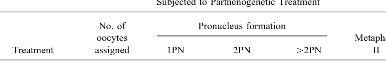
in control oocytes was 77.8%, and the
propor-GV oocytes were frozen in (2) 1.8 M EG/
tions of 1PN, 2PN, ú2PN, or metaphase II
0.05 M T, (3) 1.8 M EG /0.05 M T / 5%
formation found in oocytes subjected to
par-PVP, (4) 1.8 M EG/0.05 M T/10% PVP,
thenogenetic treatment were 22.2, 44.4, 11.1,
or (5) 1.8 M EG / 0.05 M T / 20% PVP.
and 22.2%. After thawing the oocytes were assessed
visu-ally by morphological analysis under the
mi-Experiment 2 croscope.
The results of this experiment are shown in Oocyte Staining Table 2. The total fertilization was higher in
oocytes cryopreserved in 1.8 M EG/0.05 M
After 24 h of maturation and either 15 h of
parthenogenetic treatment or 15 h of insemi- T or 1.8 M EG/0.1 M T treatments than in
those frozen in EG either without trehalose or nation, the cumulus cells surrounding the
em-bryos were removed by pipetting them in me- with 0.2 M T. Polyspermy occurred in oocytes
frozen in 1.8 M EG / 0.05 M T and in 1.8
dium containing 150 U ml01
TABLE 1
Frozen-Thawed Bovine GV Oocytes (in 1.8 M EG/0.05 M T/5% PVP) Subjected to Parthenogenetic Treatment
No. of Pronucleus formation
oocytes Metaphase
Treatment assigned 1PN 2PN ú2PN II Degenerated
Control 27 0 (0.0)a 0 (0.0)a 0 (0.0)a 21 (77.8)a 6 (22.2)a
Ethanol-exposed 27 6 (22.2)b 12 (44.4)b 3 (11.1)b 6 (22.2)b 0 (0.0)b
EG, ethylene glycol; T, trehalose; PVP, polyvinylpyrrolidone.
a,bValues within columns with different superscripts are significantly different (P
õ0.01).
M EG/0.1 M T, whereas it did not occur in cryopreserved oocytes. Table 4 shows the
proportions of cryopreserved oocytes that 30 oocytes frozen in 1.8 M EG and in another
30 oocytes frozen in 1.8 M EG / 0.2 M T. cleaved and developed to the blastocyst
stage. The cleavage rate of unfrozen oo-The fertilization rate was higher in oocytes
cryopreserved in 1.8 M EG / 0.05 M T or cytes (control) was significantly higher than
that of frozen oocytes. Nevertheless, 5%
1.8 M EG/0.1 M T than in those frozen in
EG either without T or with 0.2 M T. PVP added to 1.8 M EG /0.05 M T had a
beneficial effect (with respect to blastocyst Experiment 3 production) on the cryopreserved oocytes;
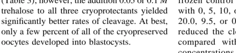
after freezing with 10% PVP or 20% PVP, There were no differences in the
propor-tions of oocytes that cleaved after being frozen fewer oocytes cleaved. The blastocyst rates,
as a percentage of cleaved oocytes, for un-in simple solutions of EG, EME, or PROH
(Table 3); however, the addition 0.05 or 0.1 M frozen control oocytes and frozen oocytes
with 0, 5, 10, or 20% PVP were 45.8, 4.4, trehalose to all three cryoprotectants yielded
significantly better rates of cleavage. At best, 20.0, 9.5, or 0.0%, respectively. Freezing
reduced the cleavage and blastocyst rates only a few percent of all of the cryopreserved
oocytes developed into blastocysts. compared with unfrozen controls. High
concentrations of PVP had a negative effect Experiment 4 on both cleavage and blastocyst formation, and the best post-thaw development oc-Addition of PVP to the freezing solution
significantly improved the development of curred with 5% PVP.
TABLE 2
Fertilization of GV Bovine Oocytes Cryopreserved Using Ethylene Glycol and Trehalose
No. (%) of oocytes fertilized No. of
Group oocytes Normal Polyspermic Total
1.8 M EG 30 10 (33.3)a 0 (0.0)a 10 (33.3)a
1.8 M EG/0.05 M T 30 12 (40.0)a 2 (6.7)a 14 (46.7)b
1.8 M EG/0.1 M T 30 15 (50.0)a 1 (3.3)a 16 (53.3)b
1.8 M EG/0.2 M T 30 8 (26.7)a
0 (0.0)a
8(26.7)a
Note. Normal and polyspermic fertilization were identified by the presence of 2PN andú2PN. EG, ethylene glycol; T, trehalose.
a,b
TABLE 3
Development Capacity of Frozen – Thawed GV Bovine Oocytes Cryopreserved Using Various Cryoprotectants
No. developed to
No. of oocytes No. cleaved blastocyst (% of
Cryoprotectant assessed (%) embryos cleaved)
1.8 M EG 288 72 (25.0)bc 0 (0.0)
EG, ethylene glycol; T, trehalose; EME, monomethyl ether; PROH, 1,2-propanediol.
a – c
Values within columns with different superscripts are significantly different (ANOVA, Scheffe F testab,bc
Põ
0.05,acP
õ0.01.
Development in Vivo of Frozen – Thawed cies diagnosed 60 days after transfer. One re-cipient delivered a single calf and a second GV Oocytes
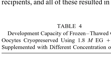
delivered twin calves on March 5th and May Six blastocysts resulting from IVM – IVF –
7th, 1995. One pregnancy resulted in abortion IVC of GV oocytes frozen in EG with 0.05
by Day 92. M T and 5% PVP were transferred into three
recipients, and all of these resulted in pregnan- DISCUSSION
In mouse oocytes, spindle microtubule or-ganization after GVBD is particularly
sensi-TABLE 4
tive to cold and is readily damaged by
expo-Development Capacity of Frozen – Thawed GV Bovine
Oocytes Cryopreserved Using 1.8 M EG/ 0.05 M T sure to low temperatures, the damage
becom-Supplemented with Different Concentration of PVPA
ing apparent only at the time of the first mitotic division (33). Parthenogenetic
activa-No. of
tion occurs after exposure to 1,2-propanediol
blastocysts
(41). In the present study, 44% of 27 frozen –
No. of No. (% of
Concentration of oocytes cleaved embryos thawed GV bovine oocytes developed to the
PVP (%) assessed (%) cleaved) 2PN stage following parthenogenetic
ment; however, without parthenogenetic
treat-Unfrozen control 100 83 (83.0)a 38 (45.8)a
ment, most frozen – thawed GV oocytes
devel-0 150 68 (45.3)b 3 (4.4)b
oped normally to the metaphase II (77.8%).
5 170 80 (47.1)b 16 (20.0)a
10 168 42 (25.0)c 4 (9.5)b No parthenogenetic activation due to freezing
20 144 18 (12.5)c 0 (0.0)c
was observed. This suggests that the treat-ments used in a successful cryopreservation
EG, ethylene glycol; T, trehalose; PVP,
polyvinylpyr-procedure do not cause irreversible damage to
rolidone.
the meiotic spindle and parthenogenetic acti-a – c
Values within columns with different superscripts are significantly different (ab,bc
Põ0.05;ac
In the second experiment, the total fertiliza- for the other. Heyman et al. (15) reported that very few (6%) bovine oocytes matured in vitro tion rates were significantly higher in oocytes
cryopreserved in EG plus low concentrations of after rapid freezing and thawing. Similarly,
Fuku et al. (10) reported that fewer than 5% trehalose (0.05 or 0.1 M) than in those frozen
in EG alone or in EG plus 0.2 M trehalose. We of frozen – thawed immature bovine oocytes
underwent GVBD and polar body formation. previously reported (38) that the cleavage rates
of GV bovine oocytes frozen in 1.6 M PROH In contrast to these observations, in the present
study the total cleavage and development rates with 0.1 or 0.2 M sucrose by a one-step
rehydra-tion procedure were higher than those of oocytes to blastocyst were 35.3 to 42.2% and 1.3 to
3.1%, respectively. These findings indicate frozen with the same cryoprotectant without
su-crose. Rayos et al. (28) also reported that su- that cleavage rate was improved, with a small
proportion developing to blastocysts in the crose or trehalose in combination with EG was
effective in the quick freezing of unfertilized presence of 0.05 or 0.1 M T.
In the fourth experiment, we used PVP to mouse oocytes. It is well known that
carbohy-drates can be used as an osmotic buffer to main- protect GV bovine oocytes against damage
caused by freezing and thawing. Although tain osmotic equilibrium between the embryonic
cells and the external environment in which the there was no improvement in the cleavage
rates, the development into blastocysts was embryo is suspended (30–32), while decreasing
the external concentration of cryoprotectant. GV significantly improved in the 5% PVP solution
compared with solutions containing 0%, 10%, bovine oocytes, however, were more sensitive
to a sucrose treatment than in vitro-matured oo- and 20% PVP. Leibo and Oda (20) reported
that when PVP is combined with a relatively cytes or zygotes (21, 25). Prolonged exposure
to sucrose had a deleterious effect on the devel- low concentration of EG, it apparently
en-hances the cryoprotective properties of EG so-opmental capacity of GV (immature) or mature
bovine oocytes (14). On the other hand, Schel- lutions, yielding high survival of zygotes and
embryos frozen either slowly or rapidly. Such lander et al. (35) indicated that the use of
carbo-hydrates during cryoprotectant removal is not a biological macromolecules are often added to
cryoprotective agent solutions either for their key element in improving oocyte
cryopreserva-tion. presumptive cryoprotective effect or as a
sur-factant (8, 31, 32). Even the use of the supple-Hernandez-Ledezma and Wright (13)
re-ported that the use of propanediol, instead of ments PVP and carbohydrates for freezing GV
bovine oocytes yielded a cleavage rate sig-glycerol or DMSO, significantly improved
sur-vival and development of cryopreserved mouse nificantly lower than that of unfrozen oocytes.
This suggests that some membrane damage oocytes to the two-cell stage. In the present
study, we used the permeable cryoprotectants and extensive disorganization of the ooplasma
may have occurred during freezing and thaw-EG, EME, and PROH to freeze GV bovine
oo-cytes (Experiment 3). There were no significant ing because of intracellular ice formation or
osmotic stress, which could have interfered in differences in cleavage and blastocyst
develop-ment rates for oocytes frozen in different cryo- the subsequent fertilization and development
of oocytes (34, 36, 47). Failure of fertilization protectants; however, to obtain a higher
propor-tion developing into blastocysts, it was neces- may also be due to the changes in the zona
pellucida during freezing and thawing, which sary to add a low concentration of a
car-bohydrate to the freezing solution. Because inhibits the entry of spermatozoa (17);
how-ever, we found that the development of two-oocytes and embryos at various stages of
de-velopment differ both physiologically and cell embryos to the blastocyst stage was
simi-lar in oocytes cryopreserved in cryoprotectant morphologically (15, 16, 19), freezing
REFERENCES
control oocytes. Schroeder et al. (37) reported
that a dramatic change in freezability of mouse 1. Al Hasani, A., Kirsch, J., Diedrich, K., Blanke, S., Van der Ven, H., and Krebs, D. Successful embryo
oocytes was associated with meiotic
matura-transfer of cryopreserved and in vitro fertilized
tion in vivo or in vitro. Moreover, the
damag-rabbit oocytes. Hum. Reprod. 4, 77 – 79 (1989).
ing effects of freeze – thawing were apparent 2. Aman, R. R., and Parks, J. E. Effects of cooling and
only up to the two-cell stage. Once past this rewarming on the meiotic spindle and
chromo-somes of in vitro-matured bovine oocytes. Biol.
hurdle, the development of oocytes to morulae
Reprod. 50, 103 – 110 (1994). or blastocysts was not affect further. The
find-3. Arav, A., Stefaneli, E., Leslie, S. J., and Crowe,
ings of Carroll et al. (8) support that
conclu-J. H. Phase transition temperature and chilling
sen-sion. Under conditions where fertilization of sitivity of immature, in vitro matured and
electro-oocytes frozen in medium with BSA or PVA fused immature and in vitro matured bovine
oo-cytes. Cryobiology 32, 570 (1995) (Abstract).
was low, substitution of FCS for BSA in the
4. Boediono, A., and Suzuki, T. Pregnancies after
trans-freezing medium restored fertilization to
fer of aggregated parthenogenetic bovine activated
within 15% of control levels. In the present
oocytes. Theriogenology 41, 166 (1994)
(Ab-study, when we used the higher concentrations stract).
of PVP (10 to 20%), we obtained significantly 5. Boediono, A., Takagi, M., Saha, S., and Suzuki, T. Influence of Day-0 and Day-7 superovulated cow
lower cleavage rates (25.0% and 12.5%) than
serum during development of bovine oocytes in
with a low concentration of PVP (45.3 and
vitro. Reprod. Fertil. Dev. 6, 261 – 264 (1994).
47.1% for 0 and 5%). The reason for this is
6. Brackett, B. G., and Oliphant, G. Capacitation of
rab-unclear, but it might be that high concentra- bit spermatozoa in vitro. Biol. Reprod. 12, 260 –
tions of PVP cause problems in seeding. 274 (1975).
7. Candy, C. J., Wood, M. J., Whittingham, D. G.,
Mer-Careful attention to temperature control
riman, J. A., and Choudhury, N. Cryopreservation
throughout oocyte retrieval, examination,
han-of immature mouse oocytes. Hum. Reprod. 9,
dling, and replacement should help to overcome
1738 – 1742 (1994).
the problems associated with cooling. Cooling 8. Carroll, J., Wood, M. J., and Whittingham, D. G.
promotes disassembly of microtubules, whereas Normal fertilization and development of frozen –
thawed mouse oocytes: Protective action of certain
cryoprotectants promote their uncontrolled
as-macromolecules. Biol. Reprod. 48, 606 – 612
sembly. Thus, a delicate balance results in which
(1993).
sufficient cryoprotectant enters to counteract the
9. Chen, C. Pregnancy after human oocyte
cryopreser-destabilizing effect of cooling. Manipulating the vation. Lancet 1, 884 – 886 (1986).
periods of exposure to cooled cryoprotectant 10. Fuku, E., Kojima, T., Shioya, Y., Marcus, G. J., and Downey, B. R. In vitro fertilization and
develop-prior to freezing and immediately after thawing
ment of frozen – thawed bovine oocytes.
Cryobiol-should enable an optimal regime for spindle
sta-ogy 29, 485 – 492 (1992). bilization to be developed (45).
11. Glass, K. W., and Voelkel, S. A. Loss of viability in
In conclusion, this study suggests that per- frozen bovine oocytes associated with specific
meating cryoprotective agents in combination steps in the cryopreservation process. Biol.
Re-prod. 42(Suppl. 1), 52 (1990). with a low concentration of a carbohydrate
12. Hamano, S., Koikeda, A., Kuwayama, M., and Nagai,
and PVP can be used effectively in the slow
T. Full-term development of in vitro-matured,
vit-freezing and thawing of GV bovine oocytes.
rified and fertilized bovine oocytes.
Theriogenol-More understanding regarding GVBD and nu- ogy 38, 1085 – 1090 (1992).
clear maturation will be helpful (24). 13. Hernandez-Ledezma, J. J., and Wright, R. W., Jr.
Deep freezing of mouse one-cell embryos and oo-ACKNOWLEDGMENTS cytes using different cryoprotectants.
Theriogenol-ogy 32, 735 – 743 (1989).
This research was supported by Grant-In-Aid for
Scien-tific Research 07556123 from the Ministry of Education, 14. Herrler, A., Rath, D., and Niemann, H. Effects of cryoprotectants on fertilization and cleavage of bo-Science and Culture, Japan. We thank Dr. S. P. Leibo,
University of Guelph, Ontario, Canada, for critical read- vine oocytes in vitro. Theriogenology 35, 212 (1991) (Abstract).
15. Heyman, Y., Smorag, Z., Katska, L., Vincent, C., 29. Richardson, R. R., and Parks, J. E. Effects of chilling on the meiotic spindle and chromosomes of bovine Garneir, V., and Cognie, Y. Influence of
carbohy-drates, cooling and rapid freezing on viability of ova. Theriogenology 37, 284 (1992) (Abstract). 30. Saha, S., Takagi, M., Boediono, A., and Suzuki, T. bovine nonmatured oocytes or 1-cell fertilized
eggs. Cryo-Letters 7, 170 – 183 (1986). Direct rehydration of in vitro fertilised bovine em-bryos after vitrification. Vet. Rec. 134, 276 – 277 16. Jackowski, S., Leibo, S. P., and Mazur, P. Glycerol
permeabilities of fertilized and unfertilized mouse (1994).
31. Saha, S., Otoi, T., Takagi, M., Boediono, A., Suman-ova. J. Exp. Zool. 212, 329 – 341 (1980).
17. Johnson, M. H., Pickering, S. J., and George, M. A. tri, C., and Suzuki, T. Normal calves obtained after direct transfer of vitrified bovine embryos using The influence of cooling on the properties of the
zona pellucida of the mouse oocyte. Hum. Reprod. ethylene glycol, trehalose and polyvinylpyrroli-done. Cryobiology, in press.
3, 383 – 387 (1988).
18. Ko, Y., and Threlfall, W. R. The effect of 1,2-pro- 32. Saha, S., Rajamahendran, R., Boediono, A., Suman-tri, C., and Suzuki, T. Viability of bovine blasto-panediol as a cryoprotectant on the freezing of
mouse oocytes. Theriogenology 29, 987 – 995 cysts after 7, 8 or 9 days of culture in vitro follow-ing vitrification and one-step rehydration. Therio-(1988).
19. Leibo, S. P., McGrath, J. J., and Cravalho, E. G. genology, in press.
33. Sathananthan, A. H., Ng, S. C., Trounson, A., Bong-Microscopic observations of intracellular ice
for-mation in unfertilized mouse ova as a function of so, A., Ratnam, S. S., Ho, J., Mok, H., and Lee, M. N. The effects of ultrarapid freezing on meiotic cooling rate. Cryobiology 15, 257 – 271 (1978).
20. Leibo, S. P., and Oda, K. High survival of mouse and mitotic spindles of mouse oocytes and em-bryos. Gamete Res. 21, 385 – 401 (1988). zygotes and embryos cooled rapidly or slowly in
ethylene glycol plus polyvinylpyrrolidone. Cryo- 34. Sathananthan, A. H., Trounson, A., and Freeman, L. Morphology and fertilizability of frozen human
Letters 14, 133 – 144 (1993).
21. Lim, J. M., Fukui, Y., and Ono, H. Developmental oocytes. Gamete Res. 16, 343 – 354 (1987). 35. Schellander, K., Peli, J., Schmoll, F., and Brem, G. competence of bovine oocytes frozen at various
maturation stages followed by in vitro maturation Effects of different cryoprotectants and carbohy-drates on freezing of matured and unmatured bo-and fertilization. Theriogenology 37, 351 – 361
(1992). vine oocytes. Theriogenology 42, 909 – 915 (1994).
36. Schmidt, M., Hyttel, P., Greve, T., and Avery, B. 22. Matsuoka, K., Sakata, S., Ichino, K., Shimaya, Y.,
and Suzuki, T. Effect of superovulated cow serum Ultrastructure of frozen/thawed bovine in vitro matured oocytes. Theriogenology 39, 304 (1993) for culture of bovine oocytes to the blastocyst
stage. Theriogenology 37, 254 (1992) (Abstract). (Abstract).
37. Schroeder, A. C., Champlin, A. K., Mobraaten, 23. Moor, R. M., and Crosby, I. M. Temperature-induced
abnormalities in sheep oocytes during maturation. L. E., and Eppig, J. J. Developmental capacity of mouse oocytes cryopreserved before and after
J. Reprod. Fertil. 75, 467 – 473 (1985).
24. Motlik, J., Koefoed-Johnsen, H. H., and Fulka, J. maturation in vitro. J. Reprod. Fertil. 89, 43 – 50 (1990).
Breakdown of the germinal vesicle in bovine
oo-cytes cultivated in vitro. J. Exp. Zool. 205, 377 – 38. Suzuki, T., and Nishikata, Y. Fertilization and cleav-age of frozen thawed bovine oocytes by one step 384 (1978).
25. Niemann, H. Cryopreservation of ova and embryos dilution method in vitro. Theriogenology 37, 306 (1992) (Abstract).
from livestock: Current status and research needs.
Theriogenology 35, 109 – 124 (1991). 39. Suzuki, T., and Shimohira, I. Cultivation in vitro of
bovine embryos in a medium supplemented with 26. Otoi, T., Tachikawa, S., Kondo, S., and Suzuki, T.
Developmental capacity of bovine oocytes cryo- bovine serum collected 7 – 8 days after superovula-tion. Jpn. J. Anim. Reprod. 31, 1 – 4 (1985). preserved after maturation in vitro and of frozen –
thawed bovine embryos derived from frozen ma- 40. Takagi, M., Boediono, A., Saha, S., and Suzuki, T. Survival rate of frozen – thawed bovine IVF em-ture oocytes. Theriogenology 38, 711 – 719 (1992).
27. Parks, J. E., and Ruffing, N. A. Factors affecting bryos in relation to exposure time using various cryoprotectants. Cryobiology 30, 306 – 312 (1993). low temperature survival of mammalian oocytes.
Theriogenology 37, 59 – 73 (1992). 41. Van der Elst, J., Van den Abbeel, E., Nerinckx, S.,
and Steirteghem, A. C. Parthenogenetic activation 28. Rayos, A. A., Takahashi, Y., Hishinuma, M., and
Kanagawa, H. Quick freezing of unfertilized pattern and microtubular organization of the mouse oocyte after exposure to 1,2-propanediol. mouse oocytes using ethylene glycol with sucrose
or trehalose. J. Reprod. Fertil. 100, 123 – 129 Cryobiology 29, 549 – 562 (1992).
A. C. In vitro maturation of mouse germinal vesi- 45. Vincent, C., and Johnson, M. H. Cooling, cryoprotec-tants, and the cytoskeleton of the mammalian oo-cle-stage oocytes following cooling, exposure to
cryoprotectants and ultrarapid freezing: Limited cyte. Oxford Rev. Reprod. Biol. 14, 73 – 100 (1992).
effect on the morphology of the second meiotic
spindle. Hum. Reprod. 7, 1440 – 1446 (1992). 46. Whittingham, D. G. Fertilization in vitro and devel-opment to term of unfertilized mouse oocytes 43. Van Uem, J. F. H. M., Siebzehnrubl, E. R., Schuh,
B., Koch, R., Trotnow, S., and Lang, N. Birth after stored at 01967C. J. Reprod. Fertil. 49, 89 – 94 (1977).
cryopreservation of unfertilized oocytes. Lancet 2,
752 – 753 (1987). 47. Yang, Q. Z., Sun, Q. Y., Liu, Q. Y., Qin, P. C., and Feng, H. L. Developmental competence and 44. Vincent, C., Garnier, J., Heyman, Y., and Renard,
J. P. Solvent effects on cytoskeletal organization ultrastructure damage of cryopreserved GV-stage bovine oocytes. Theriogenology 41, 342 (1994) and in-vitro survival after freezing of rabbit
Fertilization and Development of Frozen –Thawed Germinal Vesicle
Bovine Oocytes by a One-Step Dilution Method
in Vitro
T. SUZUKI,* A. BOEDIONO,† M. TAKAGI,‡ S. SAHA,* AND C. SUMANTRI*
*United Graduate School of Veterinary Sciences, Yamaguchi University, Yamaguchi 753, Japan; †Faculty of
Veterinary Medicine, Bogor Agriculture University, Bogor 16151, Indonesia; and ‡Obihiro University of Agriculture and Veterinary Medicine, Hokkaido 080, Japan
E-mail: [email protected]
The objective of this study was to evaluate in vitro fertilization and cleavage rates of frozen – thawed bovine oocytes at the germinal vesicle (GV) stage. In mouse oocytes, spindle microtubule reorganization after GV breakdown is particularly sensitive to cold and readily damaged by exposure to low temperatures, the damage becoming apparent only at the time of the first mitotic division. The effects of various permeating cryoprotective agents [1.8 M ethylene glycol (EG), 1.3 M ethylene glycol monomethyl ether (EME), and 1.6 M 1,2-propanediol (PROH)] and different concentrations of trehalose (T) and polyvinylpyrrolidone (PVP) on post-thaw developmental capacity were examined. When bovine GV oocytes were frozen slowly in mixtures of 1.8 M EG plus 5% PVP and 0.05 M T, almost 80% developed to metaphase II; 22.2% degenerated after in vitro maturation, and none of those that had been cryopreserved underwent parthenoge-netic activation. The total fertilization rate was higher (Põ0.05) for oocytes frozen in a mixture of 1.8
M EG plus 0.05 M T or 0.1 M T than in a mixture of 1.8 M EG with or without 0.2 M T; however, there
was no difference in the number of normally fertilized or polyspermic oocytes that had been frozen in various cryoprotective solutions. No significant difference was observed in subsequent development using EG, EME, and PROH for GV oocytes. The addition of 0.05 or 0.1 M trehalose to the freezing solution yielded significantly better cleavage and blastocyst rates than the solutions containing 0.2 M or no trehalose. For unfrozen controls, GV oocytes yielded significantly higher (Põ0.01) cleavage and blastocyst rates compared with frozen – thawed GV oocytes. It was found that 5% PVP had a beneficial effect compared with 10 or 20% concentrations for the development of blastocysts. Transfer of six blastocysts derived from frozen – thawed GV oocytes into three recipient heifers resulted in three pregnancies and the birth of one set of twins and one singleton calf. q1996 Academic Press, Inc.
Oocytes are extraordinarily large cells. In cytes are barrel-shaped, with the diameter of
the metaphase plate longer than the pole-to-the germinal vesicle (GV) stage oocyte,
chro-matin is in a rather decondensed state and a pole distance. The chromosomes are clustered
in a discrete bundle at the metaphase plate. few microtubule organizing centers (MTOCs)
are found perinuclearly. At the onset of matu- Some microtubules traverse the length of the
spindle from pole to pole; others extend from ration, cytoplasmic MTOCs are recruited from
the cytoplasm. On breakdown of the nuclear the spindle poles to chromosomes. In mature
oocytes, microtubules appear to be restricted envelope and condensation of the chromatin,
MTOCs combine with nuclear pericentriolar largely to the meiotic spindle, with little
evi-dence for foci of pericentriolar material (2). material (PCM) and polymerize microtubules
toward the condensed chromosomes (42). Cooling induces chromosomal abnormalities
including disorganized metaphase plates and Also, the actin-containing microfilaments are
arranged in the perinuclear site (27). The mei- multipolar spindles in oocytes cooled at all
otic spindles of in vitro-matured bovine oo- stages of meiosis from germinal vesicle
break-down (GVBD) to metaphase II (23). Glass and Voelkel (11) observed that loss of viability in
specific step in the cryopreservation process. development rates of frozen – thawed oocytes are still low. Moreover, progress in the A limiting factor for achieving efficient
cryo-preservation of oocytes is direct chilling injury preservation of GV bovine oocytes has been
limited and production of offspring derived (DCI), which occurs to oocytes during cooling
(3). The most striking among the detrimental from them has not been reported. So more
research is needed because storing immature effects caused by cooling oocytes are on the
second meiotic spindle where microtubules oocytes is an important advance with
implica-tions for the cattle breeding industry and for are disrupted or disassembled, apparently as
a result of tubulin depolymerization (27, 29). conservation. Along with in vitro-matured
oo-cytes (12), if GV stage oooo-cytes also can be Cooling also alters the zona pellucida,
re-sulting in decreased sensitivity to chymotryp- cryopreserved successfully, the timing of in
vitro maturation – in vitro fertilization – in vitro sin and reduced fertilization rates, caused by
cortical granule exocytosis leading to a prema- culture (IVM – IVF – IVC) will be more
man-ageable and genetic resources of various spe-ture cortical granule reaction (17).
Fertiliza-tion and development rates of frozen – thawed cies or strains can be preserved more
effi-ciently. Application of these techniques pro-bovine ova are lower than those of unfrozen
ova. Mature mouse oocytes can be success- vides a means of preserving oocytes before
maturation and avoids the possible damage fully cryopreserved when cooled slowly in 1.5
M dimethyl sulfoxide (DMSO) (46). Since this that may occur in the spindle of the mature oocyte during freeze – thawing (7). Recently, observation was first made, offspring have
been produced after transfer of embryos ob- Leibo and Oda (20) reported high survival of
mouse zygotes and embryos cooled rapidly or tained from frozen – thawed rabbit (1, 44), cow
(10, 26), and human oocytes (9, 43); however, slowly in EG plus PVP. A gradual dilution of
sucrose in several steps seems to be suffi-the overall success of suffi-these procedures
re-mains low [with some exceptions (8)], primar- ciently effective to mitigate osmotic injury so
that the oocytes can develop after IVF (12). ily because of the reduced rate of fertilization
after freezing and thawing. In the mouse, ma- This study was conducted to examine the
ef-fect of trehalose and PVP on the cryopreserva-ture oocytes can be slowly frozen and
pre-served at 01967C using 1,2-propanediol tion of bovine oocytes at the GV stage with
a single step addition and removal of the cryo-(PROH) as a cryoprotectant (18). Otoi et al.
(26) obtained normal offspring from matured protectants. In addition, we examined the
nor-mality of embryos resulting from oocytes fro-bovine oocytes frozen and thawed in PROH
following fertilization, culture in vitro, and zen in cryoprotectants following maturation,
fertilization and culture in vitro (IVM – IVF – embryo transfer. Three cryoprotectants —
eth-ylene glycol (EG), etheth-ylene glycol mono- IVC), and embryo transfer.
methyl ether (EME), and PROH — are
rela-MATERIALS AND METHODS
tively nontoxic and have been used for bovine
Oocyte Collection embryos, permitting direct rehydration of
thawed embryos (40). In a previous report The method for IVM, IVF, and IVC used
in these experiments was a modification of the (38), we described the successful
cryopreser-vation of GV bovine oocytes by a combined procedure of Boediono et al. (5). Ovaries were
collected from cows at a local abattoir and process of dehydration of the oocytes with
trehalose and permeation with PROH before were brought to the laboratory in
physiologi-cal saline [0.89% (w/v) NaCl] at 25 to 307C
plunging the oocytes into liquid nitrogen. In
contrast to the many advanced studies with within 3 h. The cumulus – oocyte complexes
(COCs) in follicular fluid (5 – 10 per ovary) bovine embryos, however, data regarding
di-ameter) with a 5-ml syringe with an 18-gauge the semen was resuspended in BO supple-mented with 1% bovine serum albumin and needle. COCs were collected and washed
three times in Dulbecco’s phosphate-buffered 20mg ml01heparin (Shimizu Pharmaceutical
Co., Ltd., Shimizu, Japan) to yield a sperm saline (PBS; Gibco, Grand Island, NY)
sup-plemented with 3 mg ml01
fraction V bovine concentration of 51106
ml01
. A 100-ml
ali-quot of sperm suspension was covered with serum albumin (BSA-V, Sigma Chemical Co.,
St. Louis MO). All media used for collection mineral oil. Oocytes that had been cultured
for maturation in vitro were transferred into
and handling of oocytes were kept at 377C on
a warming block. sperm microdrops (20 – 25 oocytes per micro
drop) for insemination for 5 h at 38.57C under
Freezing and Thawing 5% CO
2in air.
COCs were suspended directly in the
cryo-In Vitro Cultures protectants for 5 min at room temperature
(207C). Following this exposure, 20 to 30 After 5 h of insemination, oocytes with
cu-mulus cells were washed and transferred into COCs were loaded into 0.25-ml plastic straws.
After loading, the straws were placed in a pro- culture medium for further development. The
culture medium consisted of TCM-199 sup-gram freezer (ET-1, Fujihira Co. Ltd, Tokyo,
Japan) maintained at 07C for 2 min. Oocytes plemented with 5% SCS, 5 mg ml01
insulin (Wako Pure Chemical Industries Ltd., Osaka,
were then cooled to 067C at a rate of 17C
min01
, seeded at067C, then held for 10 min, Japan), and 50 mg ml01
gentamicin. Forty-eight hours after insemination, the cumulus
cooled again at a rate of 0.37C min01
to
0307C, and finally plunged into liquid nitro- cells surrounding the embryos were removed,
and the cumulus cell layer attached to the
bot-gen. The straws were held at 01967C for 1
to 6 months. The cryopreserved straws were tom of the culture dish was used for coculture.
The incubation medium was replaced with placed in air for 5 s and then plunged into a
377C water bath for 10 s for thawing. new medium every 96 h.
Experiment 1 In Vitro Maturation
Frozen – thawed GV oocytes were placed in The first experiment was conducted to
de-termine the response to an activating stimulus a polystyrene culture dish and then transferred
directly into maturation medium containing 25 in GV oocytes previously frozen and then
ma-tured in vitro. Oocytes were frozen in 1.8 M mM Hepes TCM-199 with Earle’s salts
(Gibco, Grand Island, NY) supplemented with EG/5% PVP /0.05 M trehalose (T).
Parthenogenetic treatment. The procedures superovulated cow serum (SCS) (22, 39), 0.01
mg ml01
follicle-stimulating hormone (FSH, for producing diploid parthenogenetic
em-bryos have been described previously (4). Fro-Denka Pharmaceutical Co., Kawasaki, Japan),
and 50mg ml01gentamicin (Sigma Chemical zen – thawed oocytes were matured for 32 h
at 38.57C under 5% CO2 in air. To induce
Co.). After being washed three times, oocytes
surrounded by cumulus cells were kept for 22 parthenogenetic activation, these matured
oo-cytes were suspended in culture medium
con-h at 38.57C under 5% CO2 in air for
matura-tion. taining 7% ethanol for 10 min and then
cul-tured in medium containing 5 mg ml01
cyto-In Vitro Fertilization chalasin D (5 h) to suppress extrusion of the
second polar body, thus producing diploid par-Frozen semen was thawed in a water bath
(377C) and washed twice using 2.5 mM caf- thenogenotes.
As controls, frozen – thawed GV oocytes feine in Brackett and Oliphant medium (BO)
CO2in air. These matured oocytes were then (Sigma). They were then fixed in Carnoy
solu-tion (3 parts ethanol:1 part acetic acid) for 72 cultured in vitro without any fertilization
treat-ment. h and stained in 1% aceto-orcein to look for
the formation of nuclei or pronuclei. Experiment 2
Embryo Evaluation The second experiment was conducted to
determine the effects of cryopreservation at The proportions of embryos that had
devel-the GV stage on subsequent fertilization in oped to the 2-, 4-, and 8-cell stages were
re-vitro. COCs were frozen in 1.8 M EG, 1.8 M corded 48 h after insemination. Blastocyst
de-EG/0.05 M T, 1.8 M EG/0.1 M T, or 1.8 velopment was assessed up to Day 9.
M EG/0.2 M T. Frozen – thawed GV oocytes
Embryo Transfer
were matured for 22 h at 38.57C under 5%
CO2in air. These matured oocytes were then Six morphologically normal blastocysts on
incubated in vitro with frozen – thawed sperm. Day 8 of the cycle (Day 0 Åonset of estrus)
At 15 h after insemination, all oocytes were after in vitro fertilization derived from
fro-stained to evaluate fertilization. zen – thawed GV oocytes were transferred
nonsurgically (two embryos per recipient) to Experiment 3
the uteri of three heifers (18 months of age).
In this experiment, GV oocytes were frozen Pregnancy diagnosis was done by ultrasound
in (1) 1.8 M EG, (2) 1.8 M EG/0.05 M T, scan after 60 days.
(3) 1.8 M EG/0.1 M T or (4) 1.8 M EG /
0.2 M T, (5) 1.3 M EME, (6) 1.3 M EME/ Statistical Analysis
0.05 M T, (7) 1.3 M EME /0.1 M T or (8)
The data were analyzed byx2test and
AN-1.3 M EME / 0.2 M T, (9) 1.6 M PROH,
OVA. Probabilities of Põ0.05 and Põ0.01
(10) 1.6 M PROH / 0.05 M T, (11) 1.6 M
were considered to be statistically significant.
PROH/0.1 M T or (12) 1.6 M PROH/0.2
M T. After thawing the oocytes were assessed RESULTS
visually by morphological analysis under the
Experiment 1 microscope.
Two groups, each consisting of 27 matured Experiment 4 oocytes, were stained to assess the stage of meiosis. The results are shown in Table 1. The In the fourth experiment, unfrozen oocytes
proportion of metaphase II formation found were kept as (1) control (while thawing), and
in control oocytes was 77.8%, and the
propor-GV oocytes were frozen in (2) 1.8 M EG/
tions of 1PN, 2PN, ú2PN, or metaphase II
0.05 M T, (3) 1.8 M EG /0.05 M T / 5%
formation found in oocytes subjected to
par-PVP, (4) 1.8 M EG/0.05 M T/10% PVP,
thenogenetic treatment were 22.2, 44.4, 11.1,
or (5) 1.8 M EG / 0.05 M T / 20% PVP.
and 22.2%. After thawing the oocytes were assessed
visu-ally by morphological analysis under the
mi-Experiment 2 croscope.
The results of this experiment are shown in Oocyte Staining Table 2. The total fertilization was higher in
oocytes cryopreserved in 1.8 M EG/0.05 M
After 24 h of maturation and either 15 h of
parthenogenetic treatment or 15 h of insemi- T or 1.8 M EG/0.1 M T treatments than in
those frozen in EG either without trehalose or nation, the cumulus cells surrounding the
em-bryos were removed by pipetting them in me- with 0.2 M T. Polyspermy occurred in oocytes
frozen in 1.8 M EG / 0.05 M T and in 1.8
dium containing 150 U ml01
TABLE 1
Frozen-Thawed Bovine GV Oocytes (in 1.8 M EG/0.05 M T/5% PVP) Subjected to Parthenogenetic Treatment
No. of Pronucleus formation
oocytes Metaphase
Treatment assigned 1PN 2PN ú2PN II Degenerated
Control 27 0 (0.0)a 0 (0.0)a 0 (0.0)a 21 (77.8)a 6 (22.2)a
Ethanol-exposed 27 6 (22.2)b 12 (44.4)b 3 (11.1)b 6 (22.2)b 0 (0.0)b
EG, ethylene glycol; T, trehalose; PVP, polyvinylpyrrolidone.
a,bValues within columns with different superscripts are significantly different (P
õ0.01).
M EG/0.1 M T, whereas it did not occur in cryopreserved oocytes. Table 4 shows the
proportions of cryopreserved oocytes that 30 oocytes frozen in 1.8 M EG and in another
30 oocytes frozen in 1.8 M EG / 0.2 M T. cleaved and developed to the blastocyst
stage. The cleavage rate of unfrozen oo-The fertilization rate was higher in oocytes
cryopreserved in 1.8 M EG / 0.05 M T or cytes (control) was significantly higher than
that of frozen oocytes. Nevertheless, 5%
1.8 M EG/0.1 M T than in those frozen in
EG either without T or with 0.2 M T. PVP added to 1.8 M EG /0.05 M T had a
beneficial effect (with respect to blastocyst Experiment 3 production) on the cryopreserved oocytes;
after freezing with 10% PVP or 20% PVP, There were no differences in the
propor-tions of oocytes that cleaved after being frozen fewer oocytes cleaved. The blastocyst rates,
as a percentage of cleaved oocytes, for un-in simple solutions of EG, EME, or PROH
(Table 3); however, the addition 0.05 or 0.1 M frozen control oocytes and frozen oocytes
with 0, 5, 10, or 20% PVP were 45.8, 4.4, trehalose to all three cryoprotectants yielded
significantly better rates of cleavage. At best, 20.0, 9.5, or 0.0%, respectively. Freezing
reduced the cleavage and blastocyst rates only a few percent of all of the cryopreserved
oocytes developed into blastocysts. compared with unfrozen controls. High
concentrations of PVP had a negative effect Experiment 4 on both cleavage and blastocyst formation, and the best post-thaw development oc-Addition of PVP to the freezing solution
significantly improved the development of curred with 5% PVP.
TABLE 2
Fertilization of GV Bovine Oocytes Cryopreserved Using Ethylene Glycol and Trehalose
No. (%) of oocytes fertilized No. of
Group oocytes Normal Polyspermic Total
1.8 M EG 30 10 (33.3)a 0 (0.0)a 10 (33.3)a
1.8 M EG/0.05 M T 30 12 (40.0)a 2 (6.7)a 14 (46.7)b
1.8 M EG/0.1 M T 30 15 (50.0)a 1 (3.3)a 16 (53.3)b
1.8 M EG/0.2 M T 30 8 (26.7)a
0 (0.0)a
8(26.7)a
Note. Normal and polyspermic fertilization were identified by the presence of 2PN andú2PN. EG, ethylene glycol; T, trehalose.
a,b
TABLE 3
Development Capacity of Frozen – Thawed GV Bovine Oocytes Cryopreserved Using Various Cryoprotectants
No. developed to
No. of oocytes No. cleaved blastocyst (% of
Cryoprotectant assessed (%) embryos cleaved)
1.8 M EG 288 72 (25.0)bc 0 (0.0)
EG, ethylene glycol; T, trehalose; EME, monomethyl ether; PROH, 1,2-propanediol.
a – c
Values within columns with different superscripts are significantly different (ANOVA, Scheffe F testab,bc
Põ
0.05,acP
õ0.01.
Development in Vivo of Frozen – Thawed cies diagnosed 60 days after transfer. One re-cipient delivered a single calf and a second GV Oocytes
delivered twin calves on March 5th and May Six blastocysts resulting from IVM – IVF –
7th, 1995. One pregnancy resulted in abortion IVC of GV oocytes frozen in EG with 0.05
by Day 92. M T and 5% PVP were transferred into three
recipients, and all of these resulted in pregnan- DISCUSSION
In mouse oocytes, spindle microtubule or-ganization after GVBD is particularly
sensi-TABLE 4
tive to cold and is readily damaged by
expo-Development Capacity of Frozen – Thawed GV Bovine
Oocytes Cryopreserved Using 1.8 M EG/ 0.05 M T sure to low temperatures, the damage
becom-Supplemented with Different Concentration of PVPA
ing apparent only at the time of the first mitotic division (33). Parthenogenetic
activa-No. of
tion occurs after exposure to 1,2-propanediol
blastocysts
(41). In the present study, 44% of 27 frozen –
No. of No. (% of
Concentration of oocytes cleaved embryos thawed GV bovine oocytes developed to the
PVP (%) assessed (%) cleaved) 2PN stage following parthenogenetic
ment; however, without parthenogenetic
treat-Unfrozen control 100 83 (83.0)a 38 (45.8)a
ment, most frozen – thawed GV oocytes
devel-0 150 68 (45.3)b 3 (4.4)b
oped normally to the metaphase II (77.8%).
5 170 80 (47.1)b 16 (20.0)a
10 168 42 (25.0)c 4 (9.5)b No parthenogenetic activation due to freezing
20 144 18 (12.5)c 0 (0.0)c
was observed. This suggests that the treat-ments used in a successful cryopreservation
EG, ethylene glycol; T, trehalose; PVP,
polyvinylpyr-procedure do not cause irreversible damage to
rolidone.
the meiotic spindle and parthenogenetic acti-a – c
Values within columns with different superscripts are significantly different (ab,bc
Põ0.05;ac
In the second experiment, the total fertiliza- for the other. Heyman et al. (15) reported that very few (6%) bovine oocytes matured in vitro tion rates were significantly higher in oocytes
cryopreserved in EG plus low concentrations of after rapid freezing and thawing. Similarly,
Fuku et al. (10) reported that fewer than 5% trehalose (0.05 or 0.1 M) than in those frozen
in EG alone or in EG plus 0.2 M trehalose. We of frozen – thawed immature bovine oocytes
underwent GVBD and polar body formation. previously reported (38) that the cleavage rates
of GV bovine oocytes frozen in 1.6 M PROH In contrast to these observations, in the present
study the total cleavage and development rates with 0.1 or 0.2 M sucrose by a one-step
rehydra-tion procedure were higher than those of oocytes to blastocyst were 35.3 to 42.2% and 1.3 to
3.1%, respectively. These findings indicate frozen with the same cryoprotectant without
su-crose. Rayos et al. (28) also reported that su- that cleavage rate was improved, with a small
proportion developing to blastocysts in the crose or trehalose in combination with EG was
effective in the quick freezing of unfertilized presence of 0.05 or 0.1 M T.
In the fourth experiment, we used PVP to mouse oocytes. It is well known that
carbohy-drates can be used as an osmotic buffer to main- protect GV bovine oocytes against damage
caused by freezing and thawing. Although tain osmotic equilibrium between the embryonic
cells and the external environment in which the there was no improvement in the cleavage
rates, the development into blastocysts was embryo is suspended (30–32), while decreasing
the external concentration of cryoprotectant. GV significantly improved in the 5% PVP solution
compared with solutions containing 0%, 10%, bovine oocytes, however, were more sensitive
to a sucrose treatment than in vitro-matured oo- and 20% PVP. Leibo and Oda (20) reported
that when PVP is combined with a relatively cytes or zygotes (21, 25). Prolonged exposure
to sucrose had a deleterious effect on the devel- low concentration of EG, it apparently
en-hances the cryoprotective properties of EG so-opmental capacity of GV (immature) or mature
bovine oocytes (14). On the other hand, Schel- lutions, yielding high survival of zygotes and
embryos frozen either slowly or rapidly. Such lander et al. (35) indicated that the use of
carbo-hydrates during cryoprotectant removal is not a biological macromolecules are often added to
cryoprotective agent solutions either for their key element in improving oocyte
cryopreserva-tion. presumptive cryoprotective effect or as a
sur-factant (8, 31, 32). Even the use of the supple-Hernandez-Ledezma and Wright (13)
re-ported that the use of propanediol, instead of ments PVP and carbohydrates for freezing GV
bovine oocytes yielded a cleavage rate sig-glycerol or DMSO, significantly improved
sur-vival and development of cryopreserved mouse nificantly lower than that of unfrozen oocytes.
This suggests that some membrane damage oocytes to the two-cell stage. In the present
study, we used the permeable cryoprotectants and extensive disorganization of the ooplasma
may have occurred during freezing and thaw-EG, EME, and PROH to freeze GV bovine
oo-cytes (Experiment 3). There were no significant ing because of intracellular ice formation or
osmotic stress, which could have interfered in differences in cleavage and blastocyst
develop-ment rates for oocytes frozen in different cryo- the subsequent fertilization and development
of oocytes (34, 36, 47). Failure of fertilization protectants; however, to obtain a higher
propor-tion developing into blastocysts, it was neces- may also be due to the changes in the zona
pellucida during freezing and thawing, which sary to add a low concentration of a
car-bohydrate to the freezing solution. Because inhibits the entry of spermatozoa (17);
how-ever, we found that the development of two-oocytes and embryos at various stages of
de-velopment differ both physiologically and cell embryos to the blastocyst stage was
simi-lar in oocytes cryopreserved in cryoprotectant morphologically (15, 16, 19), freezing
REFERENCES
control oocytes. Schroeder et al. (37) reported
that a dramatic change in freezability of mouse 1. Al Hasani, A., Kirsch, J., Diedrich, K., Blanke, S., Van der Ven, H., and Krebs, D. Successful embryo
oocytes was associated with meiotic
matura-transfer of cryopreserved and in vitro fertilized
tion in vivo or in vitro. Moreover, the
damag-rabbit oocytes. Hum. Reprod. 4, 77 – 79 (1989).
ing effects of freeze – thawing were apparent 2. Aman, R. R., and Parks, J. E. Effects of cooling and
only up to the two-cell stage. Once past this rewarming on the meiotic spindle and
chromo-somes of in vitro-matured bovine oocytes. Biol.
hurdle, the development of oocytes to morulae
Reprod. 50, 103 – 110 (1994). or blastocysts was not affect further. The
find-3. Arav, A., Stefaneli, E., Leslie, S. J., and Crowe,
ings of Carroll et al. (8) support that
conclu-J. H. Phase transition temperature and chilling
sen-sion. Under conditions where fertilization of sitivity of immature, in vitro matured and
electro-oocytes frozen in medium with BSA or PVA fused immature and in vitro matured bovine
oo-cytes. Cryobiology 32, 570 (1995) (Abstract).
was low, substitution of FCS for BSA in the
4. Boediono, A., and Suzuki, T. Pregnancies after
trans-freezing medium restored fertilization to
fer of aggregated parthenogenetic bovine activated
within 15% of control levels. In the present
oocytes. Theriogenology 41, 166 (1994)
(Ab-study, when we used the higher concentrations stract).
of PVP (10 to 20%), we obtained significantly 5. Boediono, A., Takagi, M., Saha, S., and Suzuki, T. Influence of Day-0 and Day-7 superovulated cow
lower cleavage rates (25.0% and 12.5%) than
serum during development of bovine oocytes in
with a low concentration of PVP (45.3 and
vitro. Reprod. Fertil. Dev. 6, 261 – 264 (1994).
47.1% for 0 and 5%). The reason for this is
6. Brackett, B. G., and Oliphant, G. Capacitation of
rab-unclear, but it might be that high concentra- bit spermatozoa in vitro. Biol. Reprod. 12, 260 –
tions of PVP cause problems in seeding. 274 (1975).
7. Candy, C. J., Wood, M. J., Whittingham, D. G.,
Mer-Careful attention to temperature control
riman, J. A., and Choudhury, N. Cryopreservation
throughout oocyte retrieval, examination,
han-of immature mouse oocytes. Hum. Reprod. 9,
dling, and replacement should help to overcome
1738 – 1742 (1994).
the problems associated with cooling. Cooling 8. Carroll, J., Wood, M. J., and Whittingham, D. G.
promotes disassembly of microtubules, whereas Normal fertilization and development of frozen –
thawed mouse oocytes: Protective action of certain
cryoprotectants promote their uncontrolled
as-macromolecules. Biol. Reprod. 48, 606 – 612
sembly. Thus, a delicate balance results in which
(1993).
sufficient cryoprotectant enters to counteract the
9. Chen, C. Pregnancy after human oocyte
cryopreser-destabilizing effect of cooling. Manipulating the vation. Lancet 1, 884 – 886 (1986).
periods of exposure to cooled cryoprotectant 10. Fuku, E., Kojima, T., Shioya, Y., Marcus, G. J., and Downey, B. R. In vitro fertilization and
develop-prior to freezing and immediately after thawing
ment of frozen – thawed bovine oocytes.
Cryobiol-should enable an optimal regime for spindle
sta-ogy 29, 485 – 492 (1992). bilization to be developed (45).
11. Glass, K. W., and Voelkel, S. A. Loss of viability in
In conclusion, this study suggests that per- frozen bovine oocytes associated with specific
meating cryoprotective agents in combination steps in the cryopreservation process. Biol.
Re-prod. 42(Suppl. 1), 52 (1990). with a low concentration of a carbohydrate
12. Hamano, S., Koikeda, A., Kuwayama, M., and Nagai,
and PVP can be used effectively in the slow
T. Full-term development of in vitro-matured,
vit-freezing and thawing of GV bovine oocytes.
rified and fertilized bovine oocytes.
Theriogenol-More understanding regarding GVBD and nu- ogy 38, 1085 – 1090 (1992).
clear maturation will be helpful (24). 13. Hernandez-Ledezma, J. J., and Wright, R. W., Jr.
Deep freezing of mouse one-cell embryos and oo-ACKNOWLEDGMENTS cytes using different cryoprotectants.
Theriogenol-ogy 32, 735 – 743 (1989).
This research was supported by Grant-In-Aid for
Scien-tific Research 07556123 from the Ministry of Education, 14. Herrler, A., Rath, D., and Niemann, H. Effects of cryoprotectants on fertilization and cleavage of bo-Science and Culture, Japan. We thank Dr. S. P. Leibo,
University of Guelph, Ontario, Canada, for critical read- vine oocytes in vitro. Theriogenology 35, 212 (1991) (Abstract).