Proceeding - ICB Pharma II
Current Breakthrough in Pharmacy Materials and Analyses ISSN : 9-772476-969006
43 | P a g e
OP
B008
PHARMACOLOGY AND MICROBIOLOGY
Screening Antibacterial Potency of
Metabolite Endophytic Fungi Mangosteen
(
Garcinia mangostana
L.) Leaf
Lisa Soegianto
1*, Martha Ervina
1, Kevin Widjaja
1, Angela Violita
11Faculty of Pharmacy, Widya Mandala Catholic University Surabaya
Jl. Raya Kalisari Selatan No. 1 Laguna Pakuwon City Surabaya *E-mail: [email protected]
Abstract—Mangosteen (Garcinia mangostana L.) is a tropical plant which fruit peel is widely used as an antioxidant, anti-diarrhea, anti-inflammatory, antitumor, and as an antibacterial. The previous study found the antibacterial activities of extract and metabolites endophytic fungi of mangosteen rind. In this research endophytic fungi was isolated from leaf of mangosteen. This research was aimed to explore utilization of mangosteen leaf and to screen antibacterial potency of the mangosteen leaf endophytic fungi metabolites.
Endophytic fungi were isolated from leaf of mangosteen in the Malt Extract Agar (MEA) medium in order to get 2 colonies of endophytic fungi. Screening of potential antibacterial metabolites was assessed using diffusion method and bioautography, obtained results of the antibacterial activity against
Escherichia coli (Ec) and Staphylococcus aureus (Sa) on the metabolites endophytic fungi of leaf. Macroscopic and microscopic characteristics from fungi isolate which has antibacterial potency, was observed and fermented into Potatoes Dextrose Yeast (PDY) medium for 14 days. At 14th day,
biomass and supernatant were separated and carried out separation by liquid-liquid extraction.
The supernatant and biomass were fractionated using n-hexane, ethyl acetate, and water. Each fraction was eluated to several mobile phase and tested its antibacterial activity against Ec and Sa. The result showed that there is a potential antibacterial activity of endophytic fungi metabolites leaf ED2 against Sa. Bioautography result was observed thatthe compound has antibacterial activity and is supposed as flavonoid compounds. It was supposed that endophytic fungi ED2 was a group of Trichoderma
.
Keywords—mangosteen (Garcinia mangostana L.); endophytic fungi; antibacterial activity; Escherichia coli; Staphylococcus aureus
I. INTRODUCTION
Infectious diseases are health problem caused by pathogenic microorganisms. Antibiotics are potent treatment to infectious diseases. The use of antibiotics in the community is quite large, especially in self medication; which action is taken to address health problems by using drugs that can be used without medical supervision. Antibiotics are one of the drugs which is often used as self treatment. The use of antibiotics that are not accompanied by a rational use would lead to the microbes become resistant. This situation was attempted with some research to find new sources of natural or synthetic antimicrobial compounds. Antimicrobial compounds from natural sources can be obtained from plants and endophytic. Compounds from plants/ endophytic obtained by extracting certain parts of the plants/ endophytic with a solvent which can attract the active compounds in the form of secondary metabolites. Endophytic microbes are microbes that grow in the tissues of plants and produce bioactive compounds that act, among others, as anti-microorganism, anti-malaria, anti-cancer and also play a role in plant growth itself[1]. Production secondary bioactive metabolites from parts of the plants many times are not efficient due to time and materials spent; while from endophytic microbes isolated from these plants and then cultured secondary metabolites can be easier to obtaine [2].
44 | P a g e Proceeding - ICB Pharma II Current Breakthrough in Pharmacy Materials and Analyses
ISSN : 9-772476-969006 from Catharanthus roseus phloem produce
anticancer vinblastine [4], endophytic fungi Fusarium oxysporum isolated from the Catharanthus roseus phloem produce vincristine compound [5] and Yang et al. also identified vincristine in endophytic fungi isolated from leaves of Catharanthus roseus[6]. Pavithra, Satish and Ananda isolated and proven the antimicrobial activity of Ocimum sanctum L. endophytic fungi,
where this plant s secondary metabolites also have antimicrobial activity[7].
Mangosteen (Garcinia mangostana L.) is one of the plants that is known has antimicrobial activity. Putra[8].proven the chloroform fraction and ethyl acetate fraction of methanol extract of mangosteen rind have antibacterial activity against L. mesenteroides and L. plantarum; while research on endophytic fungi mangosteen rind was done by Elfina, Martina and Roza was proven their antimicrobial activity against Candida albicans, Staphylococcus aureus and Escherichia coli[9]. The antimicrobial activity of the mangosteen rind and its endophytic fungi was expected to be found also in other parts of this plant. As the previous hypothetic the same bioactive compound was isolated from endophytic from other parts of the plant; i.e from the leaves antibacterial potency of endophytic fungi and its metabolite so that expectations can be used as one source of the active compounds as antimicrobial.
II. MATERIALS AND METHODS A. Plant Material
Fresh leaves of mangosteen (Garcinia mangostana L.) were obtained from the area of Malang. The plant was identified and sample documented at Botany Laboratory Faculty of Pharmacy, Widya Mandala Catholic University Surabaya Indonesia.
B. Material Media
Malt Extract Agar, Potatoes Dextrose Yeast, Mueller Hinton Broth, Sodium hypochlorite, and Sodium chloride.
C. Microorganism
Escherichia coli ATCC 8739 and Staphylococcus aureus ATCC 6538.
D. Methods
This study was an experimental study with endophytic fungi isolated from mangosteen leaves. Leaves were cultured to Malt Extract Agar (MEA)
in order to obtain fungi colonies. Colonies isolated to pure colonies obtained were tested for antibacterial activity against Escherichia coli (Ec) and Staphylococcus aureus (Sa) by diffusion method and the area of growth inhibition diameter (GID) was observed. Endophytic fungi from each of the leaves that have been observed wide diameter inhibition was characterized and fermented in Potatoes Dextrose Yeast (PDY) medium for 14 days. Filtrate was separated from the biomass, and then fractionated with n-hexane and ethyl acetate. Each fraction will be tested its antibacterial potency against Ec and Sa to determine the most active fraction.
Variables in this research consist of independent variable, dependent variable and controlled variable. The independent variable was used preparation of plant and endophytic fungi, bacterial test; the dependent variable was covers an area of growth inhibition diameter; while the controlled variables was include processing the growth of endophyte, fermentation method, and time of incubation.
E. Macroscopic and Microscopic Observation of Leaves of Mangosteen (Garcinia mangostana L.) Macroscopic and microscopic observation was done by observing the characteristics of leaves and adapted to the characteristics of the leaves of the mangosteen.
F. Isolation of Endophytic Fungi Cultures of the Leaves of Mangosteen (Garcinia mangostana L.) Fresh leaf of mangosteen was cleaned under running water and then cut into smaller pieces approximately the length/width of 3 cm. Pieces of leaf were soaked in 70% ethanol for 2 minutes then followed by immersion in a solution of 5.3% sodium hypochlorite for 5 minutes and soaked in 70% ethanol for 1 minute[10]. Pieces of leaf were dried with sterile filter paper for a few minutes, then each pieces of leaf were placed on the MEA medium and incubated at room temperature for 2-14 days.
G. Purification Culture of Endophytic Fungi from Leaves of Mangosteen (Garcinia mangostana L.) and Antibacterial Potency Assay
Proceeding - ICB Pharma II
Current Breakthrough in Pharmacy Materials and Analyses ISSN : 9-772476-969006
45 | P a g e H. Fermentation Endophytic Fungi
Pure colonies endophytic fungi were inoculated into Erlenmeyer containing PDY medium and incubated at room temperature for 14 days. At 14th day, it was centrifuged at 3000 rpm for 20 minutes to separate the supernatant/ filtrate and biomass. The supernatant and biomass were separated and carried out separation by liquid-liquid extraction.The supernatant and biomass were fractionated using n-hexane, ethyl acetate, and water.
I. Bioautography
Mueller Hinton Broth was inoculated with 1.5 x 10⁶ CFU/ml bacteria. Each fraction of a filtrate/ supernatant and biomass spotted on TLC plates as much as 20 µL to silica gel F254 plates and eluated with n-hexane:ethyl acetate (2:8) mobile phase. The plates were sprayed with bacterial suspension, incubated at 37°C for 24 hours and observed of bacterial growth inhibition area. The plate was sprayed with 1% solution of triphenyl tetrazolium chloride. The existence of growth inhibition areas would be observed as colorless area in the spot contrast to the red stain on the plate.
J. Thin Layer Chromatography (TLC) Screening Bioactive Compounds
Determination of compounds that have antibacterial activity against Sa and Ec was done by spraying some specific reagents to the compound to TLC of the extract and fractions; for example FeCl3, AlCl3, Dragendorff, and Liberman Burchard. Observations were also made on the bioautography spot test results which were give positive result as areas of growth inhibition.
III.RESULT AND DISCUSSION
A. Macroscopic and Microscopic of Leaves and Endophytic Fungi Cultures of Mmangosteen (Garcinia mangostana L.)
Macroscopic and microscopic observation revealed that materials which were used have the same characteristics as mangosteen (Garcinia mangostana L.) (Fig. 1).
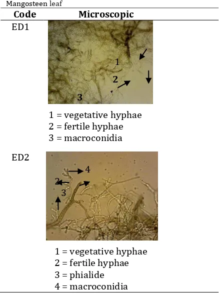
Endophytic fungi was isolated from leaf begin to be observed growth in the 2nd day of incubation and the longer will be more widespread. Each of these fungi has different incubation period so as to easier the purification of the culture of fungi observations made every day and if the observation has been able to differentiate fungi then continue the purification process without waiting for the time of incubation for 14 days. Isolates endophytic fungi that grow on MEA was observed macroscopic and microscopic (Fig.2, Table 1, Table 2).
Table 1. Macroscopic Endophytic fungi isolated from Mangosteen leaf
Fig 1. Hypha of an Endophyte in epidermal cells of Mangosteen leaf.
Fig 2.Endophytic fungi isolated from Mangosteen leaf
B. Assay for Antibacterial Activities Against Staphylococcus aureus and Escherichia coli Screening of antibacterial activity showed that ED2 isolate has antibacterial activity against Staph. aureus but not against E. coli. This antibacterial activity can be observed due to the presence of compounds that have antibacterial activity against Staph. aureus and E. coli diffuses from endophytic fungi isolates to medium and inhibit the growth of bacteria so that the assay remains colorless clear medium in the surroundings (Table 3).
Code Macroscopic
ED1 Filamentous colony
orange
diameter ± 9 cm (Fig 2., A)
Filamentous colony Grey
46 | P a g e Proceeding - ICB Pharma II Current Breakthrough in Pharmacy Materials and Analyses
ISSN : 9-772476-969006
Table 2. Microscopic Endophytic fungi isolated from
Mangosteen leaf
Code Microscopic ED1
1
2
3
1 = vegetative hyphae 2 = fertile hyphae 3 = macroconidia
ED2
4 2 3
1 = vegetative hyphae 2 = fertile hyphae 3 = phialide 4 = macroconidia
Table 3. Result screening antibacterial potency of endophytic fungi isolated from Mangosteen leaf
( - ) = no activity , ( + ) = activity
Bioautography with TLC (indicate compounds that can inhibit the growth of Staph. aureus. Results of screening such compounds with FeCl3 blue black coloration, this color indicates phenol group in the metabolites. Screening with AlCl3 yellow coloration, this color indicates the flavonoids.
IV.CONCLUSION
Mangosteen leaf was obtained 2 kinds of endophytic fungi; which were 1 have the potential as a producer of antibacterial compounds against Staph. aureus; which were predicted to be the class of compounds is flavonoids. Endophytic fungi isolates that have characteristic of macroscopic and microscopic is supposed a group of Trichoderma.
Acknowledgment
This work was supported by the grants from Faculty of Pharmacy Widya Mandala Catholic
University Surabaya. We thanks to Aprilia Karolin and Anastasia Yessy for their assistance in working.
References
[1] Strobel, G.A., Natural products from endophytic
microorganism , Journal of Natural Products, , , pp. 257-268.
[2] Nugroho, N.B. dan B., Sukmadi, )solasi dan seleksi jamur
endofit penghasil antibiotika , Pertemuan )lmiah Tahunan
Perhimpunan Mikrobiologi Indonesia, 1998, pp. 1-2.
[3] Tan, R.X. and Zou, W.X., Endophytes : a Rich source of
functional metabolites, Natural Products Report, 18, 2001, pp. 448-459
[4] Guo, B., Li, H.and Zhang, L., Isolation of the fungus
producing vinblastine, Journal of Yunnan University (Natural Science Edition), 20, 1998, pp. 214-215
[5] Yang, X., Zhang, L., Guo, B and Guo, S., Preliminary study of
a vincristine-producing endophytic fungus isolated from leaves of Catharanthus roseus, Chinese Ttraditional and Herbal Drug, 34, 2004, pp. 79-81.
[6] Zhang, L., Guo, B., Li., H., Zheng, S., Shao , H., Gu, S. and Wei, R., Preliminary Study on the isolation of endophytic
fungus of Catharanthus roseus and its fermentation to
produce products of therapeutic value, Chinese
Ttraditional and Herbal Drug, 31 2000, pp. 805-807.
[7] Pavithra, N., Sathish, L. and Ananda, K., Antimicrobial and
Enzyme Activity of Endophytic Fungi Isolated from Tulsi,
Journal of Pharmaceutica and Biomedical Sciences, Vol. 16, Issue 16, 2012, pp. 1-6.
[8] Putra, I.N.K, Aktivitas antibakteri ekstrak kulit buah
manggis (Garcinia mangostana L.) serta kandungan
senyawa aktifnya, Jurnal Teknologi dan Industri Pangan, Vol. XXI, No.1, 2010, pp. 1-5.
[9] Elfina, D., Martina, A. Dan Roza, R.M., Isolasi dan
karakterisasi fungi endofit dari kulit buah manggis (Garcinia mangostana L.) sebagai antimikroba terhadap
Candida albicans, Staphylococcus aureus dan Escherichia
coli, Skripsi, FMIPA, Universitas Riau, 2014.
[10] Sugiharto, C., Isolasi, identifikasi dan profil KLT
Densitometri metabolit jamur endofit pada tanaman Solanum wrightii Benth, Tesis, Universitas Airlangga, 2006.
Code Antibacterial potency
Staph. aureus E. coli
ED1 - -