Vol 10, No 2, April
-
June 2001 Exposure to UVp of Jluorescent light on the bone remodelingThe
influence of
exposure
to UVB of fluorescent
light
on the bone
remodeling
of
hypoestrogeni
c
macsc& fas
cicularis
Abstrak
Pencegah
merupakansuatu
sia harapan hidup wanita.Penuruna
esûogenyang
mineralisasi tulang (olehkalsitriol)
koLagen tipeI
(o
bentukan kalsitriol"de:nganbahnn dasar utama berasal dari vitamin
Di
kuLil yang dibentuk dengan bantuan paparan IJVp matahari. Saatini
dengan adanyaperubahan gaya hidup, khususnya kaum wanita yang bekerja aktif dan selalu terhindar dari
piparan
UVB sepanjang halri dan akan mengaLami Penurunan vitamin Djdi
kaLsitriol dan memasuki usia menopause okon*nigànram
teiiadinya
oieoporosis dini. Pemberian paParan UVp Lampu fluoresen yang panjang gelombangnya sàma denganmatihari
290-320 nm, telah l^ama dikenaLsebagai pengobatan penyakit kulit dengan harapan produksi vitamin D
j
kulit naik. Telah dilakut<an paparcm tJVp lampu fluoresen terhadapkulit
Macaca fascicularis yang kadar estrogennyct normal, estrogen mulai rendah dan estrogen mulai sangat rendah. t kenaikan osteokalsin dan DpD tetap, yang berarti remodering tulangr
UVp baikyang kadar estrogen normal, mulai rendah nnupun sangat kadar DPD yang berarti terjadi perubahan remodeLing tulang kearahAbstract
dealt with by increasing the women's
lift
expectation. Theosteoblast, wilL resuh in bone mineralization (due to calcitriol) tion of
calcitiol
with the main basic materialsfrom
vitaminDj
is achieved with the aid of sunray UV p, The changes in the lifestyle of women, which make them now accustomed to performing indoor activities and prevent them from being exposedto
UV B alt day, have resulted in the decrease of vitamin Dj
in calcitrioL in women.In
addition'when
e
theywill
be threatened with early ost"oporosis. The exposureto
the oVB
oJfluorescent light
w
sunof
290-320 nm has long been knàwn as'a modality Jor treating skin diseases inthe
hope thatthe
be increased.w"
"xpor"i
Macaca fascicularis, whose esÛogen levels were set arnormal' beginning low, beginning very low levels, to IJV B of fluore:,scent Light.
It
showed thatthe
Macaca fascicularis that were exposedto
UVP
experiencedan
increasein
osteocalcinwith
unchang)dDPD
which means that bone remodeling remains unchanged' By contrast, Macaca fascicularis with normal, beginninglow,\nd
beginning very low estrogen levels which were not exposedto
UVp were found to exPeriencea
decreasein
osteocalcin and unchangedDPD
levels- This means thata
change has occurred in the bone remodeling toward bone resorption. (MedJ
rilones 2001;I0:
63_g)Keywords: (JVp, osteoporosis, estrogen, vitamin D.1, calcitriol, osteocalcin, DpD
The exposure
to
IJVp of
sunray stimulates theskin
toform
vitamin
Dr
as the
basic
material
for
theformation
of
calcitriol.t-o
Calcit.iol and
osteoblasthave
a
receptor
in
the
osteoblast
which
causes the osteoblastto
undergo bone mineralization
andtype
I
Depart nent of Obstetics antl Gytecology, Facuhy of Medicine University
of
Indonesin/DrCipto
Mangunhtsuma General Ho spital, J akarta, In^do ne siacollagen responsible
for
of
thebones.''o
In
the
tropica
ray
isabundant throughout
the
avoid
the
heat of
the
sun
for
fearing
that
their skin
woulclturn
darker.
In addition,
they
are
accustomed
toworking
all
day
in
the buildings
covered
with
anti-UVB
glass
walls.
The pervasive use of air-condition
system
in
cars has also
prevented
them from
being
exposedsufficiently ro UVP
of
sunray.
This lifestyle
64 Rachman
premenopausal
age when
estrogen
is
starting
todecline they will encounter the problem
associatedwith the
decline in
the bone remodeling
which
will
result
in
bone resorption
andprecipitate
the
processof
bone osteoporosii. l'2'3Fluorescent
light
which
emanatesUVp
rays (equivalentto
UVp
of
sunraywith
awave length
of
290-320 nm) haslong
been used as a phototherapyfor
psoriasisby
making use
of
vitamin
Dj
produced
by
the
skin.oWould the
exposure
to
UVB
rays
from
fluorescentlight,
apartfroin
increasing the production
of'vitamin
D:
by
the skin,
increase
the calcitriol
level
which
would
activate the
osteoblastand bone formation?
A
number
of
studies
have
suggestedthat
combinedtreatment
of
conjugated
estrogen+
progesterone
+calcium
and
weight
lifting
exercise
may
be
able
toincrease bone
mass
from
1l
to
22Voin
one year periodof
treatment,T-Il
The
experimental
study
on
theprevention
of
osteoporosisin
human
cannotbe
easilyperformed.
For
this
reason,
we
used
Macacafascicularis
as a modelfor
the present study.The objective
of
this
studyis
to
evaluate the influenceof the
exposure
to fluorescent
light
UVp
on
boneGrouping EO (n=40),
El
(n=40), E2 (n=40)Med J Indones
remodeling
in
hypoestrogenic Macaca
fascicularis
in
the
effort for
early preventionof
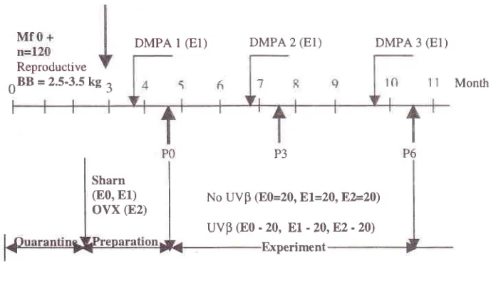
osteoporosis.METHODS
The
study was pedormed
in
120 female
Macacafascicularis
aged5-8
years
with body
weight
rangingfrom
2.5to
3.5kg.
These animals were quarantinedfor
a period
of
three monthsbefore
undergoing treatment.After
being quarantined they were randomly dividedinto
three groups, i.e. normal estrogen group @0), beginning
low
esfrogen
group
@1),
and beginning
very
low
estrogengroup @2).
During
the
preparationperiod
of
one and
a
half month,
a
sham surgery (open
closedlaparotomy) was
performed
in
groupsE0
andEl,
and an intervention ofOVX
(removal of both ovaries)
weredone
in
group
E2.
After
sham
surgery Depomedroxyprogesterone acetate
of
5 mg was administeredfor
groupEl
every three month.Each
group
was thendivided
into two
subgroups andreceived the
following
treatment:à.
Non-UVB
exposureduring
a periodof
six months.b.
Exposure.to
fluorescent
light
WB
at a
dose
of
l2.mJlcrr:.
duringfour
hours a dayfor
six months.DMPA
l
(El)
DMPA 2(El)
DMPA 3(EI)
P3
No UVp (E0=20,
El=20,E,È20)
uvp
(Eo - 20,Et
- 20,82 - 20)Month
Sharn (80,
El)
ovx
(E2)Experiment Reproductive
[image:2.612.84.432.446.645.2]nBB = 2.5-3.5 kC a
Vol 10, No 2, April
-
June 2001The
changesin calcitriol,
osteocalcin
andDpD
levelsafter
three months
and
six
months
of
UVp
light
exposure and
non-UVB
exposurewere recorded
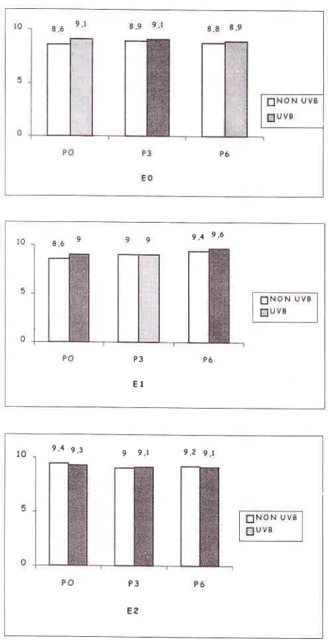
(p0, P3, P6).The
fluctuation of DPD level in
both
UVp exposure
group
and
non-Wp
exposure
group
after
threemonths and
six
months
of
exposure
was
within
thenormal
limits.
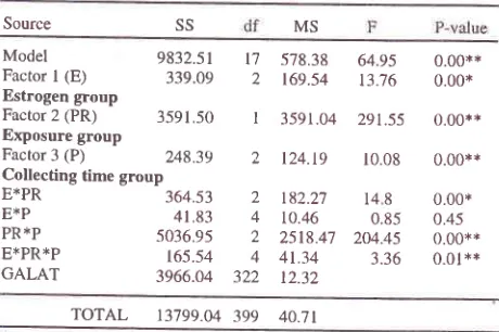
Factorial design analysis
of
meanvariant
osteocalcin (osteoblast
activity) and
DpD
(osteoblastactivity)
was presentedin
Table
1.Table
l.
Factorial design analysisof
mean variant osteocalcin level against the factorsSource
Exposure to UVB of Jluorescent light on the bone
remodeling
65estrogen
levels in
the
threeexperimental
groups did
[image:3.612.305.539.152.321.2]not affect
osteocalcin level.Table 2. Factorial designs analysis
of
mean variantDpD
level against the factors
MS
SS P-value
Model Facror I (E) Estrogen group Factor 2 (PR) Exposure group Factor 3 (P) Collecting time grouP E*PR E*P PR*P E*PR*P GALAT
o.23
0.630.05
0.950.86
0.920.92
0.451.00
0.370.40
0.80Model
9832.51Factor
I
(E)
339.09Estrogen group
Factor 2 (PR) 3591.50 Exposure group
Factor 3 (P) 24939 Collecting time group
364.53 41.83 5036.95 t65.54 3966.O4
578.38
64.95
0.00**169.54
13.76
0.00*3591.04
291.55
0.00** MS ss 14.47 4.95 o.23 1.10t.7 |
3.66 1.98 1.59 295.66 0.85 2.48 o.23 0.05 0.85 0.91 0.99 0.40 0.98 0.86
2.5t 008062 t7 2 I
)
2 4 2 4 299TOTAL
13799.04399 40.7t
** Significant at < 0.05 Note: SS = sum of squares; df = degree of freedom; MS
= man squares; F= F= testl P>f = F- value
Table
I
showed
that
there was also
a
significant
difference between osteocalcin
level and estrogen
factor
(p=0.00),
exposure
factor
(p=0.00),
andcollecting
time factor
(p=0.00). There was also
asignificant difference (p=0.00) between
osteocalcinlevel and
the
interaction of
estrogen-exposure factor.Similarly,
a
significant
difference
(p=O.OO)
wasobserved
in the
interaction
of
exposure
factor
andcollecting time factor, interaction
of
estrogen-exposurefactor and
factor-collecting
time.
However,
nosignificant
difference was
found
in
the
interaction
of
the estrogen factor and
collecting time
factor (p=0.4).Therefore,
it
may
beconcluded
that
osteocalcinlevel
was substantially influenced
by
estrogen
factor
andexposure
factor, as
well
as collecting
time factor.
However, beginning
low
and
beginning very low
TOTAL
310.13 316Note: SS = sum of squares; df = degree of frædom; MS = rean squæs;
F = F = test; P > f= F - value
There was
no significant relationship
between
meanDPD level
and
estrogenfactor,
exposure
factor,
andcollecting time factor.
UVp
light
exposure
increasedcalcitriol and
osteocalcin levels
in
the
normal
estrogen
level (80), beginning
low
estrogenlevel (E1)
and beginning
very
low
estrogen
level.
However,
DPD
wasstill
at
thenormal
level
which
means thatbone
remodeling was
still in
balanced
condition. In
non-UVp
exposuregroup a
decreasein calcitriol
andosteocalcin
levels was
observed,
while
DpD
level
was
still
within
the
normal
limits
in
the
threeexperimental
groups. This means that
boneremodeling was
not
in
balanced
condition
with
low
boneformation
andnormal
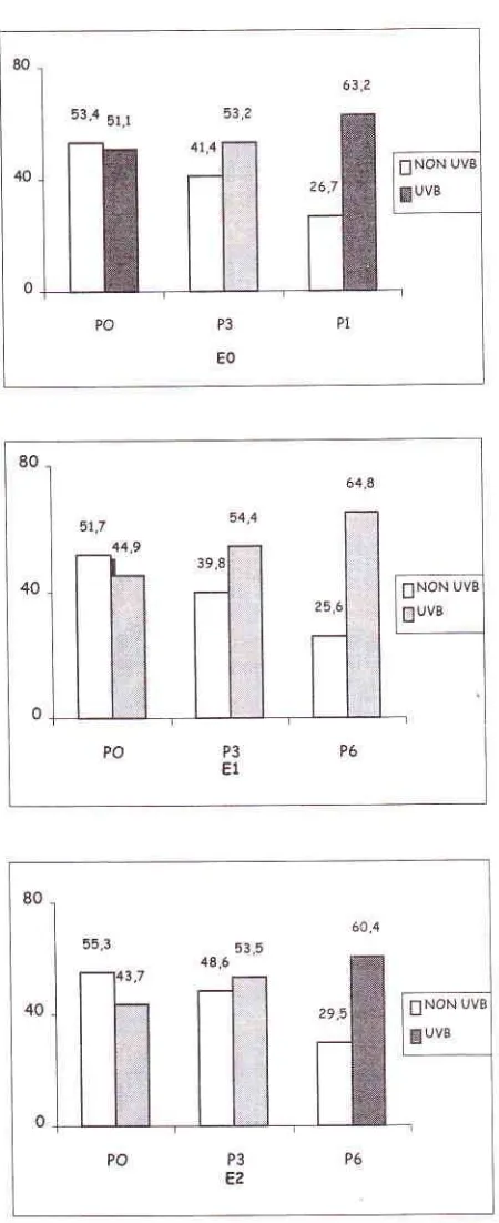
resorption.RESULTS AND DISCUSSION
In
this
study
we found
that
hormone calcitriol
level
and osteocalcin
level
decreasedto
the lowest normal
level
in the
three experimental groups after
threemonths
and
six
months
without
LIVB
exposure(Figure
2
and3).
t7 2 I ? 2 4 2 4 322 098 E*PR E*P PRXP E*PR*P GALAT
t24.t9
10.08t82.27
14 810.46
0.852518.47 204.45
[image:3.612.47.277.257.410.2]Med J Indones
30
20
10
[image:4.612.59.284.83.634.2] [image:4.612.324.546.84.631.2]0
Figure 3. Mean osteocalcin level
in
groups 80, 81, and E2with
UVB
exposureand
without
UVp
erytosure (normal osteocalcin level rangedfrom
15to
32ng/ml
Z
non-UV|exposure, WUV
p exposure).
RachmanFigure 2. Mean
calcitriol
level in groups E0,EI,
and E2 with IJVB exposure and without UVB exposure (normal calcitrioLtev et 4 3.7 -7 5. 5
ng/ml,l
non- UV B exp o sur e,@U VB
exp o sure ).25'1 zz,a 30
20
10
0
PO
P3
P6EO
30
20
10
0
P3
Vol 10, No 2,
April-
June 2001An
increase
in
calcitriol and
osteocalcin
levels
wasfound
in UVp
exposure
group after three and
six monthsof
UVp
exposure. However, this
changes wasstill
within the
normal
limits
in
the
experimental
groups (Figure
2 and3). It
meansthat
theactivity
of
osteoblast
in
bone formation
was
without
UVB
exposure.
Nevertheless, theactivity
of
osteoblast wasstill normal in
UVp
exposurewhich
meansthat
boneformation
was
in
normal.
Regression
graph
of
[image:5.612.307.545.78.543.2] [image:5.612.40.285.271.417.2]diff'erent osteocalcin
level which functions
atcalcitriol
level
is presentedin
Figure
4.Figure 4. Regression equation graph of
dffirent
calcitriol level anddffirent
osteocalcin levelThe regression equation between osteocalcin
level
as afunction
of
different
calcitriol.
level
is
:Osteocalcin =
0.35x calcitriol
+ 0.42Exposure to UVB offluorescent light on the bone remodeling 67
g,+ 9,6
P3
EI
Figure
5.
Mean DPD level in groups 80,EI,
and E2 with and without UVp exposure (normal DPD level was 6.64-
125 nM .Znon
IJV\ exposure,fi UVp exposure) nM creatinine,j 10
: -1O
Ostcrclcin = 035 x Calciriol + 0.12
9,4 gj c 91 92
P3
2.
68 Rachman
CONCLUSIONS
1.
Exposure
to
fluorescent
light
UVp
during
theperiod
of
six
months proved
to
maintain
theequilibrium
of
bone remodeling (calcitriol
andosteocalcin content was
within
the normal
limits,
and
deoxypiridinoline
content
remainedunchanged).
An
equilibrium
was
also maintainedat
normal
estrogen
level,
beginning
low
andbeginning very
low
estrogen levels.Normal, beginning
and
beginning
very
low
estrogen
levels during the
first
period
of three
months
did not
affect bone
formation directly.
It
would be best
to
get
exposed
to UVp
rays
to prevent the changein
boneremodeling during
thisperiod.
REFERENCE
Fieldman D, Molloy P, Gross C. Vitamin D, Metabolism and
action.
In: MarkusL,
FieldmanD,
Kelsey J, Editor. Osteoporosis. San Diego: Academic Press Inc; 1996,205-25.Holock MF, Chen
TC.
Vitamin D3 synthesis and biologic functionin skin.
Boca Raton-Florida: CRC Press Inc;1992,183-202.
Deluca
lIF.
The vitamin D story.A
Collaborative effectpfbasic science and clinical Medicine Faseb J; 1988;2: 224-56. Subcomitte on the Tenth edition
of
the RDA'S, National ResearchCouncil,
RecommendedDietry
allowances.Washington DC: National Academic Press 1989.
Mongiska RJ, Rysky JT, Einhom
JA.
Direct modulation of osteoblastic activitywith
estrogen.J
Bone Joint Surg;Med J Indones
1994;76:715-28.
6. Aloir
JF, Vaswani A, Yeh JK, Ellis K, Yosumura S, CohnSH.
Calcitriol
in
the
treatment of postmenopausalosteoporosis. Am J Med 1988;84: 401-8.
7.
RachmanIA,
HestiantoroA,
SurjanaEI,
Kampono N,Helmy
CH.
Comparativestudy
between hormonal replacementtherapy
plus
calcium
and
hormonal replacement therapy, calcium andvitamin
D.
on
the treatmentin
postmenopausal osteoporosis Indonesiawoman. World
Congresson
Osteoporosis, Netherland, May 18-23, 1996.8.
RachmanIA,
HestiantoroA, Surjana
EJ,
Baziad A, MoeloekFA,
SumosardunoS.
Evaluationof
certain clinical and laboratory changesin
the treatmentof
post-menopausal osteoporosis between hormon replacement+ calcium and hormon replacement+calcium+ vitamin D3and only
vitamin
D3
with
weight
bearing exercise.Proceedings
of
thefirst
Asian European Congress on the Menopause; Bangkok, Thailand 1998, 193-98.9.
Tylard WL, Speors GF, Thomsom J, DoveyS.
Treatment of postmenopausal osteoporosis with calcitriol or calcium. N Engl J Med 1992;326:357-62.10. Falch
JA,
OdegroundOR,
FinanngerM,
Mathesson I. Postmenopause osteoporosis:No
effect
of
three yearstreatment
with
1,25 dihydroxy cholecalciferol. Acta MedScand 1987 ;22 I :199
-2V.
11. Gallangher JC, Riggs
BL,
Recker RR, GoldgerD.
Theeffect
of
calcitriol
on
patient
with
postmenopausalosteoporosis with special reference to fracture. Proc Soc
Exp Med 1989;19l-287.
l2.Gallagher JC, Goldger
D.
Treatmentof
postmenopausalosteoporosis
with high
doseof
syntheticcalcitriol.
A randomized controlled study,Am
InternMed
1990;ll3: 649-655.I
4.