EISSN: 2086-4094 DOI: 10.4308/hjb.18.1.1
_________________ ∗
∗ ∗ ∗
∗Corresponding author. Phone: +62-251-8628080, Fax: +62-251-8628181, E-mail: [email protected]
Motion Mode and Two Dimensional Echocardiographic
Measurements of Cardiac Dimensions of Indonesian Mongrel Dogs
DENI NOVIANA∗∗∗∗∗, DEVI PARAMITHA, RETNO WULANSARI
Departement of Clinic, Reproduction and Pathology, Faculty of Veterinary Medicine, Bogor Agricultural University, Darmaga Campus, Bogor 16680, Indonesia
Received November 20, 2009/Accepted March 8, 2011
Prevalence of heart disease in dogs was very high and required early diagnosis through physical examination, electrocardiogram, and echocardiography. Normal reference values of echocardiography are highly breedspecific and need for comparison and evaluation of dogs suspected with heart disease. Therefore the aim of this study was to establish normal reference echocardiographic values for Indonesian mongrel dogs, specifically to find out intracardiac dimensions, wall thickness, and fractional shortening. Motion-mode and two-dimensional echocardiography from right parasternal short axis and long axis view were performed on nine clinically healthy dogs consisting of five males and four males. The results showed that wall thickness and fractional shortening of Indonesia mongrel dogs were higher compared with those in the other breed that have the same average weight. As opposite, the intracardiac dimensions and lumen dimensions of aorta and left atrial diameter were smaller. These differences might occur due to factors other than the dog’s habits and functions such as working and hunting, but can also be caused by the existence of breed differences. There was no significant difference between male and female dogs in terms of intracardiac dimension systole (P = 0.53), diastole (P = 0.38), fractional shortening (P = 0.053), and the ratio of aorta and left atrial diameter (P = 0.06).
Key words: echocardiography, mongrel dog, motion-mode, cardiac
___________________________________________________________________________
INTRODUCTION
In dogs heart disease present with clinical symptoms that vary from an invisible symptom until the very dramatic appereances (Martin et al. 2009). Heart disease is potentially lethal to dogs if not treated properly, and requires early diagnosis through history, physical examination, thorax radiographs, and an electrocardiogram (Kraetschmer et al. 2008; Prieto et al. 2009; Crosara et al.
2010; Falk et al. 2010). In line with the findings of echocardiography techniques in cardiology clinical sciences, veterinarian can observe abnormalities associated with heart disease (Schaer 2008; Gillings et al.
2009; Schober et al. 2010; Wess et al. 2010a). Two
dimensional M-mode echocardiography records the amplitude and the rate of motion of moving objects, such as cardiac muscles and valves, along a single line with great accuracy therefore enables veterinarian to see and measure range of motion (Penninck & d’Anjou 2008). Pathological changes that can be found such as dilated cardiomyopathy, mitral or tricuspidalic valve regurgitation, ventricular septal defect and persistent ductus arteriosus (Barr 1990; Bonagura & Schober 2009; Tidholm et al. 2009;
García-Rodríguez et.al 2009; Wess et al. 2010b).
Normal reference value of echocardiographic is needed for comparison and evaluation of dogs suspected heart disease (Voros et al. 2009). Some references values of echocardiographic for dogs based on breed and body
size have been published (Gooding et al. 1986; Cornell et al. 2004; Kayar et al. 2006). However, these references range obtained from several dogs are highly specific for their breeds and could lead to inaccuracy when applied to the other breed (Kayar et al. 2006).
The definition of Indonesian mongrel (kampong) dog is a dog that has long been known to exist but not observed of their offspring strain. Dogs of this type are common in Indonesia as a pet, hunter and also laboratory animal for advance research. In correlation with the electrical activity of the heart, research on the electrocardiogram (ECG) of Indonesian mongrel dogs has been done (Ngabdusani 2006; Nurulhuda 2007). However, echocardiography of Indonesian mongrel (kampong) dog has never been observed. Therefore, the aim of this study was to establish normal reference echocardiographic values for Indonesian mongrel dogs breeds, specificallyto find out intracardiac dimensions, wall thickness, lumen dimensions, and calculating the derivatives. The results of this study can be used as referencesvalue for heart disease diagnostic.
MATERIALS AND METHODS
D300). Animal used in the study was nine healthy Indonesian mongrel dogs. They were four males, five females and within 2-5 years old (average 3.5 + 0.8). Physical and ECG examination were carried out prior to echocardiography examination. Echocardiography examination was performed in right lateral recumbency within right chest area on the medial-ventral of the 3rd to
6th intercostae. Two dimensional motion mode (m-mode)
echocardiography standard scanning view were performed i.e. short axis view and long axis view. The direction of short axis view was conducted to obtain imaging at the level of left ventricle and base of aorta. Long axis view was conducted to obtain imaging of the mitral valve level (Penninck & d’Anjou 2008).
Left ventricular internal dimension diastole (LVIDd) was measured at end diastole coinciding with the internal dimension of the largest in the left ventricle chamber, immediately after the onset of the QRS complex on ECG. LVIDs measured at end systole coinciding with the internal dimension of the smallest left ventricle chamber and the by the end of the T wave of ECG. Fractional shortening (FS) values obtained from the calculation formula FS = (LVIDd - LVIDs)/LVIDd. Left ventricular wall at diastole (LVWd) and left ventricular wall at systole (LVWs) parameters measured at the same line with the measurement LVIDd and LVIDs, but carried out on the wall located at the bottom part of the LVwall. Interventricular septal at diastole (IVSd) and interventricularseptal at systole (IVSs) parameters measured at the same line with LVID and LVW, but measured on the wall located at the upper of the LV. Measurement of aortic dimension (AOD) obtained at end diastole and measurement of left atrial dimension (LAD) obtained at the time of maximum dimensions occurs, coinciding with the end of systole. Mitral valve end point to septal separation (EPSS) was performed when the anterior mitralic leaflet (AML) of mitral valve opened, and the space between opened AML and IVS was measured. Correlation of sex differenceson these measurements were analyzed by using t-test. The results of the study also compared with previous echocardiography studies from another breed dogs weighing nearly the same with Indonesian Mongrel dog i.e. English Cocker Spaniel (Gooding et al. 1986) and Dachshund (Cornell et al. 2004).
RESULTS
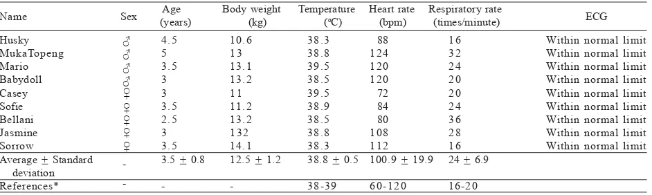
Preliminary examination which includes physical examination and ECG showed all animals used in this study were healthy dogs (Table 1). The results of intra cardiac dimension measurements and fractional shortening calculation of Indonesia mongrel dogs were summarized in Table 2. The average value of LVID during diastole was 27.22 + 3.30 mm, whereas LVIDd in male dogs were higher than female dogs (P = 0.38). Further measurementof the LVIDs showed the average number 16.31 + 2.78 mm. In contrast with LVIDd, in male dogs the value of LVIDs were lower than female dogs (P = 0.53). Fractional shortening (FS) was defined as a difference between end diastolic and end systolic dimensions divided by end diastolic dimension. Overall FS in Indonesian mongrel dog was 0.41 + 0.06 and the value in male dogs werehigher than female dogs (P = 0.053). Myocardium contractility of heart was reflected with the FS value. Therefore myocardium contractility in Indonesian mongrel male dogs were stronger than female dogs.
The heart wall thickness measurements were presented in Table 3. Both left ventricular and inter ventricular wall thickness during diastole and during systole of male dogs were higher than the female dog. Of the four parameters of heart wall thickness, only left ventricular wall thickness during systole was higher in female dogs (P = 0.71).
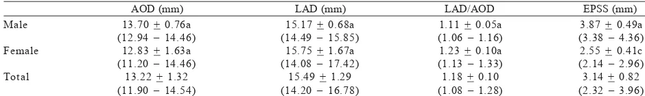
Subsequent measurements of lumen dimensions in mongrel dogs i.e. AOD, LAD, and ratio of LAD/AOD were
Table 1. Physical and electrocardiography (ECG) examination results of Indonesian mongrel dogs Age Body weight Temperature Heart rate Respiratory rate
Average + Standard deviation
*Source Birchard and Sherding (2000).
13.22 + 1.32, 15.49 + 1.29, and 1.18 ± 0.10 mm respectively (Table 4). Aortic dimension in male dogs were higher, in contrast with LAD and ratio of LAD/AOD that were higher in female dogs (P = 0.06). The last parameter measured was the EPSS. The EPSS is a measurement of the perpendicular distance between the most posterior point of the interventricular septum during systole and the end point of the anterior mitral valve leaflet in the same cardiac cycle. Overall EPSS value for mongrel dogs were 3.14 + 0.82 mm (Table 4). In the male dogs EPSS value was greater than the female dog (P = 0.003).
DISCUSSION
In healthy Indonesian mongrel dogs, value of LVID were smaller compared with those in Dachshund and English Cocker Spaniel that have the same average weight with Indonesian mongrel dogs (Gooding et al. 1986; Cornell
et al. 2004). However eventhough they have the same average body weight, dachshund dogs used for hunting, while Home Cocker Spaniel is a working hyperactive and also used for hunting (Sayer 1994). Thus, these two dogs have higher body activity compared to Indonesian mongrel. According to Vander et al. (1990), in working and active condition, the body requires much more blood supply to the organ. Meanwhile, the expansion of the left ventricle associated with increased of blood volume that occur secondary along with increasing of skeletal muscle activity (Stepien et al. 1998). Therefore, LVID in both dogs is greater than the value measured in Indonesian mongrel dog. Left ventricular internal dimensions emerged as the most important echocardiographic predictors of congestive heart failure among several echocardiographic variables evaluated (Vasan et al. 1997; Bonagura & Schober 2009).
Fractional shortening of Indonesian mongrel dog was higher when compared with the English Cocker Spaniel (Gooding et al. 1986) and Dachshund (Cornell et al. 2004). Meanwhile, according to Goddard (1995), the range of FS values in normal dogs without distinction of breed ranges between 0.28-0.50. According to Kayar et al. (2006), for determining the FS, the number of dog samples should be noted because will affect the precision of the range. Fractional shortening is the most common echocardiography parameter performed to see myocardium contractility and to estimate capability of the left ventricles (Nelson & Couto 1998; Voros et al. 2009). Meanwhile, according to Cornell et al. (2004), FS is widely used as an indicator of left ventricular systolic function, and the value of less than 0.25 is usually associated with heart disease i.e. hypovolemia. Fractional shortening is also an important parameter to distinguish between hypertrophic and dilated cardiomyopathy (Schille & Skrodzi 1999; Borgarelli et al. 2007).
In Indonesian mongrel dog, the thickness of the heart muscle both LVW and IVS during systole is much higher than diastole. This is be due to systole is the phase where the heart is contracting to pump blood, hence the heart wall thickness greater than that measured during diastole. According to Fox (2004), at the end of diastole, the left ventricle filled with blood therefore push down the walls of the heart so that when measured the value is smaller than during systole. Both LVW and IVS values during systole and distole in Indonesian mongrel were higher compared with Dachshund. This might occur due to factors other than the dog’s habits and functions such as working and hunting, but can also be caused by the existence of breed differences. According to Kayar et al. (2006), dogs with the same weight but different breeds have the results of different echocardiographic
Table 3. Heart wall thickness of Indonesian Mongrel Dogs. Values expressed as average + standard deviation
LVWd (mm) LVWs (mm) IVSd (mm) IVSs (mm) Male
Female Total
7.83 + 1.46a (6.37 – 9.29) 7.28 + 0.59a (6.69 – 7.87) 7.50 + 1.03 (6.47 – 8.53)
10.50 + 1.57a (8.93 – 12.07) 11.12 + 0.23a (10.89 – 11.35)
10.84 + 2.02 (8.82 – 12.86)
6.36 + 0.21a (6.15 – 6.57) 6.15 + 0.39a (5.76 – 6.54) 6.39 + 0.29 (6.10 – 6.68)
9.10 + 0.47a (8.63 – 9.57) 8.43 + 0.35b (8.08 – 8.78) 8.73 + 0.52 (8.21 – 9.25) Left ventricle wall at end-diastole (LVWd), left ventricle wall at end-systole (LVWs), interventricular septa at end-diastole (IVSd), interventricular septa at end-systole (IVSs). Different superscript letters in the same column stated the significant difference with confidence interval a-b 95% and a-c 99%. Exact P value between male and female dogs for LVWd P = 0.47, LVWs P = 0.71, IVSd P = 0.27, and IVSs P = 0.04.
Table 4. Lumen dimension measurements and mitral valve end point to septal separation of Indonesian Mongrel Dogs. Values expressed as average + standard deviation
AOD (mm) LAD (mm) LAD/AOD EPSS (mm) Male
Female Total
13.70 + 0.76a (12.94 – 14.46)
12.83 + 1.63a (11.20 – 14.46)
13.22 + 1.32 (11.90 – 14.54)
15.17 + 0.68a (14.49 – 15.85)
15.75 + 1.67a (14.08 – 17.42)
15.49 + 1.29 (14.20 – 16.78)
1.11 + 0.05a (1.06 – 1.16) 1.23 + 0.10a (1.13 – 1.33) 1.18 + 0.10 (1.08 – 1.28)
measurements. If the data from different breeds are combined to estimate the size of the heart based on dog weight, the results can be wrong and misleading. Meanwhile, according Penninckand and d’Anjou (2008), differences in the evaluation of heart structure and function can be caused by body size, species variation, breed variation and individual variation.
AOD and LAD of Indonesia mongrel dogs were smaller than the Dachshund dog. This is also related to the activity of Dachshund dogs which is more active so that more blood volume and produced a larger space or dimension (Vander et al. 1990). Measurement of aortic dimensions was carried out to see abnormalities of the heart stroke volume, which decreased lumen dimension of the aorta is an indication of a low stroke volume (Goddard 1995; Cunningham 2002). Whereas the left atrial measurements in dogs are relevant to guide the diagnosis and prognosis of heart disease, but the calculation is problematic due to three-dimensional shape of the left atrium still unclear. According to Nelson and Couto (1998), it is important to understand that the M-mode image of the left atrium is usually acquired in the area between the left atrium and left auricle, especially in dogs. So that these measurements often do not represent the maximal atrial size. The ratio of left atrial diameter and aortic (LAD / AOD) in Indonesian mongrel dog is 1.18 + 0.10 mm. According to Nelson and Couto (1998), in all dogs the average value of the ratio LAD/AOD based on M-mode measurements is approximately 1.0 with the maximum value is 1.4 depending on the breed (Penninck & d’Anjou 2008). The value of ratio LAD/AOD will increase if there is dilatation of the left atrium (Goddard 1995). At the time of the observation of left atrial dimension, ultrasound beam waves will pass through the left atrial appendage (LAA) or the cranial part of the left atrium in most M-mode echocardiography examination of dogs. Therefore, increasing the ratio of LAD/AOD may also indicate the existence of left ventricular dilatation (Nelson & Couto 1998).
There is a significant difference of EPSS value between male and female of Indonesian mongrel dogs, which EPSS value in male dogs were greater. It is also proportional to the greater value of LVIDd in male dogs. According to Nyland and Mattoon (2002), regardless of gender, EPSS value for dogs weighing 10-15 kg is 2 mm. However, all M-mode measurements and calculations derivatives changes according to body weight, body surface area, breed and other variables. The EPSS value is usually measured to see any indication of left ventricular dilatation, when the value is more than 6 mm is very important to note because it indicates a dilatation of the left ventricle (Goddard 1995). EPSS value will increase in animals with poor myocardium contractility (Nelson & Couto 1998). EPSS value is calculated and associated with ejection fraction (EF) that is the fraction of end diastolic volume released during the phase of ventricular systole (Cunningham 2002) and measured to assess the efficiency of the heart in pumping blood (King et al. 2002). EPSS parameters very trustworthy to view the left ventricular function in aortic stenosis, but
its usefulness is limited to valve diseases such as chronic mitral and aortic regurgitation (Lehmann et al. 1983).
The results showed the different characteristics of Indonesian mongrel dogs in terms of intracardiac and lumen dimensions, wall thickness and cardiac contractility when compared with the other breed that have the same average weight. These results in line with previous research that the differences might occur due to factors other than the dog’s habits and functions such as working and hunting, but can also be caused by the existence of breed differences. Indonesian mongrel dogs included in the medium sized breeds and workers dog with mild to moderate intensity.
Through this research has defined several characteristics of heart in Indonesian mongrel dogs which is very useful as a normal parameter for the diagnosis of cardiac diseases. In the future, the development of this research is not only specific for canine cardiac characteristics, more importantly can be extend to study the pathogenesis of some cardiac diseases in humans such as the narrowing of blood vessels and valve abnormalities, which is a degenerative disease with high incidence frequency.
REFERENCES
Barr F. 1990. Diagnostic Ultrasound in The Dog and Cat. Oxford: Blackwell Sci Publ.
Birchard SJ, Sherding RG. 2000. Saunders Manual of Small Animal Practice. 2nd ed. USA: WB Saunders Company.
Bonagura JD, Schober KE. 2009. Can ventricular function be assessed by echocardiography in chronic canine mitral valve disease? J Small Animal Pract 50:12-24. doi:10.1111/j.1748-5827.2009.00803.x
Borgarelli M, Tarducci A, Zanatta R, Haggstrom J. 2007. Decreased systolic function and inadequate hypertrophy in large and small breed dogs with chronic mitral valve insufficiency. J Vet Intern Med 21:61-67. doi:10.1111/j.1939-1676.2007.tb02929.x
Cornell CC, Kittleson MD, Della Torre P, Haggstrom J, Lombard CW, Pedersen HD, Vollmar A, Wey A. 2004. Allometricscalling of m-mode variables in normal adult dogs. J Vet Intern Med
18:311-321. doi:10.1111/j.1939-1676.2004.tb02551.x
Crosara S, Borgarelli M, Perego M, Häggström J, La Rosa G, Tarducci A, Santilli RA. 2010. Holter monitoring in 36 dogs with myxomatous mitral valve disease. Aust Vet J 88:386-392.
doi:10.1111/j.1751-0813.2010.00628.x
Cunningham JG. 2002. Textbook of Veterinary Physiology. USA: Saunders.
Falk T, Jönsson L, Olsen LH, Tarnow I, Pedersen HD. 2010. Associations between cardiac pathology and clinical, echocardiographic and electrocardiographic findings in dogs with chronic congestive heart failure. Vet J 185:68-74.
doi:10.1016/j.tvjl.2010.04.016
Fox SI. 2004. Human Physiology. 8th Ed. USA: MCGraw-Hill. p
353-384.
García-Rodríguez MB, Granja MA, García CC, Gonzalo Orden JM, Cano Rábano MJ, Prieto ID. 2009. Complex cardiac congenital defects in an adult dog: an ultrasonographic and magnetic resonance imaging study. Can Vet J 50:933-935.
Gillings S, Johnson J, Fulmer A, Hauck M. 2009. Effect of a 1-hour IV infusion of doxorubicin on the development of cardiotoxicity in dogs as evaluated by electrocardiography and echocardiography. Vet Ther 10:46-58.
Gooding JP, Robinson WF, Mews GC. 1986. Echocardiographic assessment of left ventricular dimensions in clinically normal English Cocker Spaniels. Am J Vet Res 47:296-300. Kayar A, Gonul R, Or ME, Uysal A. 2006. M-mode
Echocardiographic Parameters and Indices in The Normal German Shepherd Dog. Vet Radio Ultrasound 47:482-486.
doi:10.1111/j.1740-8261.2006.00166.x
King DL, Coffin LE, Maurer MS. 2002. Myocardial Contraction Fraction. A volumetric index of myocardial shortening by freehand three-dimensional echocardiography. J Am Coll Cardiol 40:325-329. doi:10.1016/S0735-1097(02)01944-7
Kraetschmer S, Ludwig K, Meneses F, Nolte I, Simon D. 2008. Vertebral heart scale in the beagle dog. J Small Animal Pract
49:240-243. doi:10.1111/j.1748-5827.2007.00531.x
Lehmann KG, Johnson AD, Goldberger AL. 1983. Mitral valve end point-septal separation as an index of left ventricular function with valvular disease. Chest 83:102-108. doi:10.1378/ chest.83.1.102
Martin MW, Stafford Johnson MJ, Celona B. 2009. Canine dilated cardiomyopathy: a retrospective study of signalment, presentation and clinical findings in 369 cases. J Small Animal Pract 50:23-29. doi:10.1111/j.1748-5827.2008.00659.x
Nelson RW, Couto CG. 1998. Small Animal Internal Medicine. 2nd
ed. USA: Mosby Inc.
Ngabdusani I. 2006. Physiological Values of Cardiovascular, Respiration and Body Temperature in Indonesian Adult and Puppy Mongrel Dogs. [scriptum]. Bogor: Faculty of Veterinary Medicine, Bogor Agricultural University.
Nurulhuda BS. 2007. Physiological Comparation Values of Cardiorespiratory and Body Temperature in Various Ages of Indonesian Puppy Mongrel Dogs. [scriptum]. Bogor: Faculty of Veterinary Medicine, Bogor Agricultural University. Nyland TG, Mattoon JS. 2002. Small Animal Diagnostic Ultrasound.
Philadelphia: WB Saunders Company.
Penninck D, d’Anjou MA. 2008. Atlas of Small Animal Ultrasonography. 1st Ed. Iowa: Blackwell Publ.
Prieto DI, Rodriguez GB, Granja RA, Rabano CM, Penabad PM, Garcia CP. 2009. Cardiac conotruncal malformations in a family of beagle dogs. J Small Animal Pract 50:597-603.
doi:10.1111/j.1748-5827.2009.00815.x
Sayer A. 1994. The Complete Dog. UK: Multimedia Books Limited.
Schaer M. 2008. Clinical Signs in Animal Medicine. UK: Manson Publishing.
Schille S, Skrodzki M. 1999. M-mode echocardiographic reference value in cats in the first three months of life. Vet Radio Ultrasound 40:491-500. doi:10.1111/j.1740-8261.1999. tb00381.x
Schober KE, Hart TM, Stern JA, Li X, Samii VF, Zekas LJ, Scansen BA, Bonagura JD. 2010. Detection of congestive heart failure in dogs by Doppler echocardiography. J Vet Intern Med
24:1358-1368. doi:10.1111/j.1939-1676.2010.0592.x
Stepien RL, Hinchcliff KW, Constable PD, Olson J. 1998. Effect of endurance training on cardiac morphology in Alaskan Sled Dogs. J Appl Physiol 85:1368-1375.
Tidholm A, Ljungvall I, Hoglund K, Westling AB, Haggstrom J. 2009. Tissue Doppler and strain imaging in dogs with myxomatous mitral valve disease in different stages of congestive heart failure. J Vet Intern Med 23:1197-1207.
doi:10.1111/j.1939-1676.2009.0403.x
Vander AJ, Sherman JH, Luciano DS. 1990. Human Physiology. 5th Ed. USA: Oxford Illustrators Limited.
Vasan RS, Larson MG, Benjamin EJ, Evans JC, Levy D. 1997. Left ventricular dilatation and the risk of congestive heart failure in people without myocardial infarction. N Engl J Med
336:1350-1355. doi:10.1056/NEJM199705083361903
Voros K, Hetyey C, Reiczigel J, Czirok GN. 2009. M-mode and two-dimensional echocardiographic reference values for three Hungarian dog breeds: Hungarian Vizsla, Mudi and Hungarian Greyhound. Acta Vet Hung 57:217-227. doi:10.1556/AVet. 57.2009.2.3
Wess G, Mäurer J, Simak J, Hartmann K. 2010a. Use of Simpson’s method of disc to detect early echocardiographic changes in Doberman Pinschers with dilated cardiomyopathy. J Vet Intern Med 24:1069-1076. doi:10.1111/j.1939-1676.2010.0575.x