YUSTINA YUSUF
DEPARTMENT OF BIOLOGY
FACULTY OF MATHEMATIC AND NATURAL SCIENCE
BOGOR AGRICULTURAL UNIVERSITY
ABSTRACT
YUSTINA YUSUF. Embryonic Development of Attacus atlas L. (Lepidoptera; Saturniidae). Supervised by DEDY DURYADI SOLIHIN and ARIEF BOEDIONO.
Attacus atlas was one of wild silkworm species that has been developed in Indonesia as an alternative silk source to domestic silkworm Bombyx mori. A. atlas was spread in tropical and subtropical rain forest and can be found almost in entire Indonesian islands. It has complete metamorphosis stage, including egg (embryo), larvae, pupa, and imago (adult). Embryogenesis was an important stage to know the physiology characteristic of insect such as diapauses. The research aimed to know the embryogenesis of A. atlas. The observation of embryonic development was done in two ways, observation within the eggshell and without the eggshell. The embryogenesis in A. atlas was done in seven days. Morphological observation on A. atlas embryo can be divided as pre-organogenesis and organogenesis stage. Pre-organogenesis stage occurred in first to fourth day after oviposition. Organogenesis stage can be observed in fifth to seventh day after oviposition. In the fifth day, body segmentation was completed. In the sixth day, cephal section and patterning of appendages was completed. Besides, the pigmentation of cephal also occurred in sixth day. In the seventh day, the pigmentation of entire body was completed and embryo was ready to hatch. In the eight day after oviposition embryo was hatching by cracking the eggshell.
Key words: Attacus atlas, embryogenesis, development.
ABSTRAK
YUSTINA YUSUF. Perkembangan Embrio Attacus atlas L. (Lepidoptera; Saturniidae). Dibimbing oleh DEDY DURYADI SOLIHIN and ARIEF BOEDIONO.
Attacus atlas adalah salah satu species ulat sutera liar yang dikembangkan di Indonesia sebagai alternatif penghasil sutera dari ulat sutera domestik Bombyx mori. A. atlas tersebar di daerah tropis dan dapat ditemukan di seluruh wilayah Indonesia. A. atlas memiliki metamorfosis sempurna mencakup fase telur, larva, pupa, dan imago. Perkembangan embrio adalah tahapan penting untuk mengetahui karakteristik fisiologi serangga, seperti diapause. Penelitian ini bertujuan untuk mengetahui perkembangan embrio A. atlas. Pengamatan perkembangan embrio dilakukan dengan dua cara, yaitu pengamatan embrio dalam cangkang telur dan tanpa cangkang telur. Tahapan perkembangan embrio pada A. atlas terjadi selama 7 hari. Pengamatan terhadap perkembangan morfologi embrio A. atlas hanya dapat dibagi menjadi tahap, pre-organogenesis dan organogenesis. Tahap pre-organogenesis terjadi pada 1 sampai 4 hari setelah oviposisi dan tahap organogenesis dapat diamati pada hari ke-5 sampai 7. Pembentukan segmentasi tubuh telah selesai pada hari ke-5 setelah oviposisi. Perkembangan embelan dan segmentasi kepala telah selesai pada hari ke-6 setelah oviposisi, pigmentasi pada kepala juga terjadi di hari yang sama. Pigmentasi keseluruhan tubuh terjadi pada hari 7 setelah oviposisi. Larva menetas pada hari ke-8 setelah oviposisi dengan memecah kulit telur.
EMBRYONIC DEVELOPMENT OF
Attacus atlas
L.
(LEPIDOPTERA: SATURNIIDAE)
YUSTINA YUSUF
Skripsi
In partial fulfillment of the requirement for the Sarjana of Science
In Department of Biology
DEPARTMENT OF BIOLOGY
FACULTY OF MATHEMATIC AND NATURAL SCIENCE
BOGOR AGRICULTURAL UNIVERSITY
Title : Embryonic Development of
Attacus atlas
L. (Lepidoptera: Saturniidae)
Name : Yustina Yusuf
NRP : G34104072
Approved by,
Supervisor I
Dr. Ir. Dedy Duryadi Solihin, DEA
NIP 19561102 198403 1 003
Supervisor II
Prof. Dr. drh. Arief Boediono
NIP 19640305 198803 1 002
Endoresed by,
Dean of Faculty of Mathematic and Natural Sciences
Bogor Agricultural University
Dr. drh. Hasim, DEA
NIP 19610328 198601 1 002
Acknowledgments
All praise and gratitude to Allah SWT the Almighty for His bless and mercy to all human kind so that this minithesis could be written.
I would like to send the acknowledgment to Dr. Ir. Dedy Duryadi Solihin, DEA and Prof. Dr. drh. Arief Boediono as their advices, knowledge, and supports during the research. Thanks also to Dr. Anja Meryandini for the help and suggestion in my skripsi. Deepest thanks to Mama, Papa, Teteh, and Bibi for their support, love, and sacrifices. Deepest appreciation to Mrs. Hara and Goodwill Committee, Australia-New Zealand Association, PT. Hutchincon, and dr. Charter for the trust, motivation, and financially support during my last two years.
Thanks to Ibu Endang Sri Ratna, Pak Heri, Pak Thomas, Mbak Tini, all staff of Department Biology for the help and suggestion. Special thanks to Disti and family, Ifa, Iday, Achid, Bu Septi, Mbak Andri, Kiki, Eti, Desi, Lia, Indri, Dyna, Riana, Pampam, Mas Andik, all Friends in Biology 41 and Bio-moleculer Laboratory for the support, cheerfulness, and friendship that they given to me this far.
Hopefully this skripsi could be useful.
Bogor, Juni 2009
Curriculum Vitae
Author was born in Sukabumi, August 21st 1986 as the last child of Muhammad Yusuf Wahyudin and Titi Kurniati.
Author was graduated from MA Husnul Khotimah Kuningan in 2004 and accepted in Department of Biology, Faculty of Mathematic and Natural Science, Bogor Agricultural University (IPB) through Seleksi Penerimaan Mahasiswa Baru (SPMB) in the same year.
During the college, author assisted the practical class for Biology in 2007 to 2009, Plant Anatomy in 2007 and 2008, General Botany in 2008, Environmental Science in 2008, and Micro technique in 2008. Author also was the active member of Himabio for 2005 to 2007, Department of Science BEM MIPA 2006-2007, and Public Relation for Pesta Sains Nasional 2006.
List of Contents
Pages
List of Tables ... viii
List of Figures ... viii
INTRODUCTION ... 1
Background ... 1
Objective ... 2
MATERIALS AND EQUIPMENTS Time and Place ... 1
Materials ... 2
Equipments ... 2
Methods ... 2
Moth copulation and egg selection ... 2
Observation of embryonic development ... 2
Observation of embryonic development within the eggshell ... 2
Observation of embryonic development without the eggshell ... 2
Temperature effect on embryogenesis ... 2
RESULT Pre-organogenesis stages ... 3
Organogenesis stages ... 3
Hatching ... 3
Temperature effect on embryogenesis ... 3
DISCUSSION Pre-organogenesis stages ... 5
Organogenesis stages ... 6
Hatching ... 6
Temperature effect on embryogenesis ... 6
CONCLUSION ... 6
SUGGESTION ... 7
REFERENCES ... 7
List of Tables
Pages
1 Copulation time of A. atlas ... 2
2 Total number of egg produced by A. atlas couple... 2
3 Percentage of egg hatching ... 5
4 Seven days old egg preserved in 10oC for 1 week ... 5
5 Seven days old egg preserved in 10oC for 2 weeks ... 5
6 Seven days old egg preserved in 10oC for 3 weeks ... 5
7 Seven days old egg preserved in 10oC for 4 weeks ... 5
List of Figures
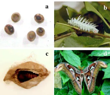
Pages 1 The developmental stage of A. atlas ... 1
2 A. atlas having copulation ... 4
3 The embryonic development of A. atlas (1 to 4 day old) within the eggshell after being immersed by NaOH 5 M overnight ... 4
4 The embryonic development of A. atlas (5 to 7 day old) ... 4
5 The first day larva (instar 1) ... 5
INTRODUCTION
Background
The sericulture of silkworm in Indonesia has begun since 10th century (Atmosoedarjo et
al. 2000). So far, the cocoons of domestic silkworm Bombyx mori have been used as the source of silk fiber. According to BKPPMD (Bureau of Local Region Promotion and Infestation Coordination) (2006), almost 70% of raw silk material (cocoons) has been imported from China and India. Meanwhile Indonesia has many species of wild silkworm that can produce silk fiber. Therefore, the Yogyakarta royal family initiated the wild silk moth development project in Indonesia in 1994. The project aimed to introduce new material of wild silk as an alternative to the domestic silk. The wild silk worms in Indonesia, on which the project focused, are Attacus atlas and Cricula trifenestrata (Nurmalitasari & Kuroda 2002).
Based on its taxonomy A. atlas is classified into kingdom Animalia, phylum Arthropoda, subphylum Atelocherata, class Hexapoda (insecta), order Lepidoptera, superfamily Bombycoidea, family Saturniidae, subfamily Saturniinae, genus Attacus, species A. atlas L. (Triplehorn & Johnson 2005). It is known as giant silk moth or “si rama-rama” and it is considered to be the largest moth in Asia with the wingspans about 250 mm. A. atlas is primarily found in tropical forest and spread around South East Asia, Southern China, and India. It can be found almost in entire Indonesian islands (Peigler 1989).
A. atlas has a perfect metamorphosis stage that included egg (embryo), larval, pupae, and adult stage (Figure 1). According to its annual breeding, A. atlas is polyvoltine, which means that it has more than two generation in a year while B. mori has two characteristic polyvoltine and bivoltine. Bivoltine here means that it has only two generation in a year. Therefore, A. atlas can be found all over year (Chapman 1971). The larva of A. atlas is polyphage that means it has more than one host plant, such as cinchona, cinnamon, tea, sour soup, and avocado (Kalshoven 1981). The adult female lay about 200 to 250 eggs that oviposited in 3 to 5 days in stage and will hatch 8 days after oviposition. A. atlas eggs are oval in shape, 0.008 gram in weight, 2 mm in length, and 1.5 mm in width (Peigler 1989).
Insect embryogenesis is an interesting field of study. The embryonic development of insect occurs in the egg directly after fertilization ended. The stage of embryogenesis in each insect can be different, depends on temperature of environment and species characteristics of development (Yamashita & Hasegawa 1985). For example, B. mori (Bombycidae) has 17 developmental stage in 9 days (Tazima 1978) while another Lepidoptera Nemophora albiantennella (Adelidae) has 12 developmental stage in 7 days (Kobayashi 1997).
Observation of insect embryogenesis is important in order to know the characteristic of each embryonic stage. One of the important physiological characteristic that is used to be studied at the embryonic stage is diapauses. Diapause is a condition in which animal’s cell growth and development are reversibly stopped or slowed (Yamashita & Hasegawa 1985). In sericulture this condition was very useful. Because, by stopping the development the egg hatching also was postponed. These diapauses egg could be used as egg stock for further sericulture program. Although much of insect embryology is still a mystery, there are remarkable progresses in some insect like B. mori. Nevertheless, the embryonic development of A. atlas has not been observed yet.
a
b
c
d
2
Table 1 Copulation time of A. atlas Objective
The research aims to know the embryonic development of Attacus atlas in order to know the possibility of artificial hibernation and accurate time to postpone hatching.
MATERIALS AND METHODS
Materials and Equipments
Materials that were used for this research were eggs A. atlas, NaOH 5M, glycerol 70%, lacto phenol, and alcohol 70%. Equipments that were used for the research were tweezers, ependorf tube, serum bottle, stereomicroscope, digital camera Nikon COOLPIX S210, petridish, and pipette.
Time and Place
This research was conducted in April 2008 to March 2009 in Laboratory of Molecular BiologyResearch Center for Bioresources and Biotechnology (PPSHB) and Laboratory of Embryology Faculty of Veterinary Medicine, Bogor Agricultural University (IPB).
Methods
Moth copulationand egg collection
A couple of A. atlas moth was isolated from the population and separated into different cage to have breeding. After copulation ended, eggs from each couples was collected in petridish. Egg collection was done everyday at 11 to 12 am. From 13 couples that has been breed, 4 couples produced the highest amount of egg. The fertile egg from these 4 couples then was used for observation of embryogenesis and treatment of temperature effect on embryogenesis.
Observation of embryonic development The embryonic development of A. atlas was observed in 7 days. The observation was done everyday (every 24 hours) from 1 day old to 7 day old after oviposition. It was done in two ways, observation within egg shell and observation without eggshell.
Observation of embryonic development within the eggshell. Ten eggs by the age of 1 to 7 days old after oviposition were selected from each couple. Total eggs that have been selected from each couple were 70 eggs. Those eggs then were immersed in NaOH 5 M overnight and moved into glycerol 70% to be stored. The egg then was observed by using stereomicroscope.
Observation of embryonic development without the eggshell. Five eggs by the age of 5 to 7 days old after oviposition were selected from each couple. Total eggs that have been
selected from each couple were 15 eggs. Those eggs were opened by cracking the eggshell. The embryo then was taken and moved into alcohol 70%. After that, the embryo was moved into lacto phenol overnight to be cleared and stored in glycerol 70%. The embryo then observed by using stereomicroscope.
Temperature effect on embryogenesis The egg was preserved in 5oC (in refrigerator) to know temperature effect on embryogenesis. The egg prese
rvation was done in three treatment, preservation without medium, preservation in sucrose 0.5 M, and preservation in NaCl 0.89%. Ten eggs were used for each treatment. Preservation was done in 1, 2, 3, and 4 weeks. Total eggs that have been used are 120 eggs. After preservation, eggs then were placed in the breeding room.
RESULT
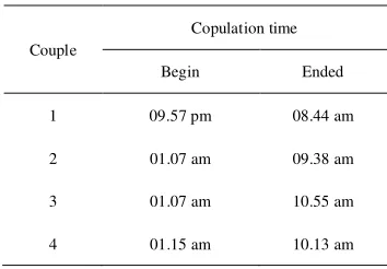
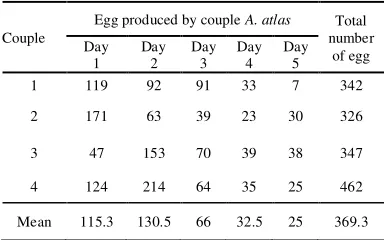
A. atlas got copulation overnight (Figure 2). The copulation begun at night, about 10 pm to 2 am and it ended at 8 am to 11 am (Table 1). Female A. atlas produced about 326 to 462 eggs in five days (Table 2). The period of embryonic stage (egg) was about 7 days in room temperature (26 to 31oC) (Appendix 1).
Couple
Egg produced by couple A. atlas Total number of egg Day 1 Day 2 Day 3 Day 4 Day 5 1 119 92 91 33 7 342
2 171 63 39 23 30 326
3 47 153 70 39 38 347
4 124 214 64 35 25 462
Mean 115.3 130.5 66 32.5 25 369.3 Couple
Copulation time
Begin Ended
1 09.57 pm 08.44 am
2 01.07 am 09.38 am
3 01.07 am 10.55 am
4 01.15 am 10.13 am
Eggs that were used for observation of embryogenesis were selected from: day 1 of 1st couple (119), day 1 of 2nd couple (171), day 2 of 3rd couple (153), and day 2 of 4th couple (214). Meanwhile, eggs that were used for the treatment of temperature effect on embryogenesis were selected from: day 2 of couple1 (91), day 2 of couple 2 (63), day 1 of couple 3 (47), and day 1 of couple 4 (124).
Pre-organogenesis stages.
The 1st day egg after oviposition of A.
atlas showed the large number of yolk fulfilling the entire egg. The 2nd,3rd, and 4th day eggs also showed the same thing. In these days, the embryo could not be seen because it was covered by a large amount of yolk (Figure 3).
Organogenesis stages.
In the 5th day, embryo within eggshell that was immersed by NaOH 0.5M showed that the embryo was migrated to the peripheral surface of egg (surrounding the yolk). The 5th day embryo was 3.5 to 4 mm in length. It has reached the organogenesis stage and performed 11 segments. In the 6th day old embryo within eggshell, it can be seen that the amount of yolk was decreasing while the body size of embryo increasing. The 6th day embryo
reached 4.5 to 5.5 mm in length and it showed the complete patterning of segmentation and appendages.
The 7th day old embryo within the eggshell showed the smaller amount of yolk than before. 7th day old embryo without eggshell showed that it has 5.5 to 6 mm length. After 7 days from oviposition, the formation of embryo is completed, as shown in Figure 4.
Hatching
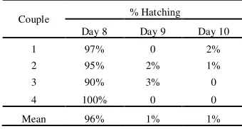
The larva hatched in 8 to 10 days after oviposition (Table 3). It has light green color with seta in entire body. The body length was about 6 to 7 mm, it has 3 thoracic legs and 5 prolegs (abdominal legs) (Figure 5). A. atlas hatched by cracking the eggshell.
Temperature effect on embryogenesis The cold temperature treatment caused the unsuccessful hatching to A. atlas embryo. The control eggs for 1 week preservation hatched 97 %, for 2 weeks preservation hatched 93%, for 3 weeks preservation hatched 89%, while for 4 weeks preservation hatched 100% (Table 4 to 7).
C : Cold treatment without medium S : in medium sucrose 0.5 M N : in medium NaCl 0.89%
Treatment
Preservation time
Treatment
Preservation time
0 week
(control) 3 weeks
0 week
(control) 4 weeks
C
89%
0 C
100%
0
S 0 S 0
N 0 N 0
Couple % Hatching
Day 8 Day 9 Day 10
1 97% 0 2%
2 95% 2% 1%
3 90% 3% 0
4 100% 0 0
Mean 96% 1% 1%
Preservation time
Treatment
Preservation time
0 week
(control) 1 week
0 week
(control) 2 weeks
C
97%
0 C
93%
0
S 0 S 0
N 0 N 0
Table 3 Percentage of egg hatching
Table 4 Seven day old A. atlas egg preserved in 10oC for 1week
Table 6 Seven day old A. atlas egg preserved in 10oC for 3 weeks
Table 5 Seven day old A. atlas egg preserved in 10oC
4
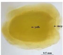
Figure 3 The embryonic development of A. atlas. (1 to 4 day old) within the eggshell after being immersed by NaOH 5 M
overnight. mcp: micropyle.
a
b
c
d
e
f
a
b
c
d
e
f
Figure 4 The embryonic development of A. atlas (5 to 7 day old). 5th day embryo within eggshell (a); 5th day embryo without eggshell (b); 6th day embryo within eggshell (c); 6th day embryo without eggshell (d); 7th day embryo within eggshell (e); 7th day embryo without eggshell (f); ant: anterior; cep: cephal; post: posterior; prl: prolegs; trl: thoracic leg; T1-T3: thoracic segments; A1-A8: abdominal segments Figure 2 A. atlas having copulation. Female
DISCUSSION
Insect embryogenesis occurs inside the egg directly after the fertilization. It is divided into four main processes, cleavage (formation of blastoderm), gastrulation, differentiation, and growth (Tazima 1978; Sander et al 1985). In the stereomicroscopic observation of A. atlas embryo, the only embryogenic stages that can be seen are pre-organogenesis stage and organogenesis stage.
Pre-organogenesis stages.
The 1st to 4th day old embryo of A. atlas
didn’t show a specific difference that could marked it into one of embryo developmental stage. The large amount of yolk covered the zygote, so that the stereomicroscopic observation could not be performed. The 1st to 4th day old embryo only can be categorized as the pre-organogenesis stage. Oppenheimer (1944) mentioned that insect has centrolecithal egg with a large amount of yolk. In the eggs with highly concentrated regions of yolk, cleavage occurs only in the non-yolky cytoplasm. The cleavage begins with mitotic division of zygote nuclei (without cytokinesis) in yolk that occurred in several cycle. This process performs hundred nuclei that often called as energids. The energids then migrated from the yolk to the peripheral surface of the egg and continued the mitotic division. This division performs one-cell-thick layer of cells surrounding the yolk in the peripheral surface that is called blastoderm, while the central yolky part enclosed by blastoderm is known as yolk sac or yolk system (Tazima 1978). In this time, cell separate from the yolk and use the yolk for its nourishment. This process is known as superficial cleavage (cleavage over the yolk surface). The first day of A. atlas embryo may
reach the formation of blastoderm comparing with B. mori that reached blastoderm formation in 12 hours after oviposition (Tazima 1978).
According to Tazima (1978), a dense embryonic primordium will be performed as soon as the blastoderm completed. The cells inside the presumptive embryonic primordium area increase in thickness and become concentrated. This area in the blastoderm extends broadly on the ventral to ventrolateral side of egg and known as germ band. The second day observation of A. atlas embryo may has the complete germ band formation compare with B. mori that completed germ band formation right after blastoderm was established in 20 hours after oviposition (Tazima 1978).
After germ band was establish, it was grow to transform three germ layers. Gastrulation is the process when the germ band enlarges and begins to lengthen and fold into a sausage shape. The one layer of cells that is located on the outside forms the ectoderm and another layer of cells on the inside form the mesoderm.
Ectoderm cells grow and differentiate to form the epidermis, the brain and nervous system, and most of the insect's respiratory (tracheal) system. In addition, the ectoderm invaginates (folds inward) at the front and rear of the embryo's body to create front and rear portions of the digestive system (foregut and hindgut). Mesoderm cells differentiate to form other internal structures such as muscles, glands, heart, blood, fat body, and reproductive organs. The midgut develops from a third germ layer (the endoderm) that arises near the fore and hindgut invaginations and eventually fuses with them to complete the alimentary canal (Sander et al 1985). Embryo of B. mori begins the gastrulation in 25 hours after oviposition (Tazima 1978). Electron microscopic scanning of the developing Manduca sexta shows the gastrulation and differentiation begins at 33 hours after oviposition (Richard et al 1988). Embryo of A. atlas may begins the gastrulation stage in the 2nd day
After completing the gastrulation and differentiation of three germ layers, the embryo moves to the surface (peripheral) of egg, this movement is a part of blastokinesis. Tazima (1978) reported that B. mori takes this stage in two days (4th and 5th day), in A. atlas it may be occurred in the 3rd or 4th day. Figure 5 The first day larva (instar 1). ant: anterior;
6
Organogenesis stages.
Organogenesis in insect embryo was including differentiation of body segments and growth of embryo. The fifth day embryo of A. atlas has reached the organogenesis stage, and the organogenesis continued to the 7th day. According to Sander et al (1985),
organogenesis could be started during late germ band stage and it takes several days or even weeks depends on specific mechanism and characters of each insect egg. The fifth day embryo of A. atlas showed the complete segmentation body, but the formation of appendages and differentiation of cephal section was not complete yet. The embryo has 3 thoracic segments and 8 abdominal segments. The embryo of B. mori reached the complete segmentation body in 6th day (Tazima 1978).
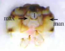
In the sixth day, the embryo of A. atlas reached the stage in which all appendages are formed, the thoracic legs are completely formed and so did the prolegs (abdominal legs). Three pairs of thoracic legs grow in three thoracic segments, while the prolegs are found in the 3rd, 4th, 5th, 6th, and last segment of abdominal segments. The section of cephal has also performed well. The cephal was consisted of 3 segments that performed maxilla, mandible, and labium (Figure 6). In this stage, the pigmentation at the cephal was beginning, while B.mori embryo begins pigmentation of the cephal in 8th day (Tazima 1978).
The seventh day old embryo has the same morphological structure with the larva, which make this stage of embryo is known as prelarva embryo or mature embryo. Besides pigments are formed in entire body (including epithelium and seta), the embryo also grow bigger than the 6th day. After reaching this stage the embryo is ready to hatch.
Hatching
A. atlas larva hatched in 8 days after oviposition by cracking the eggshell. Some insect embryo is used to crack the egg shell to
hatch or hatch through the hatching line (Sander et al. 1985). Different from B. mori that hatched in 9 or 10 days after oviposition (Tazima 1978).
Temperature effect on embryogenesis Insect embryogenesis was affected by many factors, such as: temperature, humidity, genetic, and oxygen consumption (Sander et al. 1985). As poichilotherm organism, temperature has a big effect to insect embryo development. The exposure of cold temperature could caused abnormal development even death to the embryo (Yamashita & Hasegawa 1985). But some insect in temperate region has ability to survive from the winter by suppressing the metabolism through diapauses. The univoltine B. mori was the common model to study the diapause in Lepidotera (Umeya 1950). While the polyvoltine one was the common model of artificial hibernation. The embryogenesis of polyvoltine B. mori can be postponed in a month by refrigerating it in 5oC (Katsumata 1964).
A. atlas was polyvoltine and used to life in tropical region. The cold temperature exposure on 7 days old A. atlas embryo caused the unsuccessful hatching. No embryo of A. atlas could hatch after being stored in 10
OC for several times (Appendix 2). In
polyvoltine B. mori, the egg that would be preserved should reach the mature stage (ready to hatch) that was indicated by the appearance of dark blue dot in the eggshell (Katsumata 1964).
This unsuccessful hatching might be caused by the lack oxygen demand or dehydration because the embryo was wrinkle (Appendix 3). Although some embryo was preserved in Sucrose 0.5 M and NaCl 0.89% medium, it did not prevent the embryo from dehydration.
CONCLUSION
The embryonic development of Attacus atlas occurred in 7 days. The morphological observation showed that the embryogenesis could be divided as pre-organogenesis and organogenesis stage. The 1st to 4th day old embryo could not be observed by the stereo microscope and it may be occurred the cleavage to gastrulation stage, while the 5th to 7th day old has reached the organogenesis stage. The exposure of cold temperature caused unsuccessful hatching in A. atlas. Figure 6 Cephal section of A. atlas embryo.
SUGGESTION
To observe the details of embryonic stage, observations are recommended to be done in every 3 to 6 hours. The serial slicing specimen with histological staining is recommended to observe the complete process of embryogenesis. The using of SEM (Scanning Electron Microscope) is recommended to study the embryo movement.
REFERENCES
Atmosoedarjo, Katsubrata J, Kaomini M, Saleb W, Moerdoko W. 2000. Sutera Alam Indonesia. Jakarta: Yayasan Sarana Wana Jaya.
[BKPPMD] Badan Koordinasi Promosi dan Penanaman Modal Daerah. 2006. Pengembangan Sutera Alam. http//:www.westjava.com [Apr 6th 2009]. Chapman RF. 1971. The Insects, Structure and Function 2nd Ed. New York: Elsevier Publishing comp, Inc.
Kalshoven LGE. 1981. The Pets of Crops in Indonesia. Laan PA van der., penerjemah; Jakarta: Ichtiar BarutoVan Hoeve. Terjemahan dari : De Plagen van de Cultuurgewassen in Indonesie. Katsumata F. 1964. Petunjuk Sederhana bagi
Pemeliharaan Ulat Sutera. Bogor: Balitbang Kehutanan.
Kobayashi Y. 1997. Embryogenesis of the fairy moth, Nemophora albiantennella issiki (Lepidoptera, Adelidae), with special emphasis on its phylogenetic implications. J Insect Morphol Embryol 27:157to166: abstract [terhubung berkala]. http://www.sciencedirect.com. [March 12th 2009].
Nurmalitasari GRAj, Kuroda F. 2002. Indonesia Progress in The Development of Wild Silkmoths. [terhubung berkala]. www.jogja.com/images/WildsilkEnglish 02.pdf. [March 20th 2009].
Oppenheimer SB. 1944. An Introduction to Embryonic development. Massachusetts: Allyn & Bacon,Inc.
Peigler RS. 1989. A revision of the IndotoAustralian genus Attacus. California:The Lepidoptera research Foundation, Inc.
Richard CD, Stanley DC, Walter GG. 1988. A scanning electron microscope study of the developing embryo of Manduca sexta (L.) (Lepidoptera : Sphingidae). Int J Insect Morphol Embryol 17: 231to242: abstract [terhubung berkala]. http://www.sciencedirect.com. [March 12th 2009].
Sander K, Goutzeit HO, Jäckel H. 1985. Insect Embryogenesis: Morphology, Physiology, Genetical and Molecular Aspects. In: Kerkut GA, Gilbert LI, editors. Comprehensive Insect Physiology, Biochemistry and Pharmacology. Vol. I. Embryogenesis and Reproduction. Oxford: Pergamon Press. 407–434.
Tazima Y. 1978. The silkworm; an important laboratory tool. Tokyo: Kodansha LTD. Triplehorn CA, Johnson NF. 2005. Borror and
De Long’s Introduction to the study of Insects. 7th Ed. California: Thomson
Learning, Inc.
Umeya Y. 1950. Studies on embryonic hibernation and diapause in insects. Bul Sericul Exper Station Tokyo 26:1–9. Yamashita O, Hasegawa K. 1985. Embryonic
8
Appendix 1 Temperature and humidity in breeding room
Date Morning (08.00) Afternoon (16.00)
T (°C) Rh T (°C) Rh
120209 27 80 27 70
130209 26 78 28 70
140209 28 65 28 65
150209 28 75 27 70
160209 27 80 27 82
170209 26 79 28 72
180209 26 81 27 80
190209 28 77 29 75
200209 29 68 28 70
210209 28 84 27 70
220209 26 75 27 71
230209 26 81 27 70
240209 26 85 26 73
250209 27 78 29 62
260209 26 85 30 60
270209 27 75 31 65
280209 27 85 27 80
EMBRYONIC DEVELOPMENT OF
Attacus atlas
L.
(LEPIDOPTERA: SATURNIIDAE)
YUSTINA YUSUF
DEPARTMENT OF BIOLOGY
FACULTY OF MATHEMATIC AND NATURAL SCIENCE
BOGOR AGRICULTURAL UNIVERSITY
ABSTRACT
YUSTINA YUSUF. Embryonic Development of Attacus atlas L. (Lepidoptera; Saturniidae). Supervised by DEDY DURYADI SOLIHIN and ARIEF BOEDIONO.
Attacus atlas was one of wild silkworm species that has been developed in Indonesia as an alternative silk source to domestic silkworm Bombyx mori. A. atlas was spread in tropical and subtropical rain forest and can be found almost in entire Indonesian islands. It has complete metamorphosis stage, including egg (embryo), larvae, pupa, and imago (adult). Embryogenesis was an important stage to know the physiology characteristic of insect such as diapauses. The research aimed to know the embryogenesis of A. atlas. The observation of embryonic development was done in two ways, observation within the eggshell and without the eggshell. The embryogenesis in A. atlas was done in seven days. Morphological observation on A. atlas embryo can be divided as pre-organogenesis and organogenesis stage. Pre-organogenesis stage occurred in first to fourth day after oviposition. Organogenesis stage can be observed in fifth to seventh day after oviposition. In the fifth day, body segmentation was completed. In the sixth day, cephal section and patterning of appendages was completed. Besides, the pigmentation of cephal also occurred in sixth day. In the seventh day, the pigmentation of entire body was completed and embryo was ready to hatch. In the eight day after oviposition embryo was hatching by cracking the eggshell.
Key words: Attacus atlas, embryogenesis, development.
ABSTRAK
YUSTINA YUSUF. Perkembangan Embrio Attacus atlas L. (Lepidoptera; Saturniidae). Dibimbing oleh DEDY DURYADI SOLIHIN and ARIEF BOEDIONO.
Attacus atlas adalah salah satu species ulat sutera liar yang dikembangkan di Indonesia sebagai alternatif penghasil sutera dari ulat sutera domestik Bombyx mori. A. atlas tersebar di daerah tropis dan dapat ditemukan di seluruh wilayah Indonesia. A. atlas memiliki metamorfosis sempurna mencakup fase telur, larva, pupa, dan imago. Perkembangan embrio adalah tahapan penting untuk mengetahui karakteristik fisiologi serangga, seperti diapause. Penelitian ini bertujuan untuk mengetahui perkembangan embrio A. atlas. Pengamatan perkembangan embrio dilakukan dengan dua cara, yaitu pengamatan embrio dalam cangkang telur dan tanpa cangkang telur. Tahapan perkembangan embrio pada A. atlas terjadi selama 7 hari. Pengamatan terhadap perkembangan morfologi embrio A. atlas hanya dapat dibagi menjadi tahap, pre-organogenesis dan organogenesis. Tahap pre-organogenesis terjadi pada 1 sampai 4 hari setelah oviposisi dan tahap organogenesis dapat diamati pada hari ke-5 sampai 7. Pembentukan segmentasi tubuh telah selesai pada hari ke-5 setelah oviposisi. Perkembangan embelan dan segmentasi kepala telah selesai pada hari ke-6 setelah oviposisi, pigmentasi pada kepala juga terjadi di hari yang sama. Pigmentasi keseluruhan tubuh terjadi pada hari 7 setelah oviposisi. Larva menetas pada hari ke-8 setelah oviposisi dengan memecah kulit telur.
1
INTRODUCTION
Background
The sericulture of silkworm in Indonesia has begun since 10th century (Atmosoedarjo et
al. 2000). So far, the cocoons of domestic silkworm Bombyx mori have been used as the source of silk fiber. According to BKPPMD (Bureau of Local Region Promotion and Infestation Coordination) (2006), almost 70% of raw silk material (cocoons) has been imported from China and India. Meanwhile Indonesia has many species of wild silkworm that can produce silk fiber. Therefore, the Yogyakarta royal family initiated the wild silk moth development project in Indonesia in 1994. The project aimed to introduce new material of wild silk as an alternative to the domestic silk. The wild silk worms in Indonesia, on which the project focused, are Attacus atlas and Cricula trifenestrata (Nurmalitasari & Kuroda 2002).
Based on its taxonomy A. atlas is classified into kingdom Animalia, phylum Arthropoda, subphylum Atelocherata, class Hexapoda (insecta), order Lepidoptera, superfamily Bombycoidea, family Saturniidae, subfamily Saturniinae, genus Attacus, species A. atlas L. (Triplehorn & Johnson 2005). It is known as giant silk moth or “si rama-rama” and it is considered to be the largest moth in Asia with the wingspans about 250 mm. A. atlas is primarily found in tropical forest and spread around South East Asia, Southern China, and India. It can be found almost in entire Indonesian islands (Peigler 1989).
A. atlas has a perfect metamorphosis stage that included egg (embryo), larval, pupae, and adult stage (Figure 1). According to its annual breeding, A. atlas is polyvoltine, which means that it has more than two generation in a year while B. mori has two characteristic polyvoltine and bivoltine. Bivoltine here means that it has only two generation in a year. Therefore, A. atlas can be found all over year (Chapman 1971). The larva of A. atlas is polyphage that means it has more than one host plant, such as cinchona, cinnamon, tea, sour soup, and avocado (Kalshoven 1981). The adult female lay about 200 to 250 eggs that oviposited in 3 to 5 days in stage and will hatch 8 days after oviposition. A. atlas eggs are oval in shape, 0.008 gram in weight, 2 mm in length, and 1.5 mm in width (Peigler 1989).
Insect embryogenesis is an interesting field of study. The embryonic development of insect occurs in the egg directly after fertilization ended. The stage of embryogenesis in each insect can be different, depends on temperature of environment and species characteristics of development (Yamashita & Hasegawa 1985). For example, B. mori (Bombycidae) has 17 developmental stage in 9 days (Tazima 1978) while another Lepidoptera Nemophora albiantennella (Adelidae) has 12 developmental stage in 7 days (Kobayashi 1997).
Observation of insect embryogenesis is important in order to know the characteristic of each embryonic stage. One of the important physiological characteristic that is used to be studied at the embryonic stage is diapauses. Diapause is a condition in which animal’s cell growth and development are reversibly stopped or slowed (Yamashita & Hasegawa 1985). In sericulture this condition was very useful. Because, by stopping the development the egg hatching also was postponed. These diapauses egg could be used as egg stock for further sericulture program. Although much of insect embryology is still a mystery, there are remarkable progresses in some insect like B. mori. Nevertheless, the embryonic development of A. atlas has not been observed yet.
a
b
c
d
Table 1 Copulation time of A. atlas Objective
The research aims to know the embryonic development of Attacus atlas in order to know the possibility of artificial hibernation and accurate time to postpone hatching.
MATERIALS AND METHODS
Materials and Equipments
Materials that were used for this research were eggs A. atlas, NaOH 5M, glycerol 70%, lacto phenol, and alcohol 70%. Equipments that were used for the research were tweezers, ependorf tube, serum bottle, stereomicroscope, digital camera Nikon COOLPIX S210, petridish, and pipette.
Time and Place
This research was conducted in April 2008 to March 2009 in Laboratory of Molecular BiologyResearch Center for Bioresources and Biotechnology (PPSHB) and Laboratory of Embryology Faculty of Veterinary Medicine, Bogor Agricultural University (IPB).
Methods
Moth copulationand egg collection
A couple of A. atlas moth was isolated from the population and separated into different cage to have breeding. After copulation ended, eggs from each couples was collected in petridish. Egg collection was done everyday at 11 to 12 am. From 13 couples that has been breed, 4 couples produced the highest amount of egg. The fertile egg from these 4 couples then was used for observation of embryogenesis and treatment of temperature effect on embryogenesis.
Observation of embryonic development The embryonic development of A. atlas was observed in 7 days. The observation was done everyday (every 24 hours) from 1 day old to 7 day old after oviposition. It was done in two ways, observation within egg shell and observation without eggshell.
Observation of embryonic development within the eggshell. Ten eggs by the age of 1 to 7 days old after oviposition were selected from each couple. Total eggs that have been selected from each couple were 70 eggs. Those eggs then were immersed in NaOH 5 M overnight and moved into glycerol 70% to be stored. The egg then was observed by using stereomicroscope.
Observation of embryonic development without the eggshell. Five eggs by the age of 5 to 7 days old after oviposition were selected from each couple. Total eggs that have been
selected from each couple were 15 eggs. Those eggs were opened by cracking the eggshell. The embryo then was taken and moved into alcohol 70%. After that, the embryo was moved into lacto phenol overnight to be cleared and stored in glycerol 70%. The embryo then observed by using stereomicroscope.
Temperature effect on embryogenesis The egg was preserved in 5oC (in refrigerator) to know temperature effect on embryogenesis. The egg prese
rvation was done in three treatment, preservation without medium, preservation in sucrose 0.5 M, and preservation in NaCl 0.89%. Ten eggs were used for each treatment. Preservation was done in 1, 2, 3, and 4 weeks. Total eggs that have been used are 120 eggs. After preservation, eggs then were placed in the breeding room.
RESULT
A. atlas got copulation overnight (Figure 2). The copulation begun at night, about 10 pm to 2 am and it ended at 8 am to 11 am (Table 1). Female A. atlas produced about 326 to 462 eggs in five days (Table 2). The period of embryonic stage (egg) was about 7 days in room temperature (26 to 31oC) (Appendix 1).
Couple
Egg produced by couple A. atlas Total number of egg Day 1 Day 2 Day 3 Day 4 Day 5 1 119 92 91 33 7 342
2 171 63 39 23 30 326
3 47 153 70 39 38 347
4 124 214 64 35 25 462
Mean 115.3 130.5 66 32.5 25 369.3 Couple
Copulation time
Begin Ended
1 09.57 pm 08.44 am
2 01.07 am 09.38 am
3 01.07 am 10.55 am
4 01.15 am 10.13 am
2
Table 1 Copulation time of A. atlas Objective
The research aims to know the embryonic development of Attacus atlas in order to know the possibility of artificial hibernation and accurate time to postpone hatching.
MATERIALS AND METHODS
Materials and Equipments
Materials that were used for this research were eggs A. atlas, NaOH 5M, glycerol 70%, lacto phenol, and alcohol 70%. Equipments that were used for the research were tweezers, ependorf tube, serum bottle, stereomicroscope, digital camera Nikon COOLPIX S210, petridish, and pipette.
Time and Place
This research was conducted in April 2008 to March 2009 in Laboratory of Molecular BiologyResearch Center for Bioresources and Biotechnology (PPSHB) and Laboratory of Embryology Faculty of Veterinary Medicine, Bogor Agricultural University (IPB).
Methods
Moth copulationand egg collection
A couple of A. atlas moth was isolated from the population and separated into different cage to have breeding. After copulation ended, eggs from each couples was collected in petridish. Egg collection was done everyday at 11 to 12 am. From 13 couples that has been breed, 4 couples produced the highest amount of egg. The fertile egg from these 4 couples then was used for observation of embryogenesis and treatment of temperature effect on embryogenesis.
Observation of embryonic development The embryonic development of A. atlas was observed in 7 days. The observation was done everyday (every 24 hours) from 1 day old to 7 day old after oviposition. It was done in two ways, observation within egg shell and observation without eggshell.
Observation of embryonic development within the eggshell. Ten eggs by the age of 1 to 7 days old after oviposition were selected from each couple. Total eggs that have been selected from each couple were 70 eggs. Those eggs then were immersed in NaOH 5 M overnight and moved into glycerol 70% to be stored. The egg then was observed by using stereomicroscope.
Observation of embryonic development without the eggshell. Five eggs by the age of 5 to 7 days old after oviposition were selected from each couple. Total eggs that have been
selected from each couple were 15 eggs. Those eggs were opened by cracking the eggshell. The embryo then was taken and moved into alcohol 70%. After that, the embryo was moved into lacto phenol overnight to be cleared and stored in glycerol 70%. The embryo then observed by using stereomicroscope.
Temperature effect on embryogenesis The egg was preserved in 5oC (in refrigerator) to know temperature effect on embryogenesis. The egg prese
rvation was done in three treatment, preservation without medium, preservation in sucrose 0.5 M, and preservation in NaCl 0.89%. Ten eggs were used for each treatment. Preservation was done in 1, 2, 3, and 4 weeks. Total eggs that have been used are 120 eggs. After preservation, eggs then were placed in the breeding room.
RESULT
A. atlas got copulation overnight (Figure 2). The copulation begun at night, about 10 pm to 2 am and it ended at 8 am to 11 am (Table 1). Female A. atlas produced about 326 to 462 eggs in five days (Table 2). The period of embryonic stage (egg) was about 7 days in room temperature (26 to 31oC) (Appendix 1).
Couple
Egg produced by couple A. atlas Total number of egg Day 1 Day 2 Day 3 Day 4 Day 5 1 119 92 91 33 7 342
2 171 63 39 23 30 326
3 47 153 70 39 38 347
4 124 214 64 35 25 462
Mean 115.3 130.5 66 32.5 25 369.3 Couple
Copulation time
Begin Ended
1 09.57 pm 08.44 am
2 01.07 am 09.38 am
3 01.07 am 10.55 am
4 01.15 am 10.13 am
3
Eggs that were used for observation of embryogenesis were selected from: day 1 of 1st couple (119), day 1 of 2nd couple (171), day 2 of 3rd couple (153), and day 2 of 4th couple (214). Meanwhile, eggs that were used for the treatment of temperature effect on embryogenesis were selected from: day 2 of couple1 (91), day 2 of couple 2 (63), day 1 of couple 3 (47), and day 1 of couple 4 (124).
Pre-organogenesis stages.
The 1st day egg after oviposition of A.
atlas showed the large number of yolk fulfilling the entire egg. The 2nd,3rd, and 4th day eggs also showed the same thing. In these days, the embryo could not be seen because it was covered by a large amount of yolk (Figure 3).
Organogenesis stages.
In the 5th day, embryo within eggshell that was immersed by NaOH 0.5M showed that the embryo was migrated to the peripheral surface of egg (surrounding the yolk). The 5th day embryo was 3.5 to 4 mm in length. It has reached the organogenesis stage and performed 11 segments. In the 6th day old embryo within eggshell, it can be seen that the amount of yolk was decreasing while the body size of embryo increasing. The 6th day embryo
reached 4.5 to 5.5 mm in length and it showed the complete patterning of segmentation and appendages.
The 7th day old embryo within the eggshell showed the smaller amount of yolk than before. 7th day old embryo without eggshell showed that it has 5.5 to 6 mm length. After 7 days from oviposition, the formation of embryo is completed, as shown in Figure 4.
Hatching
The larva hatched in 8 to 10 days after oviposition (Table 3). It has light green color with seta in entire body. The body length was about 6 to 7 mm, it has 3 thoracic legs and 5 prolegs (abdominal legs) (Figure 5). A. atlas hatched by cracking the eggshell.
Temperature effect on embryogenesis The cold temperature treatment caused the unsuccessful hatching to A. atlas embryo. The control eggs for 1 week preservation hatched 97 %, for 2 weeks preservation hatched 93%, for 3 weeks preservation hatched 89%, while for 4 weeks preservation hatched 100% (Table 4 to 7).
C : Cold treatment without medium S : in medium sucrose 0.5 M N : in medium NaCl 0.89%
Treatment
Preservation time
Treatment
Preservation time
0 week
(control) 3 weeks
0 week
(control) 4 weeks
C
89%
0 C
100%
0
S 0 S 0
N 0 N 0
Couple % Hatching
Day 8 Day 9 Day 10
1 97% 0 2%
2 95% 2% 1%
3 90% 3% 0
4 100% 0 0
Mean 96% 1% 1%
Preservation time
Treatment
Preservation time
0 week
(control) 1 week
0 week
(control) 2 weeks
C
97%
0 C
93%
0
S 0 S 0
N 0 N 0
Table 3 Percentage of egg hatching
Table 4 Seven day old A. atlas egg preserved in 10oC for 1week
Table 6 Seven day old A. atlas egg preserved in 10oC for 3 weeks
Table 5 Seven day old A. atlas egg preserved in 10oC
Figure 3 The embryonic development of A. atlas. (1 to 4 day old) within the eggshell after being immersed by NaOH 5 M
overnight. mcp: micropyle.
a
b
c
d
e
f
a
b
c
d
e
f
Figure 4 The embryonic development of A. atlas (5 to 7 day old). 5th day embryo within eggshell (a); 5th day embryo without eggshell (b); 6th day embryo within eggshell (c); 6th day embryo without eggshell (d); 7th day embryo within eggshell (e); 7th day embryo without eggshell (f); ant: anterior; cep: cephal; post: posterior; prl: prolegs; trl: thoracic leg; T1-T3: thoracic segments; A1-A8: abdominal segments Figure 2 A. atlas having copulation. Female
5
DISCUSSION
Insect embryogenesis occurs inside the egg directly after the fertilization. It is divided into four main processes, cleavage (formation of blastoderm), gastrulation, differentiation, and growth (Tazima 1978; Sander et al 1985). In the stereomicroscopic observation of A. atlas embryo, the only embryogenic stages that can be seen are pre-organogenesis stage and organogenesis stage.
Pre-organogenesis stages.
The 1st to 4th day old embryo of A. atlas
didn’t show a specific difference that could marked it into one of embryo developmental stage. The large amount of yolk covered the zygote, so that the stereomicroscopic observation could not be performed. The 1st to 4th day old embryo only can be categorized as the pre-organogenesis stage. Oppenheimer (1944) mentioned that insect has centrolecithal egg with a large amount of yolk. In the eggs with highly concentrated regions of yolk, cleavage occurs only in the non-yolky cytoplasm. The cleavage begins with mitotic division of zygote nuclei (without cytokinesis) in yolk that occurred in several cycle. This process performs hundred nuclei that often called as energids. The energids then migrated from the yolk to the peripheral surface of the egg and continued the mitotic division. This division performs one-cell-thick layer of cells surrounding the yolk in the peripheral surface that is called blastoderm, while the central yolky part enclosed by blastoderm is known as yolk sac or yolk system (Tazima 1978). In this time, cell separate from the yolk and use the yolk for its nourishment. This process is known as superficial cleavage (cleavage over the yolk surface). The first day of A. atlas embryo may
reach the formation of blastoderm comparing with B. mori that reached blastoderm formation in 12 hours after oviposition (Tazima 1978).
According to Tazima (1978), a dense embryonic primordium will be performed as soon as the blastoderm completed. The cells inside the presumptive embryonic primordium area increase in thickness and become concentrated. This area in the blastoderm extends broadly on the ventral to ventrolateral side of egg and known as germ band. The second day observation of A. atlas embryo may has the complete germ band formation compare with B. mori that completed germ band formation right after blastoderm was established in 20 hours after oviposition (Tazima 1978).
After germ band was establish, it was grow to transform three germ layers. Gastrulation is the process when the germ band enlarges and begins to lengthen and fold into a sausage shape. The one layer of cells that is located on the outside forms the ectoderm and another layer of cells on the inside form the mesoderm.
Ectoderm cells grow and differentiate to form the epidermis, the brain and nervous system, and most of the insect's respiratory (tracheal) system. In addition, the ectoderm invaginates (folds inward) at the front and rear of the embryo's body to create front and rear portions of the digestive system (foregut and hindgut). Mesoderm cells differentiate to form other internal structures such as muscles, glands, heart, blood, fat body, and reproductive organs. The midgut develops from a third germ layer (the endoderm) that arises near the fore and hindgut invaginations and eventually fuses with them to complete the alimentary canal (Sander et al 1985). Embryo of B. mori begins the gastrulation in 25 hours after oviposition (Tazima 1978). Electron microscopic scanning of the developing Manduca sexta shows the gastrulation and differentiation begins at 33 hours after oviposition (Richard et al 1988). Embryo of A. atlas may begins the gastrulation stage in the 2nd day
After completing the gastrulation and differentiation of three germ layers, the embryo moves to the surface (peripheral) of egg, this movement is a part of blastokinesis. Tazima (1978) reported that B. mori takes this stage in two days (4th and 5th day), in A. atlas it may be occurred in the 3rd or 4th day. Figure 5 The first day larva (instar 1). ant: anterior;
6
Organogenesis stages.
Organogenesis in insect embryo was including differentiation of body segments and growth of embryo. The fifth day embryo of A. atlas has reached the organogenesis stage, and the organogenesis continued to the 7th day. According to Sander et al (1985),
organogenesis could be started during late germ band stage and it takes several days or even weeks depends on specific mechanism and characters of each insect egg. The fifth day embryo of A. atlas showed the complete segmentation body, but the formation of appendages and differentiation of cephal section was not complete yet. The embryo has 3 thoracic segments and 8 abdominal segments. The embryo of B. mori reached the complete segmentation body in 6th day (Tazima 1978).
In the sixth day, the embryo of A. atlas reached the stage in which all appendages are formed, the thoracic legs are completely formed and so did the prolegs (abdominal legs). Three pairs of thoracic legs grow in three thoracic segments, while the prolegs are found in the 3rd, 4th, 5th, 6th, and last segment of abdominal segments. The section of cephal has also performed well. The cephal was consisted of 3 segments that performed maxilla, mandible, and labium (Figure 6). In this stage, the pigmentation at the cephal was beginning, while B.mori embryo begins pigmentation of the cephal in 8th day (Tazima 1978).
The seventh day old embryo has the same morphological structure with the larva, which make this stage of embryo is known as prelarva embryo or mature embryo. Besides pigments are formed in entire body (including epithelium and seta), the embryo also grow bigger than the 6th day. After reaching this stage the embryo is ready to hatch.
Hatching
A. atlas larva hatched in 8 days after oviposition by cracking the eggshell. Some insect embryo is used to crack the egg shell to
hatch or hatch through the hatching line (Sander et al. 1985). Different from B. mori that hatched in 9 or 10 days after oviposition (Tazima 1978).
Temperature effect on embryogenesis Insect embryogenesis was affected by many factors, such as: temperature, humidity, genetic, and oxygen consumption (Sander et al. 1985). As poichilotherm organism, temperature has a big effect to insect embryo development. The exposure of cold temperature could caused abnormal development even death to the embryo (Yamashita & Hasegawa 1985). But some insect in temperate region has ability to survive from the winter by suppressing the metabolism through diapauses. The univoltine B. mori was the common model to study the diapause in Lepidotera (Umeya 1950). While the polyvoltine one was the common model of artificial hibernation. The embryogenesis of polyvoltine B. mori can be postponed in a month by refrigerating it in 5oC (Katsumata 1964).
A. atlas was polyvoltine and used to life in tropical region. The cold temperature exposure on 7 days old A. atlas embryo caused the unsuccessful hatching. No embryo of A. atlas could hatch after being stored in 10
OC for several times (Appendix 2). In
polyvoltine B. mori, the egg that would be preserved should reach the mature stage (ready to hatch) that was indicated by the appearance of dark blue dot in the eggshell (Katsumata 1964).
This unsuccessful hatching might be caused by the lack oxygen demand or dehydration because the embryo was wrinkle (Appendix 3). Although some embryo was preserved in Sucrose 0.5 M and NaCl 0.89% medium, it did not prevent the embryo from dehydration.
CONCLUSION
The embryonic development of Attacus atlas occurred in 7 days. The morphological observation showed that the embryogenesis could be divided as pre-organogenesis and organogenesis stage. The 1st to 4th day old embryo could not be observed by the stereo microscope and it may be occurred the cleavage to gastrulation stage, while the 5th to 7th day old has reached the organogenesis stage. The exposure of cold temperature caused unsuccessful hatching in A. atlas. Figure 6 Cephal section of A. atlas embryo.
Organogenesis stages.
Organogenesis in insect embryo was including differentiation of body segments and growth of embryo. The fifth day embryo of A. atlas has reached the organogenesis stage, and the organogenesis continued to the 7th day. According to Sander et al (1985),
organogenesis could be started during late germ band stage and it takes several days or even weeks depends on specific mechanism and characters of each insect egg. The fifth day embryo of A. atlas showed the complete segmentation body, but the formation of appendages and differentiation of cephal section was not complete yet. The embryo has 3 thoracic segments and 8 abdominal segments. The embryo of B. mori reached the complete segmentation body in 6th day (Tazima 1978).
In the sixth day, the embryo of A. atlas reached the stage in which all appendages are formed, the thoracic legs are completely formed and so did the prolegs (abdominal legs). Three pairs of thoracic legs grow in three thoracic segments, while the prolegs are found in the 3rd, 4th, 5th, 6th, and last segment of abdominal segments. The section of cephal has also performed well. The cephal was consisted of 3 segments that performed maxilla, mandible, and labium (Figure 6). In this stage, the pigmentation at the cephal was beginning, while B.mori embryo begins pigmentation of the cephal in 8th day (Tazima 1978).
The seventh day old embryo has the same morphological structure with the larva, which make this stage of embryo is known as prelarva embryo or mature embryo. Besides pigments are formed in entire body (including epithelium