Expression of hypoxia inducible factor-1a (HIF-1a) gene and apoptosis in
the heart induced by systemic hypoxia
Siufui Hendrawan,1 Sri W.A. Jusman2, Frans Ferdinal1, Ani R. Prijanti2, Septelia I. Wanandi2, Mohamad Sadikin2
1 Department of Molecular Biology and Biochemistry, Faculty of Medicine, Tarumanegara University, Jakarta, Indonesia 2 Department of Molecular Biology and Biochemistry, Faculty of Medicine, University of Indonesia, Jakarta, Indonesia
Abstrak
Tujuan Studi ini adalah untuk mengetahui pengaruh hipoksia terhadap pola ekspresi gen HIF-1α pada jantung tikus serta mengamati timbulnya apoptosis pada kardiomiosit akibat hipoksia sistemik.
Metode Hewan coba (tikus Sprague-Dawley) dibagi secara acak menjadi 7 kelompok (n= 4 per kelompok): kelompok
kontrol normoksia (oksigen atmosir), dan beberapa kelompok hipoksia yang ditempatkan dalam sungkup-hipoksik
(kadar O2 8%) selama 1, 3, 7, 14, 21, dan 28 hari. Pemeriksaan ekspresi gen HIF-1α dilakukan dengan real-time PCR dan apoptosis dengan metode TUNEL.
Hasil Dibandingkan dengan kelompok normoksia, ekspresi gen HIF-1α meningkat secara bertahap sejalan dengan lamanya hipoksia dan mencapai puncak pada hari ke-21. Tidak ada sel yang terlabel dengan cara TUNEL pada
kelompok kontrol. Dibandingkan dengan kontrol, indeks apoptotik meningkat sejalan dengan lamanya hipoksia. Tidak
ada hubungan bermakna antara peningkatan ekspresi HIF-1a dengan peningkatan indeks apoptotik.
Kesimpulan Hipoksia sistemik kronik mengakibatkan peningkatan ekspresi mRNA HIF-1α dan apoptosis pada kardiomiosit. (Med J Indones 2009; 18: 97-101)
Abstract
Aim This study explored the expression of HIF-1α in hypoxic cardiac muscle in mice, and observed the evidence of apoptosis in hypoxia induced cardiomyocyte.
Methods Male Sprague-Dawley rats, were randomized into 7 groups (n= 4 per group): control normoxia group that was exposed to atmospheric oxygen and hypoxia groups that were housed in hypoxic chambers (O2 level 8%) for 1,
3, 7, 14, 21, and 28 days respectively. Animals were sacriiced, hearts were rapidly excised, total RNA was extracted with an mRNA isolation kit and the expression of HIF-1α mRNA was then detected by real-time RT-PCR. Apoptosis was assessed by TUNEL method.
Results For rat in hypoxia group, the expression of HIF-1α mRNA in cardiac myocytes was clearly up-regulated compared to the control normoxia group. Further, HIF-1α expression level elevated gradually and reached a peak at 21 days of hypoxia. No cell labeled by the TUNEL method was detected in the control group. Compared with the control group, the apoptotic index was signiicantly increased in the hypoxia group (P < 0.05). There was no signiicant correlation between the elevation of HIF-1α mRNA and the elevation of apoptotic index.
Conclusion Systemic chronic hypoxia caused the elevation of HIF-1α mRNA and apoptosis in cardiac myocytes. (Med J Indones 2009; 18: 97-101)
Key words: TUNEL, RT-PCR, mRNA, apoptotic index.
The mammalian heart is an obligate aerobic organ. A constant supply of oxygen is indispensable for cardiac viability and function. Myocardial hypoxia occurs as a consequence of a mismatch in the relationship between myocardial mass, myocardial oxygen demand, and oxygen delivery.1
availability and cardiac energy metabolism. A deiciency in high-energy phosphate (ATP) stores is responsible for the transition from adaptive hypertrophy to maladaptive heart failure.1,2
It is crucial for a living organism to be able to detect and respond to hypoxia. Systemic hypoxia is sensed by central and arterial chemoreceptors. These chemoreceptors induce acute systemic cardio-respiratory responses to hypoxia, such as increased respiratory rate and elevation in cardiac pulse rate.3 In addition to the acute responses, chronic adaptations occur at molecular and cellular level. Oxygen availability changes gene expression in cells, including regulation of gene transcription by hypoxia-inducible factor 1 (HIF-1). Hipoxia-inducible factor-1 is a transcriptional activator that functions as a master regulator for cellular oxygen homeostasis. It consists of two constitutively produced subunits: HIF-1α and HIF-1β. Under normoxic conditions, HIF-1α is rapidly degraded, whereas under hypoxia, HIF-1α becomes stabilized, translocates to the nucleus, dimerizes with HIF-1β, and binds hypoxia response element (HRE) to induce certain gene expression. A broad range of HIF-1 actived genes facilitates adaptation to low oxygen conditions, including those that are involved in angiogenesis, erythropoiesis, glucose metabolism, and apoptosis.4,5
Apoptosis can be induced in response to hypoxia. The severity of hypoxia determines whether cells become apoptotic or adapt to hypoxia and survive. In addition to energy deprivation, radical formation, in particular reactive oxygen species (ROS) generation contributes to hypoxia-induced apoptosis. Hipoxia inducible factor-1 can initiate apoptosis by inducing pro-apoptotic protein, such as BNIP3, and can cause stabilization of p53. The p53 can induce the Bax and Bak proteins, which regulate the release of cytochrome-c from mitochondria, thereby initiating the cascade that leads to apoptosis. Cytochrome-c binds to the apoptotic protease activating factor-1 (Apaf-1) and activates caspase-9, which in turn cleaves caspase-3 and 6, and leads to cell death.6
It has generally been believed that apoptosis does not occur in terminally differentiated adult cells such as cardiac myocytes. The aims of this study were to induce hypoxia in an animal model to show that apoptosis could be induced in cardiomyocytes by hypoxia and to observe whether there was any correlation between the expression level of HIF-1α gene and apoptosis during hypoxia.
METHODS
Animals
A total sample of 28 male rats, (10-12 weeks old, weight: 200-250 g), were used in this study. The rats were randomized into 7 groups (n= 4 per group) i.e.: control normoxia group that was exposed to atmospheric oxygen and hypoxia groups that were housed in hypoxic chambers (O2 level 8%) for 1, 3, 7, 14, 21, and 28 days respectively. Control rats were raised in atmospheric oxygen for 28 days before being sacriiced. All animals had free access to water and standard rat chow. Water and food consumption were assessed every 2 days..
Study design
Experimental, in vivo, completely randomized design
was used in this study.
The design of the cages that were used for the hypoxia group in this study prevented exposure of the animals to room air during the entire stay, feeding, cleaning operations, and sacriice. The cages were composed of two types of chambers. The bigger chamber (each has a capacity for six animals) was lushed with 8% O2 and 92% N2 containing gas. The O2 tension inside the chamber was monitored by an O2 meter. When a cage opening was required for regular cleaning, replenishment of food and water, or sacriicing an animal, the compensation (smaller) chamber was irst lushed with hypoxic gas before the animal was then transferred into it.
Each rat was weighed and then anesthetized with ether. After induction of general anesthesia, the chest was opened through median sternotomy. The heart were rapidly excised and weighed. The ventricle was cut horizontally into two slices. The half basal portion was ixed in 10% phosphate buffered formaldehyde (formalin). Formaldehyde-ixed tissues were processed for apoptosis studies. The apical remaining portion was kept frozen at -80oC for further total RNA isolation.
Real-time RT PCR
on the rat HIF-1α sequence (GenBank Accession no. AF057308) [forward 5’-ACA-GTG-GTA-CTC-ACA-GTC-GG, reverse 5’-CCC-TGC-AGT-AGG-TTT-CTG-CT]. The β-actin were based on the rat β-actin sequence (GenBank Accession no. NM_031144) [forward 5’-ACC-ACA-GCT-GAG-AGG-GAA-ATC-G, reverse 5’-AGA-GGT-CTT-TAC-GGA-TGT-CAA-CG]. The predicted size for real-time PCR products for each primer pairs was 466 bp for HIF-1α and 277 bp for β-actin. Real-time PCR reaction mix consisted of 25 µL 2X SYBR Green RT-PCR Reaction Mix, 1.5 µL of each primer, and 19 µL nuclease-free H2O, 2 µL RNA template, and 1 µL iScript Reverse Transcriptase. Real-time PCR conditions were: cDNA synthesis at 50oC for 10 minute; inactivation of iScript reverse transcriptase at 95oC for 5 minute, 39 cycles of 95oC for 10 seconds, 60oC for 30 seconds, and 72oC for 30 seconds. Real-time PCR data analysis used the 2-∆∆CT relative quantiication (Livak) method.
Determination of Apoptosis
Parain-embedded myocardial sections were mounted on silanized slides. Immunohistochemical procedures for detecting apoptotic cardiomyocytes were performed by TUNEL (terminal deoxynucleotidyl transferase-mediated deoxy-UTP nick end labeling) method. This method involves the addition of deoxyuridine triphosphate (dUTP) labeled with luorescein to the 3’-OH ends of DNA by the catalytic action of terminal
deoxyribonucleotidyl transferase (TdT). In situ cell death detection kit, POD was used according to the manufacturer’s (Roche, USA) instructions. The apoptotic index was determined by counting a total of at least 1000 nuclei that were subdivided in ± 10 randomly chosen ields at 400x magniication.
Statistical Analysis
The reported values were means ± SD. SPSS 16.0 was used in this study. The differences among groups were tested by Mann Whitney-U Test. The correlation between continuous variables was calculated by the method of Pearson. P< 0.05 was considered statistically signiicant.
RESULTS
Real-time RT PCR
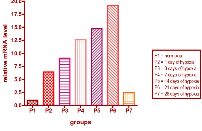
The cDNA of HIF-1α was synthesized by reverse transcription of total RNA from ventricle tissue, using β-actin as an internal control. The result of the current study showed that the expression levels of HIF-1α in hypoxia groups were clearly higher than that of the control group. The expression level elevated gradually and reached a peak at 21 days of hypoxia, that resulted in more than 19-fold increase compared to normoxia group (Figure 1).
Figure 1. The HIF-1α mRNA level in each group, using Livak’s real-time PCR data analysis
a
P1 P2 P3 P4 P5 P6 P7 0.0
2.5 5.0 7.5 10.0 12.5 15.0 17.5 20.0
P1 = normoxia P2 = 1 day of hypoxia P3 = 3 days of hypoxia P4 = 7 days of hypoxia P5 = 14 days of hypoxia P6 = 21 days of hypoxia P7 = 28 days of hypoxia
groups
re
la
ti
v
e
m
R
N
A
l
e
v
e
l
Į
Determination of Apoptosis
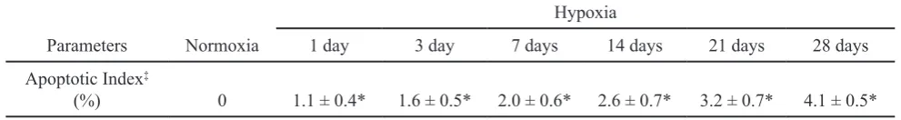
To examine the effect of chronic hypoxia on apoptosis we induced hypoxia in animal models using hypoxic-chambers. Hypoxia induced morphological alterations were typical for apoptotic cell death, as determined by TUNEL assays. Evidence of apoptosis in myocytes by nick end-labeling studies was only observed in the hypoxia groups but none in the normoxia group. Compared with the control group, the apoptotic index was signiicantly increased in hypoxia groups (table 1). However, the elevation of HIF-1α mRNA during hypoxia was only weakly and insigniicantly correlated with the apoptotic index (R= 0.464, P= 0.294).
DISCUSSION
In experimental studies on the effects of hypoxia, several variables need to be taken into consideration, including the animal species, age at entry into the protocol, the method to produce hypoxia, intensity and duration of hypoxia, and precise nature of the experimental design to evaluate the myocardial response.7
The response of myocardial cells to chronic hypoxia may differ from that to acute hypoxia and the in vivo
response may be different from the in vitro response.8 To asses the effect of chronic hypoxia on cardiomyocytes, we used an animal model to study the expression of HIF-1α gene and apoptosis at different levels of hypoxia. In this study the rats were placed in a hypoxic chamber, and oxygen level was maintained at 8%.
A recent research found that HIF-1α was a sensitive marker for hypoxia. Hypoxia and several other stimuli induced HIF-1α signaling cascade. In turn, HIF-1α would transcriptionally activate multiple genes, wich regulated metabolic cardio protective and cross-adaptive mechanisms.9,10 In this study, the real-time PCR result
showed that the expression level of HIF-1α were up-regulated at the mRNA level during hypoxia. The expression of HIF-1α mRNA in cardiac muscle was clearly up-regulated after 1 day exposure to hypoxia (O2 level 8%) and reached a peak at 21 days, and then decreased after 28 days.
Our results are consistent with those reported by Ning et al. in which short-term hypoxia elevated 1α mRNA by promoting transcription. However, HIF-1α expressions were suppressed after a more severe hypoxia occured, accompanied by ATP depletion and extensive apoptosis.10
Terminally differentiated cells such as myocardial cells are not believed to undergo apoptosis under natural conditions. However, recent evidence suggests that apoptosis can be induced in cardiomyocytes by hypoxia, ischemia, and other insults.11
This study showed that apoptotic index was signiicantly increased compared with control group. There was evidence of gradually increased apoptosis with severity of hypoxia, and the mean apoptotic index in each group was more than 1%. These results oppose those reported by Bitar et al. that studied the apoptosis in the chronically hypoxic rat hearts,8 but is in line with the result of Narula et al.11
In single cells, the process of apoptosis runs its course in a short time (on the order of hours). For this reason, identifying and quantifying apoptosis are dificult. Since apoptotic cells are present only transciently, it may be more appropriate to calculate the total number of cells lost by integrating the frequency of the event over time. With such approach, and assuming that a cell undergoing apoptosis is detectable for 24 hours, the percent loss of cells per day can be estimated.12
A higher percentage of apoptotic cardiomyocytes in the hearts studied had been reported by Narula et al. It may be related to the variability in the distribution of apoptotic cells, or to the use of myocardial samples from
Table 1. The number of apoptotic myocytes in each group
Parameters Normoxia
Hypoxia
1 day 3 day 7 days 14 days 21 days 28 days
Apoptotic Index‡
(%) 0 1.1 ± 0.4* 1.6 ± 0.5* 2.0 ± 0.6* 2.6 ± 0.7* 3.2 ± 0.7* 4.1 ± 0.5*
‡number of apoptotic myocytes/total number of myocytes x 100 (in percent),
Values given were mean ± SD
different regions of the myocardium. Apoptosis usually occurred in groups of cells, and the severities varied from extensive to mild to absent in different regions of the myocardium. Furthermore, it may also be due to the variability in the sensitivity of the staining procedures.10 The accessibility of enzymatic reactions to DNA breaks is reduced by the nuclear protein environment and impaired by cell ixation. With adequate pretreatment, reliable TUNEL can be obtained. The microwave pretreatment provided the best TUNEL sensitivity without notable loss of speciicity.13,14
In addition to its presence in myocytes, apoptosis can also be observed in vascular smooth-muscle cells of an intra myocardial arteriole as well as in rare interstitial cells. Consistent with Narula et al. study, we also found that there was no myocyte that was labeled by the TUNEL method in the control group. Narula et al. study also showed that cardiomyocytes’ apoptosis was induced by cytochrome-c release and cleavage of caspase-3 (intrinsic pathway).15
Further, our study showed that there was no signiicant correlation between the elevation of HIF-1α expression and the apoptotic index. After 28 days of hypoxia, the expression level of HIF-1α was decreased, in contrast to the apoptotic index that continued to increase. Several reports demonstrated that under more severe conditions of hypoxia, p53 inhibited HIF-1 activity by targeting HIF-1α for degradation. With prolonged hypoxia, p53 started to accumulate and competed with HIF-1α for binding to p300. It seemed that, under prolonged hypoxia, cardiomyocytes apoptosis was more likely induced by the role of p53 rather than HIF-1α.16
In conclution, systemic chronic hypoxia caused elevation of HIF-1α mRNA and apoptosis in cardiac myocytes.
Acknowledgments
We thank Tarumanagara University for funding this study, and to The Department of Molecular Biology and Biochemistry, University of Indonesia, Jakarta, Indonesia where this study was held.
REFERENCES
Giordano FJ. Oxygen, oxidative stress, hypoxia, and heart
1.
failure. J Clin Invest 2005;115:500-8.
Braunwald E, Bristow MR. Congestive hearth failure: ifty
2.
years of progress. Circulation 2000;102:IV-14−23. Sharp FR, Bernaudin M. HIF-1 and oxygen sensing in the
3.
brain. Neuroscience. 2004; 5:437-46.
Zagorska A, Dulak J. HIF-1: the known and unknowns 4.
of hypoxia sensing. Acta Biochimica Polonica. 2004;51(3):563-78.
Tan Q. Inhibition of cholesterol biosynthesis under hypoxia
5.
[thesis]. Texas: Texas A&M University; 2005.
Greijer AE, Wall E. The role of hypoxia inducible 6.
factor 1 (HIF-1) in hypoxia induced apoptosis. J Clin Pathol.2004;57:1009-14.
Corno AF, Milano G, Morel S, Tozzi P, Genton CY, Samaja
7. of apoptosis in the chronically hypoxic neonatal rat heart. Pediatr Res. 2002;51(2):144-9.
Li ZG, Wang JF, Cheng JD, Liu YW, Xing HW, Wang
9.
Y, et al. Regularity of hypoxia inducible factor 1 alpha expression in acute myocardial ischemia in rats. Chin Med J. 2007;120(2):162-5.
Ning XH, Chen SH, Buroker NE, Xu CS, Li FR, et al.
10.
Short-cycle hypoxia in the intact heart: hypoxia inducible factor 1α signaling and the relationship to injury threshold. Am J Physiol Heart Circ Physiol. 2007;292:H333-41. Narula J, Haider N, Virmani R, Disalvo TG, Kolodgie FD,
11.
Hajjar RJ, et al. Apoptosis in myocytes in end-stage heart
failure. New Engl J Med. 1996; 335(16):1182-9.
Colucci WS. Apoptosis in the heart [review]. Boston:
12.
Boston Medical Center; 2003.
Negoescu A, Lorimer P, Labat-Moleur F, Drouet C, Robert
13.
C, Guillermet C, et al. In situ apoptotic cell labeling by tunel method: improvement and evaluation on cell preparations. J Histochem Cytochem. 1996;44(9):959-68.
Negoescu A, Lorimer P, Labat-Moleur F, Azoti L, Robert
14.
C, Guillermet C, et al. Tunel: improvement and evaluation of the method for in situ apoptotic cell identiication. Biochemica. 1997;2:12-5.
Narula J, Pandey P, Arbustini E, Haider N, Narula N, Kologie
15.
FD, et al. Apoptosis in heart failure: release of cytochrome-c
from mitochondria and activation of caspase-3 in human cardiomyopathy. Proc Natl Acad Sci.1999; 96:8144-9. Schmid T, Jie Z, Brüne B. HIF-1 and p53: communication 16.