by polymerase chain reaction
C.K.
Liml, M.L.
Ling2, Y.S. Leo3, H.S. SeowlAbstrak
Sepasang primer nested-PCR telah dirancang untuk mendeteksi secara spesifik S, typhi, agen penyebab demam tifuid. DNA target untuk pasangan primer PCR tersebut adalah daerah spacer antara gen 23 S rRNA dan 5 S rRNA darl S. typhi (ATCC 167). Pasangan
primer ini telah dicoba pada 5 spesimen darah pasien tifuid yang telah dikonfirmasi dengan kultur Produk nested PCR positif yang berupa 140 bp ditemul<nn pada semua 5 spesimen. Pemeril<saan lebih dari 100 spesimenfeses yang kemudian dikonfirmasi dengan kultur S. typhi negatif tidak satupun memberikan produk PCR positif. Tingkat deteksi dalam darah yang diinokulasi dengan S. typhi daLam jumlah yang diketahui adaLah l0 cfu per ml darah. Pasangan primer ini mempunyai potensi untuk pengembangan sLuttu uji untuk
diag-nosis cepat demam tifoid.
Abstrak
A pair of nested PCR primers has been designed for the specffic detection o/ S. typhi, the causative agent of typhoid fever The
target DNA for this pair of PCR primers is the spacer region betvveen the 235 rRNA and 55 rRNA genes from S, typhi (ATCC 167 ). This pair of primers has been tested against 5 blood specimens from patients. These patients are typhoid cases, confirmed by culture. The
positive 140 bp nested PCR product was obtained from all 5 specimens. From more thatx 1 00 stool specimens, which were Later confirmed to be S. typhi negative by culture, none gave the positive PCR product. The detection Level in blood inoculated with known numbers of S. typhi, is 10 CFU per ml blood. This pair of primers, therefore, has potentialfor development into a diagnostic toolfor rapid diagnosis of typhoid fever
Suppl
I -
1998Direct
detection
of Salmonella typhi
in
blood
MATERIALS AND METHODS
Bacterial
strails
and specimensS. typhi (ATCC 167) obtained from the Singapore General Hospital was used as the type strain. Blood and stool specimens were also from SGH. Other S.
typhi and non-typhoid salmonellae were clinical iso-lates from various hospitals in Singapore.
Extraction of DNA
Direct extraction
of
genomic DNA from blood was carried out as according to manufacturer's(Boehrin-ger Mannheim mammalian blood DNA extraction
kit)
instructions. The DNA precipitate obtained from 1 ml blood was re-dissolvedin
100 pl TE buffer (10mM Tris, 1 mM EDTA, pH 7.5). Five
pl
of this wasused in each PCR. Extraction of genomic DNA from
bacteria was carried out as described in Zhu et alr.
Diagnostic
59s3-s
Extraction
of
genomicDNA from stool
specimenswas carried out as described by Kongmuang et al2.
PCR primers
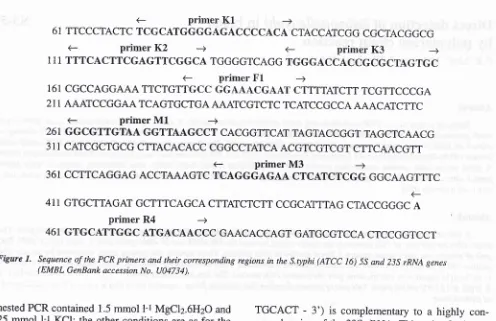
For specific detection of S. typhi, two pairs of primers
(F1/R4
& M1^43)
were used. The positions of these two pairs of primers are shown in Figure 1. For PCRribotyping, four pairs of primers were used. Primers
EIIEZ are based on Escherichia coli rRNA sequences (Kostman et aI3). Primers
Kl,
K2, K3 (see Figure 1)were used with M3.
All
primers were synthesized by Bio-Synthesis Inc. (TX, USA).PCR conditions
The optimum conditions for both primary
&
nested primers are given in Table 1.Alt
PCR were carried out using Taq polymerase (HT Biotechnology Ltd, U.K.) in
a DNA
thermal cycler (Hybaid Omnigene). The primary PCR mixture contained75
mmol l-tKCl,
10 mmol l-r Tris-HCl (pH 9,2),3.5 mmol l-1MgClz.6HzO, 2 mmol l-r of each dNTP,0.004 pg pl-t of each primer, 0.4 U pl-t Taq polymerase and 0.002 ng pl-l to 0.02 ng pl-t of template DNA. The primary PCR products were diluted 500
fold
before beingused
for
the nested PCR. The buffer usedfor
theI
Department
of
re'Department of rDepartment of
<-
primerK1
-+61 TTCCCTACTC TCGCATGGGGAGACCCCACA CTACCATCGG CGCTACGGCG
<-
primerK2
-+<-
primerK3
-+1 1 1 TTTCACTTCGAGTTCGGCA TGGGGTCAGG TGGGACCACCGCGCTAGTGC
<-
primerIrl
-+1 61 CGCCAGGAAA TTCTGTTGCC GGAAACGAAT C]TTTTATCTT TCGTTCCCGA
2 1 1 AAATCCGGAA TCAGTGCTGA AAATCGTCTC TCATCCGCCA AAACATCTTC
e
primerMl
-+26 1 GGCGTTGTAA GGTTAAGCCT CACGGTTCAT TAGTACC GGT TAGCTCAAC G
3 1
I
CATCGCTGCG CTTACACACC CGGCCTATCA ACGTCGTCGT CTTCAACGTT<-
primerM3
-+361 CCTTCAGGAG ACCTAAAGTC TCAGGGAGAA CTCATCTCGG GGCAAGTTTC
<_
411 GTGCTTAGAT GCTTTCAGCA CTTATCTCTT CCGCATTTAG CTACCGGGC A
primer
R4
-+46 1 GTGCATTGGC ATGACAACCC GAACACCAGT GATGCGTCCA CTCCGGTCCT
Figare 1. Sequence of the PCR primers and their corresponding regions in the S.typhi (ATCC 16) 55 and 235 rRNA genes
(EMBL GenBank accession No. U04734).
nested PCR contained 1.5 mmol l-t MgClz.6HzO and 25 mmol
l-t
KCI; the other conditions are as for the primary PCR. PCR conditions for ribotyping are the same as for primary PCR except that template con-centration was 0.01 pg trl-1 and the primerconcentra-tion was 0.6mM
for
E1lE2 and 0.2mMfor
KÀrI4. The PCR mixtures (20pl)
were analysed by electro-phoresisin
I-I.2
Vo aguose gel (Sigma Type 1-A). PCR patterns were compared after ethidium bromide staining.Nucleotide sequence analysis
The PCR amplicons (300 bp &.14O bp) were excised
from
agarose gels and purifiedby
phenol(Ultra-pure,Gibco-BRl) extraction and ethanol
precipita-tion. The
purified PCR amplicons were thense-I prism d-action kit)
RESUIJTS
Specificity of the PCR primers
The primary PCR primers were designated
Fl
and R4(Zht
etalt).Fl
(5' - TGCCGGAAACGAATCT - 3')is
complementaryto a
segmentin
the55
to
23SrRNA spacer region, which
is
highly variable and specific for S. typhi. R4(5'-
GGTTGTCATGCCAATGCACT
-
3')
is
complementaryto
a highly con-served region of the 23S rRNA. This pair of primary PCR primerswill
amplify a 300 bp DNA fragment (nucleotides I77-480 in Figure 1).The nested primers
Ml
(5'-
GGCGTTGTAAGGTT AAGCCT- 3')
andM3
(5'-
AGGCT'IAACCTTA-CAACGCC- 3') will
amplifya
140 bp DNA frag-ment (nucleotides 261-400 in Figure 1). The identityof both the 300 bp and 140 bp PCR amplicons were confirmed
by
sequencing. The optimum conditionsfor
both pairsof
primers were determined and aregiven in Table 1.
Table 1. PCR conditions
Primary PCR-Typing
Temp. Time
taken
Temp. Time
taken
Temp Time
taken
Pre-denaturation
Denaturation Annealing Extension No. of cycles Post-extension
95'C
5 min95"C 50 s
58'C
60 s'120c 50 s
35
72'C
5 min95'C
2 min94C
5 min95'C
30s 94C
I min52"C 30
s
53'C
I
min'72"C 30
s
72"C
I min25
3072"C
5 min72"C
5 min3h
[image:2.595.52.548.77.398.2]Suppl
I
-
1998Figure 2. Electrophoretogram of primary PCR products using DNA extracted from blood spiked with varying CFU of
S. typhi
lane
l:
103, lane 2: IÛ, lane 3: 50, lane 4: 25, lane 5: 10, Iane 6 & 1I: pGEM, lane 7 & 8: 0,Lane 9: S. typhi genome DNA, Iane
l0:
waterFigure 3. Electrophoretogram of nested PCR products using di-luted primary PCR from Figure 2 as template
lane
l:
103 CFII, Iane 2: 102 CFU, lane 3: 50 CFU, lane 4: 25 CFU, lane 5: 10 CFU, lane 6 & 7: 0 CF|J,Lane 8: pGEM, Iane 9: watetr, lane l0: S. typhi genome DNA,
Diagnostic
61Sensitivity
of the
PCRThe primary and nested amplification products are
shown in Figures 2 &.
3.In
Figure 2, only lane 5 did not show the expected 300 bp PCR product, i.e. thedetection level was 25 CFU. In Figure 3, only lane 5
showed the 140 bp nested PCR product, indicating that the detection level is 10 CFU. Therefore the use of nested primers has increased the sensitivity level
(see Table 2).
Thble 2. Detection of S. typhi in whole blood by PCR
Concentrations of S. typhi in artificially inoculated blood used for PCR
'PCR result
zNested PCR result
CFU/ml
(300 bp product) (140 bp producr) 1x1051x103 1x10² 50
25
10
+ + + + + (faint)
+
+ + + + + + (faint) 0 (uninoculated control)
Positive control for PCR
HzO control for PCR
I
I
ml of the artiflciaLly inocuLated blood was processed using Boehringer Bloorl Kit. The extracted DNA was dissolvedin 100 1tl TE buffea and (i.e., 5 1t"l) of which was used
for PCRs.
2A 300-lold dilution of the primary pCR prodttcts was carried out, and 5 1t"l of which was used for the nested pCRs.
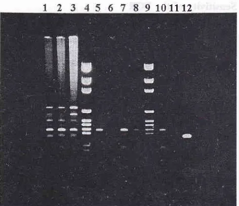
Direct detection of S. typhi
in
bloodfrom typhoid
patients
Of five blood specimens from typhoid patients which were tested, all five showed the S. typhi-specific 300 bp amplicon (see Figure 4, results from 3 specimens
are shown). The use of undiluted DNA extract from typhoid patients'blood as template, yielded the S. 4r-phi-specific 300 bp amplicon (see lanes
I,2
& 3, Fig-ure 4). However there were several non-specificam-plicons.
Dilution
of
the template ten-fold removes most of the non-specific amplicons while yielding a fairly distinct S. typhi-specific 300 bp amplicon (seelanes 5, 7
&.I0,
Figure 4).Direct detection of S. typhi
in
stool specimens One hundred stool specimens were tested with theprimary primers but no S. typhi specific PCR
ampli-con
were
obtained. These specimenswere
later shown to culture negative for S. typhi. Another tenstool specimens, from typhoid patients who had
9
1û1112Figure 4, Electrophoretogram ofprimary PCR products using
DNA extracted from bLood of typhoid patients
Iane
l:
833, Iane 2: 834, Lane 3: 835, Lane 4 & 9: pGEM,lane 5: 833 diluted l:10, Lane 6: 833 diluted l:30,
Lane 7: 834 diluted l:10, Iane 8: 834 diluted l:30,
Lane l0:835 diluted l:10, lane
ll:835
diluted I:30,lane 12: S. typhi genomic DNA
ready started on antibiotic therapy, were also tested. These also did not yield the S. typhi specific PCR
am-plicon.
PCR-ribotyping
Based on the PCR results, four pairs of primers were
designed
for
usein
ribotyping. Using the K3/M3(K3:
5'-TGGGACCACCGCGCTAGTGC-3')prim-ers, 14 non-typhoid salmonellae were differentiated
into
8
ribotypes. Usinga
mixtureof
two
pairs ofprimers @l/F2; E 1 : 5' - TTGTACACACCCCCCGTCA
-
3',82:5'
-GGTACCTTAGATGTTTCAGTTC - 3'&
K3/I43), these 14 non-typhoid salmonellae werefurlher differentiated into 9 ribotypes (see Figure 5
&
Table 3). The other two pairs of primers (K2lM3 8.
K3/I\43)
did not
increase the differentiating power when usedon
its
ownor
togetherwith
the otherpflmer palrs.
Thble 3. PCR ribotypes
Strains PCR ribotypes PCR ribotypes
(K3/M3)
(81/82 &. K3/M3) Salmonella agonaS, anatum
S. cervo
S, choleraesuis
S, enteritidis
S. give
S. haardt
S. hader
S. javiana
S. kingston
S. paratyphiA
S. paratyphi B
S. typhimurium
S. weltevreden
S. typhi (ATCC 167)
Escherichia coli TGl
a
b b h
b
b a
not tested
s
h
f
dI
.I
II
ilI
X IV
I VI
I
X
not tested
VII VIII
IX
XI
E 5 <''3
.2
E'= € ==
3A
E
t
E
s
Es
t
[c
Ëa
ÈeËe
ë
t
t
[image:4.595.50.286.89.293.2]Suppl
I
-
1998DISCUSSION
All
the blood specimens from typhoid patients testedpositive with the primary primers. The use of nested
PCR primers increased the sensitivity of detection to
10 CFUs. Therefore these two pairs of primers have
the potential to be developed into a routine diagnostic
agent for the rapid diagnosis of typhoid fever.
Conventional ribotyping, using rRNA genes to probe
bacterial genomic DNA, has been shown to be useful
in strain differentiation in a variety of bacteria
(Bin-gen et a/4). However this method is time-consuming
involving electrophoresis, blotting, hybridisation and
X-ray
film
development. PCR ribotyping,on
theother hand, is a more rapid method.
It
involves onlyPCR and electrophoresis. In this study, both the
165-23S and 23S-5S rRNA intergenic spacers were used
as targets for PCR. The current primer se| (EI|E2
&
K3A{3) differentiates 14 tested serotypes into only 9
ribotypes. Therefore the differentiation power is as
Diagnostic
63yet inadequate for use in typing salmonellae. Due to
the extreme ease and rapidity of PCR-ribotyping, this
metho{ has great potential
for
developmentinto
atyping method.
REFERENCES
1.
Zhu Q, Lim CK, Chan YN. Detection of S. typhi bypolym-erase chain reaction. J Appl Bacteriol 1996; 80 244-51.
2,
Kongmuang U, Luk JMC, Lindberg AA. Comparison ofthree stool-processing methods for detection of Salmonella
serogroups B, C2 & D by PCR. J Clinical Microbiol 1994;
32:3072-4.
3.
Kostmân JR, Edlind TD, Li Puma JJ, Stull TL. Molecular epidemiology of Pseudomonas cepacia determined by po-lymerase chain reaction. J Clinical Microbiol 1992; 30: 2084-7.4.
Bin$en EH, Denmar E, Elion J. Use of ribotyping inepide-miological surveillance of nosocomial outbreaks. Clinical