Insects on pig carcasses as a model for predictor of death interval
in forensic medicine
Keywords: Chrysomya, forensic entomology, Hermetia
pISSN: 0853-1773 • eISSN: 2252-8083 • http://dx.doi.org/10.13181/mji.v24i2.1224 • Med J Indones. 2015;24:70-8 • Received 25 Mar 2015 • Accepted 25 May 2015
Correspondence author: Sunny Wangko, [email protected]
Copyright @ 2015 Authors. This is an open access article distributed under the terms of the Creative Commons Attribution-NonCommercial-ShareAlike 4.0 International License (http://creativecommons.org/licenses/by-nc-sa/4.0/), which permits unrestricted non-commercial use, distribution, and reproduction in any medium, provided the original author and source are properly cited.
Sunny Wangko,1 Erwin G. Kristanto,2 Sonny J.R. Kalangi,1 Johannes Huijbregts,3 Dantje T. Sembel4
1 Department of Anatomy-Histology, Faculty of Medicine, University of Sam Ratulangi, Manado, Indonesia
2 Department of Forensic and Medicolegal Science, Faculty of Medicine, University of Sam Ratulangi, Manado, Indonesia 3 Naturalis Biodiversity Center, Leiden, Netherlands
4 Department of Pests and Plant Diseases, Faculty of Agriculture, University of Sam Ratulangi, Manado, Indonesia B a s i c M e d i c a l Re s e a rc h
ABSTRAK
Latar belakang: Entomologi forensik belum dimanfaatkan sebagaimana mestinya di Indonesia. Keberadaan serangga forensik di Indonesia juga belum banyak dilaporkan. Penelitian ini bertujuan untuk mendapatkan jenis-jenis serangga pada bangkai hewan coba yang dapat dipergunakan untuk perkiraan saat kematian.
Metode: Empat babi domestik yang dimatikan dengan cara berbeda digunakan sebagai model. Bangkai diamati dua kali sehari (sekitar pukul 09.00 dan 16.00) selama 15 hari untuk mendapatkan tahap-tahap dekomposisi dan koleksi serangga baik imatur maupun dewasa. Serangga imatur dipelihara dan serangga dewasa diidentifikasi di Laboratorium hama dan penyakit tanaman, Universitas Sam Ratulangi, Manado. Chrysomya megacephala dan C. rufifacies diidentifikasi baik secara morfologik maupun tehnik deoxyribose-nucleic acid
(DNA).
Hasil: Lima tahap dekomposisi didapatkan dalam penelitian
(segar, pembusukan awal, pembusukan aktif, pembusukan lanjut, dan skeletonisasi ). Serangga yang ditemukan ialah 11 spesies Diptera and delapan Coleoptera selama 15 hari suksesi. Chrysomya megacephala, C. rufifacies dan Hermetia illucens berkolonisasi pada semua bangkai.
Kesimpulan: Pada keempat ekor babi serangga yang ditemukan terutama terdiri dari Diptera dan Coleoptera. Chrysomya megacephala, C. rufifacies dan Hermetia illucens tampaknya merupakan kandidat utama untuk perkiraan saat kematian.
ABSTRACT
Background: Forensic entomology has not been acknowledged in Indonesia so far. Indonesian carrion insects are very rarely reported. The aim of this study was to obtain the types of insects on pig carcasses that could be used for the estimation of post-mortem interval.
Methods: Four domestic pigs sacrificed with different methods were used as a model. The carcasses were observed twice daily (around 9 a.m and 4 p.m) during 15 days to assess the stages of decomposition and to collect insects, both in mature and immature stages. The immature insects were reared and
the mature insects were indentified in the Laboratory of Pests and Plant Diseases, University of Sam Ratulangi, Manado. Chrysomya megacephala and C. rufifacies were identified both
morphologically and with deoxyribose-nucleic acid (DNA)
techniques.
Results: Five stages of decomposition (fresh, bloated, active decay, post-decay, and skeletonization) were observed.
A total of 11 Diptera and 8 Coleoptera species were found
during a 15-days succession study. Chrysomya megacephala,
C. rufifacies and Hermetia illucens colonized in all carcasses. Conclusion: Insects found on four different pig carcasses consisted mainly of widespread Diptera and Coleoptera.
Chrysomya megacephala, C.rufifacies and Hermetia illucens
Insects have been used in forensic investigation as early as the thirteenth century in China.1 The first serious description of insect succession on
corpses was given by Mégnin.2 Early forensic
work on insects was done by Nuorteva3 in Finland
and Leclercq4 in Belgium. After the publication of the first handbook by Smith1 in 1986, forensic
entomology became gradually more fashionable in normal police work. In the last three decades forensic entomology developed rapidly, especially in the areas with a temperate climate. More recently studies on this topic have been carried out in several tropical countries. As the carrion insects in most tropical countries are not very
well documented, thus the findings of those insects will be a great challenge. So far, no specific
forensic entomology studies have been conducted
in Indonesia. Research was done on the island of
Sulawesi, which is on the transition of the Oriental and Australian biogeographical regions. Sulawesi has a very peculiar fauna, rich in endemic species.
During the Wallace project in 19855 carrion flies
were collected by Kurahashi, et al6-9 and carrion
beetles by Hanski and Krikken10, and Hanski and
Niemela.11 These insects were studied mainly in
undisturbed rainforest habitats. Most human corpses are however found in an urban or suburban environment and the carrion fauna of this habitat type was not examined in Sulawesi on a systematic way.
According to the information from the Department of Forensic and Medicolegal Prof. Dr. R. D. Kandou
General Hospital Manado, the most common cases of violent deaths in Manado are blunt stroke on the occipital area and cardiac puncture. In order
to test whether the cause of death influenced the insect succession, four pigs were sacrificed in
different ways. The aim of the present study was to identify the insect succession on pig carcasses as a model in forensic entomology for predictor of death interval.
METHODS
Field studies were carried out at Winangun and
Batu Kota, Manado, North Sulawesi, Indonesia, between January 27th and February 15th, 2012. Winangun and Batu Kota are located at a
latitude 1°26’41”N and longitude 124°50’11”E,
respectively, at an altitude of 66 metres above sea
level. Four domestic pigs weighing approximately
20 kg each (aged two month old) were used as a
model. The 1st pig was sacrificed by using 200 g
potassium cyanide per oral designated as carcass I; the 2nd pig was sacrificed by a blunt stroke on its
occipital area, carcass II; the 3rd and 4th pig were sacrificed by cardiac punctures, carcass III and IV.
After death, three pigs were placed in protective cages at a distance of 20 m from each other at
Winangun, and the other one (carcass IV) at Batu
Kota. The cages consisted of a wooden frame
(1.6x1x1 m3) were covered with wire meshing
(diameter ± 1.7 cm).
The carcasses were observed twice daily (around 9 a.m and 4 p.m) during 15 days to assess the stages of decomposition (fresh, bloated, active decay, post-decay or skeletonization) and to collect insects.
Both mature and immature insects were collected.
Insects flying around the carcasses or pearched
on the carcasses were collected with an insect net and recorded. Immature insects including eggs, larvae in natural cavities and wounds, and larvae from other parts of the carcasses were collected
for rearing and identification in the Laboratory of Pests and Plant Diseases, Sam Ratulangi University, Manado. Soil around and underneath
the carcasses were examined to observe the presence of insects including pupae. Immature insects were reared on slices of fresh pig liver.
Adult insects were sacrificed with ethyl acetate
and mounted on entomological pins, while immature specimens were stored in 95% alcohol.
Specific literature for the identification of adult
Sulawesi carrion insects is mainly limited to
blowflies. Most other species were identified by direct comparison with identified museum material. The identification of immature insects
is based on rearing to adults. Voucher specimens
were deposited at the Sam Ratulangi University,
Manado; and Naturalis Biodiversity Center, Leiden.
Chrysomya megacephala and C. rufifacies were
identified both morphologically and with deoxyribose-nucleic acid (DNA) techniques. Samples for DNA identification were extracted with AxyPrep Multisource Genomic DNA Miniprep Kit. This kit uses column purification
technique that extracts both core and
After extraction, samples with 1270 bp
cytochrome oxidase I (COI) gene were amplified with polymerase chain reaction (PCR) using
primer for Chrysomya, as follows:12
● C1-J-1718f (5’–
GGAGGATTTGGAAATTGATTAGTTCC) ● TL2-N-3014r (5’ –
TCCAATGCACTAATCTGCCATATTA) ● TL2-N-3014MODr (5’ –
TCCATTGCACTAATCTTGCCATATTA)
Step Temperature Time
Predenaturation 95°C 2 minutes
Denaturation 95°C 30 seconds
Primary annealing 54°C 30 seconds
Extention 72°C 90 seconds
Final extention 72°C 90 seconds
Table 1. Steps of PCR of COI gene
Species Source
Coleoptera
Hybosoridae Phaeochrous emarginatus Castelnau Hanski & Krikken, 199110
Hybosoridae Phaeochrous sulawesi Kuijten Kuijten, 197813
Scarabaeidae Onthophagus aper Sharp Hanski & Krikken, 199110
Scarabaeidae Onthophagus mentaveiensis Boucomont Hanski & Krikken, 199110
Scarabaeidae Onthophagus scrutator Harold Hanski & Krikken, 199110
Silphidae Necrophilia renatae (Portevin) Ruzicka, Schneider, Qubaiová & Nishikawa, 201214
Silphidae Necrophilia charon Sikes & Madge Sikes, Madge & Trumbo, 200615
Silphidae Nicrophorus distinctus (Grouvelle) Sikes, Madge & Newton, 200216 Diptera
Calliphoridae Calliphora hasanuddini Kurahashi & Selomo Kurahashi & Selomo, 19979
Calliphoridae Chrysomya greenbergi Wells & Kurahashi Wells & Kurahashi, 199617
Calliphoridae Chrysomya nigripes Aubertin Singh, Kurahashi & Wells, 201118
Calliphoridae Chrysomya rufifacies (Macquart) James, 197719
Calliphoridae Chrysomya yayukae Kurahashi & Magpayo Kurahashi & Magpayo, 19878
Calliphoridae Hemipyrellia ligurriens (Wiedemann) James, 197719
Calliphoridae Idiella divisa (Walker) James, 197719
Calliphoridae Isomya delectans (Walker) James, 197719
Calliphoridae Lucilia adisoemartoi Kurahashi, 19886
Calliphoridae Lucilia papuensis Macquart James, 197719
Calliphoridae Phumosia abdominalis Robineau-Desvoidy James, 197719
Calliphoridae Phumosia elegans Kurahashi Kurahashi, 19897
Calliphoridae Phumosia indica (Surcouf) James, 197719
Calliphoridae Phumosia njonja Kurahashi Kurahashi 19897
Calliphoridae Phumosia promittens (Walker) James, 197719
Calliphoridae Strongyloneura prolata (Walker) James, 197719
Table 2. Sulawesi carrion insects reported in taxonomic literature
Primers were made at PT Genetika Science Indonesia located in Jakarta, Indonesia. PCR
was carried out with Biometra® T Personal, with
temperatures of each cycle as seen as Table 1.
PCR products were visualized using 1.0%
agarose gel, dyed in ethydium bromide 1.5% and
transiluminated with ultraviolet (UV) rays, and
were then sequenced with COI gene bar coding
that is commonly used in flies identification.
RESULTS
The mean daily temperature at the study sites
fluctuated between 23.4°C and 27.2°C. Almost all
days had heavy rainfalls.
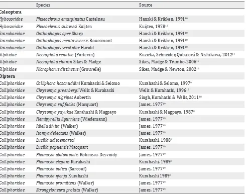
that they were collected on carrions. More recent taxonomical papers usually contain more ecological
information but it is very difficult to trace specific
carrion insects in scattered publications. Based on the carrion-taxa found in neighbouring countries, literature was searched for potential carrion insects in Sulawesi. The results of the literature search are summarized in Table 2.
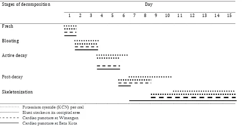
Five stages of decomposition were distinguished in all carcasses. The duration of the different stages is illustrated in Table 3. The fresh stage was observed on day one. The bloated stage was observed on days two and three for all carcasses,
but facial destruction was first observed on
carcass II. The active decay stage was observed
on days four and five in carcass II, III and IV
meanwhile in carcass I on days four to six. The post-decay stage was observed on days six to
eight in carcass II and III, and just on day six in
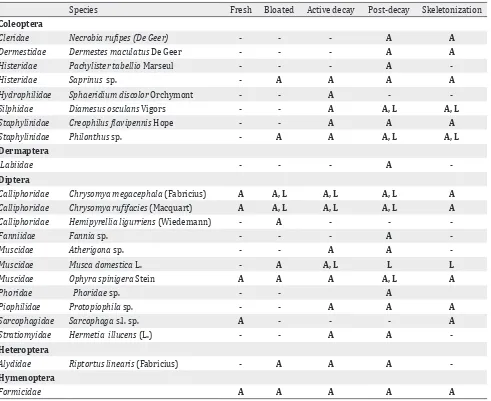
carcass IV. In carcass I, however, this stage lasted until day 10. Skeletonization was observed on day nine for carcasses II and III, on day seven for carcass IV, but on day 11 for carcass I. The different insects in relation to the decomposition stages are summarized in Table 4.
Fresh stage
C. megacephala and C. rufifacies visited the carcasses at the fresh stage from day one. Eggs
Stages of decomposition Days
1 2 3 4 5 6 7 8 9 10 11 12 13 14 15
Fresh
Bloating
Active decay
Post decay
Skeletonization
Table 3. Duration of decomposition stages in four different pig carcasses in Manado
Potassium cyanide (KCN) per oral Blunt stroke on its occipital area Cardiac puncture at Winangun Cardiac puncture at Batu Kota
of Chrysomya were foundon day one in natural
orifices (mouth, nose, ear, and anus) and the
stab wounds. Other Diptera which visited the
carcasses on the same day were Ophyra spinigera
and Sarcophagidae. Formicidae were found from
the first hour of post-mortem in the natural orifices, especially in those with fresh blood.
Bloated stage
Blowfly larvae invaded the carcasses’ faces
voraciously especially in carcasses II, III and IV. C. rufifacies and C. megacephala were
abundant. Musca domestica, Ophyra spinigera,
and Sarcophagidae were still found at this stage.
Sarcophagidae were observed only on intact skin,
and not on the natural orifices or wounds. Beetles
found at this stage were the predators Saprinus
and Philonthus on day three. Riptortus linearis
was found on intact abdominal skin on day three.
On day two, Formicidae left the natural orifices
invaded by Chrysomya maggots, but were still
found in the non-invaded areas.
Active decay stage
At the end of this stage, Diptera larvae had consumed most part of the body tissues leaving the skin, cartilages, bones, and the last part of muscles and guts. This stage lasted for two days in carcasses II, III and IV, and three days in carcass I.
Species Fresh Bloated Active decay Post-decay Skeletonization Coleoptera
Cleridae Necrobia rufipes (De Geer) - - - A A
Dermestidae Dermestes maculatus De Geer - - - A A
Histeridae Pachylister tabellio Marseul - - - A
-Histeridae Saprinus sp. - A A A A
Hydrophilidae Sphaeridium discolor Orchymont - - A -
-Silphidae Diamesus osculans Vigors - - A A, L A, L
Staphylinidae Creophilus flavipennis Hope - - A A A
Staphylinidae Philonthus sp. - A A A, L A, L
Dermaptera
Labiidae - - - A
-Diptera
Calliphoridae Chrysomya megacephala (Fabricius) A A, L A, L A, L A
Calliphoridae Chrysomya rufifacies (Macquart) A A, L A, L A, L A
Calliphoridae Hemipyrellia ligurriens (Wiedemann) - A - -
-Fanniidae Fannia sp. - - - A
-Muscidae Atherigona sp. - - A A
-Muscidae Musca domestica L. - A A, L L L
Muscidae Ophyra spinigera Stein A A A A, L A
Phoridae Phoridae sp. - - A
Piophilidae Protopiophila sp. - - A A A
Sarcophagidae Sarcophaga s.l. sp. A - - - A
Stratiomyidae Hermetia illucens (L.) - - A A
-Heteroptera
Alydidae Riptortus linearis (Fabricius) - A A A
-Hymenoptera
Formicidae A A A A A
Table 4. Insects found on four different pig carcasses in Manado in relation to the decomposition stages
Notes: A = adults; L = larvae
C. rufifacies and C. megacephala both larvae and
adults were abundant. Musca domestica, Ophyra
spinigera, and Sarcophagidae still visited the carcasses. Other Diptera such as Protopiophila,
Phoridae, and H. illucens were found at the end of
the active decay stage (day five).Beetles in this stage were Saprinus, Philonthus and Sphaeridium discolor. There were many Formicidae, predating on Chrysomya larvae which left the carcasses for pupation. R. linearis were still present.
Post-decay stage
During this stage, especially Dermestes consumed
the remaining tissue. Only very few adult C.
rufifacies and C. megacephala were observed. Other Diptera (Protopiophila, Phoridae and H. illucens) and R. linearis were still found in the early post-decay stage (day six). The beetles of the active decay stage were still present in this
stage as well as a few Formicidae. On day eight, one adult Labiidae was found in carcass II.
Skeletonization
In this stage, adult C. rufifacies and C.
megacephala of the new generation emerged from their pupae on days 12 and 13. However,
their larvae and Muscidae larvae were still
found under and around the carcasses.
Dermestes maculatus, Saprinus, Philontus, and
Necrobia rufipes were still present.
DISCUSSION
Decomposition
Generally, five stages of decomposition are
recognized.20-23 Payne24 reported six stages (fresh,
remains) in Clemson, South Carolina. Archer and Elgar25 reported only two stages (initial
decomposition and advanced decomposition)
from Victoria, Australia while Wang, et al26 in South China distinguished four stages.
In our study we observed five stages of
decomposition in all carcasses; the duration of the decomposition stages in the different carcasses was however not the same. Carcass I took 10 days for skeletonization, carcasses II and III only took eight days, and carcass IV six days. Carcass I had no wounds or external blood in contrast to carcass II (fresh blood that came out of the mouth), III and IV (stab wound); oviposition by
blowflies on carcass I was therefore much later.
The duration of decomposition (7-10 days)
resembled to those reported by Wang, et al26 in South China and Heo, et al27 at Tanjung Sepat,
peninsular Malaysia meanwhile Apichat, et al28 in
Phitsanulok, Thailand reported one day each for
fresh, bloated, and active decay stages, but three days for post-decay stage and a much longer
megacephala and C. rufifacies were abundantly
found in both adult and immature forms. C.
rufifacies maggots are predators on C. megacephala
maggots and their eggs are therefore deposited later than those of C. megacephala. These dominant
Chrysomya species are the same as those from similar experiments in other parts of tropical Asia as reported by Heo, et al27 in Malaysia, Apichat, et
al28 in Thailand and Wang, et al26 in South China.
Rearing of larvae and pupae in the laboratory
resulted in adult C. rufifacies and C. megacephala.
From both larva and pupa samples of carcasses II and III, the adults emerged one day earlier than those of carcass I. Adult Chrysomya at the study site emerged one day later than those in the laboratory. This faster development was caused by the higher
temperature in the laboratory (28°-30°C).
Heo, et al27 reported that the second generation blowflies appeared on day-12 and -13 around the
vegetation on the study sites, which are similar to our results. Larvae of C. megacephala and C.
rufifacies were still present underneath and around
the carcasses until skeletonization. According to Gunn30 adult flies also feed on the carcass during
the post-decay stage or skeletonization, but they did not initiate oviposition.
Hermetia illucens was originally restricted to the
New World but it has spread to other regions,
both tropical and temperate.29,31 This species is
not often found in corpses, and is reported to colonize carrion in an advanced stage: 20-30 days post-mortem21,30,32 In our study, adult Hermetia illucens were already found on day five. Diclaro
and Kauffman33 reported that H. illucens larvae
have six instars which needed 14 days, and a pupation period of two weeks. The rearing of soil around the carcasses collected on day 11, resulted in a second generation of Hermetia illucens on days 35-39. The growth of the larvae was in
accordance with the findings of Tomberlin, et al.34
Sarcophagidae larvae are reported from large carrion including forensic cases.29,35 In our study,
adult Sarcophagidae were found in small numbers from day 1 until the end of the study, but no maggots or pupae were observed. As these species apparently did not develop in the carcasses they cannot be used as forensic indicators. The adult
Sarcophagidae were not identified to species
level.
Larvae of Ophyra (instar two and three) are
predators of larvae Musca domestica and other Muscidae larvae.1,29,36 In our study, adult Ophyra spinigera was found from day 1-8, and their larvae on day 6-14 together with other Muscidae
larvae.
Adults of Musca domestica usually visit a corpse
after the arrival of blowflies and flesh flies in both the active and post-decay stages. House flies are
rarely attracted to fresh corpses, unless there is excreta or exposed gut present.1,29,30 In our study, Musca domestica was found from day 2-5, earlier than that observed by Byrd and Castner29, Gunn30,
and Smith1. The first adult fly emerged during
rearing in the laboratory on day seven which is similar to that of Gunn30 who stated that the life
cycle of Musca domestica takessix to eight days at a relatively high temperature.
In our study, adults of Phoridae were found on
day-5. Rearing in the laboratory of soil collected
which shows that development of the Phoridae occurred on the carcasses. Unfortunately the identification of Phoridae is notoriously difficult
which prevents their usage as a forensic indicator at the moment.
In general, adults of Protopiophila visit carcasses in an active state of decay and an early dry stage.
Larvae, as well as the adults, are necrophagous.29
In our study, adult Protopiophila were found on
day 4-6 but their larvae were not found on the
carcasses or during rearing of soil samples. Adult
Protopiophila were not identified to species level.
The beetles found in our experiment did feed on maggots or on parts of the carcass that were not consumed by maggots and therefore, they
arrived usually later than the blowflies. The
earliest Coleoptera visiting the carcasses were
Staphylinidae (day-3), followed by Saprinus sp.
(day-3, -5, -6, -10); these taxa are predators29
and their presence was in accordance with the
abundance of Diptera larvae on the carcasses.
Sphaeridium discolor was found on day-4 and -5.
Most Sphaeridium species are dung specialists
and they were probably attracted by the gut
contents of the carcasses. Dermestes maculatus
(day-6, -10) and Necrobia rufipes (day-10) were
observed later in the succession. Dermestes
species are usually found on dry carcasses in the wild. Almost all species are scavengers and feed on various types of dried animal tissue.29 Necrobia sp. have a special preference for fatty
tissues: their larvae are sometimes predacious.37
The Coleoptera larvae collected are belong to
Diamesusosculans and Staphylinidae.
Formicidae are social insects which are distributed all over the world. In tropical areas they are very abundant and can be found in all stages of decomposition as predators or omnivores
feeding on both body tissue and exudate fluids.1,30 The ants were observed in the first hour after
the start of the experiment and during the whole decomposition process in accordance with the statements of Aggarwal38 and Heo, et al39. Ants were usually found in the natural orifices, they
will leave these areas after being invaded by the
Chrysomya.
Adults of Riptortus linearis were found on day
2-6. These bugs landed on the intact abdominal skin of carcasses, and not on the natural orifices
or the open wounds, which suggest they are accidental visitors. All Alydidae, the family to which Riptortus belongs, are apparently plant-feeders1,40 but Payne41 reported three species of Alydidae feeding on a pig carcass in the USA. The
relation between Coreidae and carcasses seems
doubtful at the moment.
Labiidae are widely distributed, especially in
hot climates.42 Several species are predators on
other insects by seizing them with their forceps
or are omnivorous.29,40,42 On day eight one adult
Labiidae was found on carcass II. At this moment we consider this condition as an accidental visitor.
Literature study revealed a total of 11 Diptera
and eight Coleoptera species which were belong
to the potential carrion fauna of Sulawesi. Most of these taxa were different from those of the literature study, probably because the literature data were mainly based on the forest environments. The suburban Manado list contains mainly widespread taxa while the species from the literature list mainly endemic species in Sulawesi.
Five stages of decomposition (fresh, bloated, active decay, post-decay and skeletonization) could be
distinguished during the 15 days study. Chrysomya
megacephala and C. rufifacies dominated the other insects in all carcasses. Formicidae were the
first visitors and found during the whole study,
but abandoned areas which were invaded by
Chrysomya larvae. Although the insect succession patterns were generally similar in all carcasses, the duration of decomposition and the moment
on which the second generation of Chrysomya
adults appeared were not the same. Traumatic carcasses showed a shorter decomposition period and an earlier emergence of Chrysomya adults. Further experiments are needed to prove that these succession patterns are reliable enough to be used in court.
The morphological identification of C. megacephala
In conclusion,during our succession study on pig
carcasses in suburban Manado, 11 Diptera and 8
Coleoptera taxa were collected. Six of the collected
taxa were collected as larvae on the field location
and two additional taxa were reared to adults from
soil samples collected at the field location. The
fact that these species developed on the carcasses classify them as potential forensic indicators. From Chrysomya megacephala, C. rufifacies and
Hermetia illucens, the development from larvae to adults in relation to the ambient temperature is already known and they seem therefore to be primary candidates for the estimation of the post-mortem interval in suburban Sulawesi.
Acknowledgment
We would like to thank the International Affair Office of University of Sam Ratulangi Manado that facilitated Drs. Johannes Huijbregts from Naturalis Biodiversity Center, Leiden, Netherlands, to come to Manado, Indonesia.
Conflict of interest
The authors affirm no conflict of interest in this
study.
REFERENCES
1. Smith KGV. A manual of forensic entomology. London: The Trustees of the British Museum (Natural History);
1986.
2. Mégnin JP. La faune des cadavres: application de l’entomologie à la médecine légale, Encyclopédie Scientifique des Aide-Mémoire 101. Paris: Masson et Gauthiers-Villars; 1894. France.
3. Nuorteva P. Sarcosaprophagous insects as forensic indicators. In: Tedeschi CG, Eckert WG, Tedeshi LG, editors.
Forensic medicine, a study in trauma and environmental
hazards. Philadelphia: Saunders; 1977. p. 1072-95.
4. Leclercq M. Entomologie et médecine légale, datation de la mort. Collection de Médecine Légale et de Toxicologie
Médicale 108. Paris: Masson; 1978. France.
5. Knight WJ, Holloway JD. Insects and the rain forests of South East Asia (Wallacea). London: Royal Entomological Society of London; 1990.
6. Kurahashi H. A new species of Lucilia (Diptera,
Calliphoridae) from Sulawesi. Kontyu. 1988;56:144-7.
7. Kurahashi H. The genus Phumosia of Sulawesi, Indonesia, with descriptions of two new species (Diptera: Calliphoridae). Jpn J Sanit Zool. 1989;40(3):203-10. 8. Kurahashi H, Magpayo FR. Two new species of the genus
Chrysomya from Wallacea (Diptera, Calliphoridae).
Kontyu. 1987;55:71-9.
9. Kurahashi H, Selomo M. A new species of Calliphora
from Sulawesi, Indonesia (Diptera, Calliphoridae), Jpn J
Syst Ent. 1997;3:123-7.
10. Hanski I, Krikken J. Dung beetles in tropical forests in South-East Asia. In: Hanski I, Cambefort Y, editors. Dung beetle ecology. Princeton: Princeton University Press;
1991. p. 179-97.
11. Hanski I, Niemela J. Elevational distributions of dung
and carrion beetles in northern Sulawesi. In: Knight WJ, Holloway JD, editors. Insects and the rain forests of South East Asia (Wallacea). London: Royal Entomological
Society of London; 1990. p. 145-52.
12. Harvey ML, Gaudieri S, Villet MH, Dadour IR. A
global study of forensically significant calliphorids: implications for identification. Forensic Sci Int. 2008;177(1):66-76.
13. Kuijten PJ. Revision of the Indo-Australian species of the genus Phaeochrous Castelnau, 1840 (Coleoptera:
Scarabaeidae, Hybosorinae), with notes on the African
species. Zool Verh.1978;165:3-42.
14. Růžička J, Schneider J, Qubaiova J, Nishikawa M. Revision
of palaearctic and oriental necrophila Kirby & Spence,
part 2: subgenus Chrysosilpha Portevin (Coleoptera: Silphidae). Zootaxa. 2012;3261:33-58.
15. Sikes DS, Madge RB, Trumbo ST. Revision of Nicrophorus
in part: new species and inferred phylogeny of the nepalensis-group based on evidence from morphology
and mitochondrial DNA (Coleoptera: Silphidae:
Nicrophorinae). Invertebr Syst. 2006;20(3):305-65.
16. Sikes DS, Madge RB, Newton AF. A catalog of the
Nicrophorinae (Coleoptera: Silphidae) of the world.
Zootaxa. 2002;65:1-304.
17. Wells JD, Kurahashi H. A new species of Chrysomya (Diptera: Calliphoridae) from Sulawesi, Indonesia, with
a key to the Oriental, Australasian and Oceanian species.
Med Entomol Zool. 1996;47(2):131-8.
18. Singh B, Kurahashi H, Wells JD. Molecular phylogeny of the blowfly genus Chrysomya, Med Vet Entomol. 2011;25(2):126-34.
19. James MT. Family Calliphoridae. In: Delfinado MD, Hardy DE, editors. A catalog of the Diptera of the oriental region. Honolulu: University Press; 1977. p. 526-56. 20. Goff ML. Early postmortem changes and stages of
decomposition. In: Amendt J, Campobasso CP, Goff ML,
Grassberger M, editors. Current concepts in forensic
entomology. Dordrecht: Springer; 2010. p. 1-24. 21. Goff ML. Forensic entomology. In: Mozayani A, Noziglia
C, editors. The forensic laboratory handbook procedures
and practice. New York: Humana Press; 2011. p. 448-54.
22. Kreitlow KLT. Insect succession in a natural environment. In: Byrd JH, Castner JL, editors. Forensic entomology: The utility of arthropods in legal investigations. Boca
Raton: CRC Press; 2010. p. 251-6.
23. Sharanowski BJ, Walker EG, Anderson GS. Insect
succession and decomposition patterns on shaded and sunlit carrion in Saskatchewan in three different seasons. Forensic Sci Int. 2008;179(2-3):219-40.
24. Payne JA. A summer carrion study of the baby pig Sus scrofa Linnaeus. Ecology. 1965;46(5):592-602.
25. Archer MS, Elgar MA. Effects of decomposition on
carcass attendance in a guild of carrion-breeding flies. Med Vet Entomol. 2003;17(3):263-71.
26. Wang J, Li Z, Chen Y, Chen Q, Yin X. The succession and development of insects on pig carcasses and their
27. Heo CC, Mohamad AM, Ahmad FM, Jeffrey J, Kurahashi H, Omar B. Study of insect succession and rate of decomposition on a partially burned pig carcass in an oil palm plantation in Malaysia. Trop Biomed.
2008;25(3):202-8.
28. Apichat V, Wilawan P, Udomsak T, Chanasorn P, Saengchai N. A preliminary study on insects associated
with pig (Sus scrofa) carcasses in Phitsanulok, northern
Thailand. Trop Biomed. 2007;24(2):1-5.
29. Byrd JH, Castner JL, editors. Forensic Entomology: The utility of arthropods in legal investigations. 2nd ed. Boca
Raton: CRC Press; 2010.
30. Gunn A. Essential forensic biology. 2nd ed. Chichester
(UK): Wiley-Blackwell; 2009.
31. Tomberlin JK, Shepard DC. Lekking behavior of the black soldier fly (Diptera: Stratiomyidae). Fla Entomol.
2001;84(4):729-30.
32. Lord WD, Goff ML, Adkins TR, Haskell NH. The black soldier fly Hermetia illucens (Diptera: Stratiomyidae)
as a potential measure of human postmortem interval: observations and case histories. J Forensic Sci. 1994;39(1):215-22.
33. Diclaro JW, Kaufman PE. Black soldier fly Hermetia illucens Linnaeus (Insecta: Diptera: Stratiomyidae).
EENY. 2009;461:1-3.
34. Tomberlin JK, Sheppard DG, Joyce JA. Selected life-history
traits of black soldier flies (Diptera: Stratiomyidae) reared on three artificial diets Ann Entomol Soc Am. 2002;95(3):379-86.
35. Villet MH. African carrion ecosystems and their insect communities in relation to forensic entomology. Pest Technol. 2011;5(1):1-15.
36. Vibe-Petersen S. Development, survival and fecundity of the urine fly Scatella (Teichomyza) fusca and predation by the black dumpfly, Hydrotaea aenescens. Entomol
Exp Appl. 1998;87(2):157-69.
37. Busvine JR. Insects and Hygiene. The biology and control
of insect pests of medical and domestic importance. 2nd
ed. London: Methuen; 1966.
38. Aggarwal AD. Estimating the post-mortem interval with
the help of entomological evidence [Thesis]. Medical
College, Patiala. Baba Farid University of Health Sciences
Faridkot, India; 2005.
39. Heo CC, Mohamad AR, Rosli H, Nurul Ashikin A, Chen CD, John J, et al. Ants (Hymenoptera: Formicidae)
associated with pig carcasses in Malaysia. Trop Biomed.
2009;26(1):106-9.
40. Kalshoven LGE. Pests of crops in Indonesia. Jakarta: Ichtiar Baru; 1981.