http://www.sciencedirect.com/science?_ob=ArticleURL&_udi=B6T43-48GF2WT-Comparison of hybrid and purebred in vitro-derived cattle
embryos during in vitro culture
A. Boedionoa, b, T. Suzukib and R. A. Godke, c
a
Faculty of Veterinary Medicine, Bogor Agricultural University, Bogor, Indonesia b
United Graduate School of Veterinary Sciences, Yamaguchi University, Yamaguchi, Japan c
Department of Animal Science, Louisiana State University Agricultural Center, 105 J.B. Francioni Hall, Baton Rouge, LA 70803, USA
Frozen-thawed spermatozoa collected from a beef bull (Japanese Black) were used for in vitro fertilization (IVF) of matured oocytes obtained from dairy (Holstein) and beef (Japanese Black) females. Embryos were examined for fertilization, cleavage rate, interval between insemination and blastocyst production (experiment I), total cell number per embryo and sex ratio during blastocyst formation (experiment II), and blastocyst production rate of zygotes that developed to 2-, 4-, and 8-cell stages at 48 h post-fertilization (experiment III). Fertilized oocytes were cultured in vitro on a cumulus cell co-culture system. The fertilization and cleavage rate of oocytes groups were similar, however, the blastocyst production rate was greater (P<0.05) in hybrid than from purebred embryos (27% versus 20%). Development of blastocysts produced from hybrid embryos developed at a faster rate than blastocysts produced from the straightbred embryos. In hybrid embryos, blastocyst production was significantly greater on day 7 (56%) and gradually decreased from 20% on day 8 to 17% on day 9. In contrast, blastocyst production rate from the purebred embryos was lower on day 7 (17%), increasing on day 8 to 59% and then decreased on day 9 to 24%. The total number of cells per embryo and sex ratio of in vitro-produced blastocysts were not different between hybrid and purebred embryos. The number of blastocysts obtained from embryos at the 8-cell stage of development by 48 h post-fertilization (94%) was greater (P<0.01) than the number of zygotes producing blastocysts that had developed to the 4-cell stage (4%) and the 2-cell stage (2%) during the same interval. These results show that the blastocyst production rate and developmental rate to the blastocyst stage were different between hybrid and purebred embryos, and that almost all of the in vitro-produced blastocysts were obtained from zygotes that had developed to the 8-cell stage 48 h post-fertilization.
Author Keywords: Bovine; Embryo; In vitro fertilization; In vitro development; Sex ratio
1. Introduction survival of the conceptus. During this time, the zygote goes through a series of cellular divisions and differentiation events leading to blastocyst formation. It is well established that the developmental rate of the mammalian zygote depends on genetic and environmental factors. In the mouse, after fertilization, embryos harvested from some strains develop earlier than those from other strains (McLaren and Bowman, 1973 and Shire and Whitten, 1980). These studies and others have verified that the maternal genotype factors can affect the rate of early embryo development.
Embryo sexing can now be executed by an easy noninvasive procedure (Avery et al., 1991) and thus, the alteration of the sex ratio at birth in livestock could be of great importance to agriculture. For meat production, a high percentage of bull calves is advantageous, because males grow faster, and are efficient at carcass production. In contrast, heifer calves are a necessity for commercial milk production units and replacement females.
http://www.sciencedirect.com/science?_ob=ArticleURL&_udi=B6T43-48GF2WT-The objectives of the present study were to investigate differences in oocytes collected from dairy and beef breeds and fertilized with spermatozoa obtained from the same beef breed bull on (a) fertilization, cleavage rate and interval between fertilization (in vitro insemination procedure) and blastocyst production; (b) the total cell number per embryo and embryo sex ratio; and (c) the blastocyst rate of IVF-produced zygotes that develop to 2-, 4-, and 8-cell stages by 48 h post-fertilization.
2. Materials and methods
2.1. In vitro maturation (IVM)
Ovaries from dairy (Holstein) and beef (Japanese Black) cows were collected separately from a nearby abattoir and transported (32–35 °C) to the laboratory in 0.9% physiological saline solution immediately following collection. Oocytes were aspirated from small antral follicles (2– 5 mm) using a 5 ml syringe and a 18 g needle and washed in modified PBS (mPBS; Embryotech, Nihonsenyaku, Fukushima, Japan). The oocytes with intact cytoplasm and surrounded by cumulus cells over more than one-third of their surface area were washed two times in maturation “medium”.
This maturation medium consisted of TCM-199 (Earle‟s salt, Gibco, Grand Island, NY, USA) supplemented with 5% superovulated cow serum (SCS) collected on day 7 of the estrous cycle (Boediono et al., 1994), 0.01 mg/ml of follicle stimulating hormone (FSH; Denka Pharmaceutical, Kawasaki, Japan) and 50 μg/ml of gentamicin sulfate (Sigma, St. Louis, MO, USA). Good quality oocytes (100–200 per replicate) were then matured in maturation medium (2.5 ml) for 20–22 h in a 35 mm polystyrene culture dish (Falcon 1008, Becton Dickson, Oxnard, CA, USA) overlaid with mineral oil (E.R. Squibb & Son, Princeton, NJ, USA) at 38.5 °C under 5% CO2 in atmospheric air.
2.2. In vitro fertilization
superovulated cow serum, 5 μg/ml of insulin (Wako Pure Chemical Industries, Osaka, Japan) and 50 μg/ml of gentamicin sulfate, then was overlaid with 0.5 ml of warmed medical-grade mineral oil.
2.3. In vitro embryo culture
Adherent cumulus cells surrounding the embryos were removed by repeated pipetting 48 h post-fertilization. The cumulus cells adhering to the surface of the culture dish were not disrupted, and embryos were cultured on this somatic cell monolayer. The culture medium was replaced with fresh medium 96 h post-fertilization.
3. Experimental procedure
3.1. Experiment I
In this experiment, 10% of sperm-exposed ova were randomly selected for evidence of in vitro fertilization. At 18 h post-fertilization the ova were removed from the culture dish. The cumulus cells were completely removed by repeated pipetting in culture medium containing 150 U/ml of hyaluronidase (Sigma). The ova were then mounted, fixed for 72 h in 25% acetic-alcohol, stained with 1% aceto-orcein and cleared with aceto-glycerol. The oocytes were then examined under a phase-contrast microscope to determine the presence of pronuclei.
The number of zygotes that had cleaved (2-, 4-, and 8-cell stages) from the remaining groups of sperm-exposed ova was recorded 48 h post-fertilization (see Fig. 1). Blastocyst development was determined on days 7, 8, and 9 post-fertilization. Once the blastocysts were identified, they were removed from the culture dish to reduce the chance for counting errors. Embryo morphological assessments were made at 24 h intervals from the 1- to the 8-cell stages and then again at 24 h intervals on days 7 to 9 of in vitro culture (seven times). In this experiment, a total of 956 hybrid and 899 purebred beef cattle oocytes were harvested from seven replicates for exposure to IVF procedures.
Full-size image (103K)
http://www.sciencedirect.com/science?_ob=ArticleURL&_udi=B6T43-48GF2WT-3.2. Experiment II
In this experiment, expanded excellent quality in vitro-produced blastocysts obtained from both hybrid and purebred beef cattle were used for either embryo sexing or for evaluating the total number of cells per embryo. A total of 127 randomly selected embryos (n=66 hybrid and n=61 purebred) were processed and chromosomes plates prepared by the method described by Iwasaki and Nakahara (1990), with minor modifications. Briefly, the embryos were cultured in a medium containing 0.04 μg/ml of colcemid (Gibco) for 4 h and then suspended in 0.9% sodium citrate solution for a 20 min interval. The embryos were then fixed in a distilled water:acetic acid:methanol:sodium citrate solution (2:4:6:9) for 5 min followed by distilled water:acetic acid:methanol (1:2:3) for 1–2 min. The fixed embryos were placed on a glass slide followed by covering them with a film of acetic acid. Chromosome preparations were stained in 5% Giemsa (E. Merck, Darmstadt) at pH 6.8 for 20 min and evaluated at different magnification under a phase-contrast microscope. The X and Y chromosomes were distinguishable by morphology; the X chromosome being a large submetacentric, the Y chromosome being a small metacentric and the remaining autosomes telocentric.
3.3. Experiment III
In an effort to compare the rate of blastocyst production in the second part of this experiment, developing cleaved zygotes were divided into 2-, 4-, and 8-cell stages at 48 h post-fertilization. Each of these morphological groups was cultured separately at 38.5 °C under 5% CO2 in air to evaluate further in vitro development. Blastocyst development rates from each group were monitored until day 9 post-fertilization. A total of 1110 embryos obtained from eight replicates of IVM, IVF and in vitro culture procedures were evaluated for blastocyts development in this experiment.
3.4. Statistical analyses
Data from experiments I and II were analyzed by Chi-square analysis. The blastocyst production rates of hybrid and purebred beef cattle embryos in experiment III were analyzed by an analysis of variance (ANOVA) using a standard SAS program (SAS, 1987). In this study, mean values were considered to be significant different when P was set at the either the P<0.05 or <0.01 level.
4. Results
4.1. Experiment I
and the incidence of polyspermy (>2 pronuclei) were found not to be different (P>0.05) between the oocytes form two different genetic origins (Table 1).
Table 1. Pronuclear status of both dairy and beef cattle ova in 100 μl droplets 18 h after in vitro exposure to frozen-thawed sperm from a fertile beef bull
The in vitro development of hybrid and purebred IVF-derived beef embryos at the 2-, 4-, 8-cell, and blastocyst stages is presented in Fig. 1. The embryo developmental rates to the 2-, 4-, and 8-cell stages did not differ (P>0.05) between the hybrid and purebred beef embryos (Table 2). However, blastocyst production was significantly greater (P<0.05) for the hybrid embryos than for the corresponding purebred embryos.
Table 2. Developmental stage of hybrid vs. purebred IVF-produced beef embryos during in vitro culture
Within columns with different superscript letters (a, b) are significantly different (P<0.05).
Blastocyst production was evaluated on days 7, 8, and 9 post-fertilization (Fig. 2). Based on the total number of blastocysts produced during in vitro culture, blastocyst production on day 7 was greater (P<0.01) for hybrid than for those from purebred embryos, while on day 8, blastocyst production from purebred was greater (P<0.01) than production from hybrid embryos. Correspondingly on day 9 of in vitro culture, blastocyst production from purebred was greater (P<0.05) than blastocyst production from hybrid embryos.
http://www.sciencedirect.com/science?_ob=ArticleURL&_udi=B6T43-48GF2WT-Fig. 2. Development rate of hybrid and purebred beef in vitro-produced embryos (n=252 hybrid and n=184 purebred embryos) to blastocyst stage on days 7, 8, and 9 post-fertilization. Different letters within days of in vitro culture are significantly different at the P<0.01 level (a,b); and
P<0.05 (c,d) level.
When the hybrid embryos were evaluated by day of culture as a group, the overall blastocyst production was highest on day 7 (56%, 140/252), was lower on day 8 (27%, 69/252), and was the lowest on day 9 post-fertilization (17%, 43/252). Correspondingly, the overall blastocyst production by day of culture the purebred embryos as a group was the lowerst on day 7 (17%, 52/184) was the highest on day 8 (59%, 108/184), and then subsequently lower on day 9 of in vitro culture (24%, 44/184).
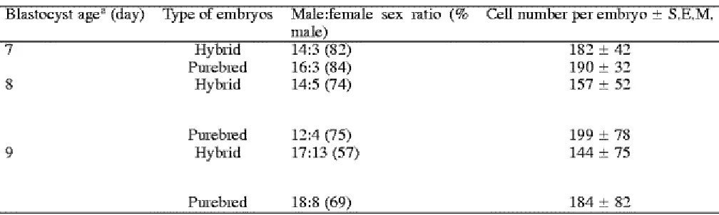
4.2. Experiment II
The sex ratio (male:female) of 127 on day 7 IVF-produced blastocysts obtained from hybrid and purebred embryos was 82:13 and 84:16 (82 and 84% males), respectively (Table 3). The sex ratio became less male-genotype dominant for the hybrid and the purebred embryos developing to blastocysts on day 8 (76:26 and 75:26) and on day 9 (57:43 and 69:31), respectively, of in vitro culture.
Table 3. Sex ratio by blastocytst age and total number of cells per embryo of hybrid and purebred beef cattle embryos developing during in vitro culture
4.3. Experiment III
The development rates of in vitro-produced embryos, classified by cell morphology (2-, 4-, and 8-cell stage embryos) 48 h post-fertilization, are shown in Table 4. At 48 h post-fertilization, 69.5% (772/1110) of the in vitro-matured oocytes had cleaved and developed to 8-cell stage in culture, 16% (181/1110) developed to the 4-cell stage and 14% (157/1110) developed to the 2-cell stage. From the total of 327 bovine blastocysts produced in this experiment, 94% (308/327) were derived from the 8-cell stage embryos at 48 h post-fertilization and only 6% (19/327) resulted from the 2- and 4-cell stages during the same interval 48 h post-fertilization.
Table 4. Blastulation rates for IVF-derived bovine embryos that had developed to 2-, 4-, or 8-cell stage at 48 h post-fertilization
Within columns with different letters (a,b) are significantly different (P<0.05).
5. Discussion
The development of in vitro-produced mammalian embryos is a complex process and clearly under the influence of genotype and environment. In the present study, the ovaries harvested from dairy and beef breed females were used as oocyte donors and frozen-thawed semen from a single same breed beef bull was used for in vitro fertilization. The proportion of oocytes reaching metaphases I and II, formation of two pronuclei and evidence of polyspermy did not differ between the two oocyte groups. These results suggest that the ability of oocytes to undergo nuclear and cytoplasmic maturation after their removal from the follicle and then fertilization in vitro, was not affected by maternal genotype.
http://www.sciencedirect.com/science?_ob=ArticleURL&_udi=B6T43-48GF2WT-In the present study, the highest blastocyst production by day of in vitro culture (the total embryos developing to blastocysts) for hybrid embryos was 56% on day 7 post-fertilization. Peak blastocyst production by day of in vitro culture of 59% was reached 1 day later (day 8 post-fertilization) than the hybrid embryos. This difference in developmental rate pattern between the hybrid and the purebred embryos was likely due to the difference in maternal genotype on pre-hatched embryonic development. These results are in agreement with those reported in a previous study with mice (Shire and Whitten, 1980), where embryos from different strains develop earlier than those from other mouse strains.
The sex ratio and total cell number per embryo of blastocysts at days 7, 8, and 9 of in vitro culture were not affected by maternal genotype. Van Soom et al. (1994) did note that sex ratio of in vitro-produced bovine embryos (on day 7) after transfer to a recipient was 71.4% the male gender. In the present study, the sex ratio of hybrid and purbred embryos on day 7 was 82 and 84% for the male genotype, respectively. The portion of the male genotype of the sex ratio was lower on days 8 and 9 of in vitro culture. In an earlier study, Boediono et al. (1995b) had reported a sex ratio of approximately 50:50 for in vitro-produced bovine embryos was obtained up to day 11 of in vitro culture, even when the total number of cells per embryo had decreased. The results in the present study, however, are in agreement with those from cattle in previous reports ( Avery et al., 1991; Avery et al., 1992; Xu et al., 1992 and Van Soom et al., 1994) that male embryos produced in vitro develop at a faster rate than female embryos. At present, the reason(s) for more rapid development of different genotypes in various in vitro-produced embryos is not clearly understood.
Sexual differences in embryo development appear starting either at the time of in vitro fertilization or cell divisions thereafter, with males progressing more rapidly than females (Mittwoch, 1989 and Boediono et al., 1995b). These results suggest that maternal genotype of the oocyte would not be expected to have a primary influence on sex-mediated development of the embryo in vitro. It should be noted that Grisart et al. (1995) reported that bovine blastocysts produced in serum-free oviduct cell-conditioned medium did not exhibit an altered sex ratio pattern, as reported herein. We propose that it should not be overlooked that the properties and/or components of the culture medium could influence the sex ratio of IVF-derived bovine embryos.
The cell number per embryo of in vitro-produced embryos on day 9 post-fertilization in this study was higher than expected (144 cells for the hybrid embryos and 184 cells for the purebred embryos). Although it would be tempting to use “good quality” day 9 in vitro-produced blastocysts for embryo transfer, preliminary results from our laboratory indicates that the pregnancy rates will be very low.
likely attributed to difference in laboratory procedures and the influence of culture conditions, such as the serum supplementation (type, source and/or the amount) added to the culture medium and whether or not a monolayer co-culture is used to enhance embryo development.
It should be noted that both superovulated cow serum and a homologous cumulus cell monlayer were used during the culture the IVF-produced embryos in the present study. Boediono et al. (1994) has previously reported the use of superovulated cow serum in the maturation and the culture media was more effective than the use of fetal calf serum for producing IVF-derived bovine embryos. Also, culturing of in vitro-produced bovine embryos with homologous cumulus cells has been reported to be excellent environment for embryonic development ( Goto et al., 1988; Kajihara et al., 1990 and Zhang et al., 1995).
In conclusion, the maternal genotype of the oocytes had no obvious effect on early in vitro development of bovine embryos, but had an effect on rate of blastocyst production. The sex ratio and the total number of cells per embryo of the groups of in vitro-produced blastocysts in this study were not different between hybrid and purebred beef cattle embryos. Finally, it should not be overlooked that most of blastocysts produced by in vitro fertilization in this study resulted from zygotes that had developed to the 8-cell stage by 48 h post-fertilization.
Acknowledgements
Approved for publication by the Director of the Louisiana Agricultural Experiment Station as Manuscript No. 03-18-0903. This research was funded, in part, by Federal Regional Project W-171.
References
Aoyogi, Y., Fukui, Y., Iwazumi, Y., Urakawa, M. and Ono, H., 1990. Effect of culture system on development of in vitro fertilized bovine ova into blastocysts. Theriogenology 34, pp. 749–759. Avery, B., Madison, V. and Greve, T., 1991. Sex and development in bovine in vitro fertilized embryos. Theriogenology 35, pp. 953–963. Abstract | PDF (631 K) | View Record in Scopus | Cited By in Scopus (75)
Avery, B., Jorgensen, C.B., Madison, V. and Greve, T., 1992. Morphological development and sex of bovine in vitro-fertilized embryos. Mol. Reprod. Dev. 32, pp. 265–270. Full Text via CrossRef | View Record in Scopus | Cited By in Scopus (63)
Boediono, A., Takagi, M., Saha, S. and Suzuki, T., 1994. The influence of day-0 and day-7 superovulated cow serum during in vitro development of bovine oocytes. Reprod. Fertil. Dev. 6, pp. 261–264. Full Text via CrossRef | View Record in Scopus | Cited By in Scopus (16)
Boediono, A., Rajamahendran, R., Saha, S., Sumantri, C., Suzuki, T., 1995a. Effect of the presence of a CL in the ovary on oocyte number, cleavage rate and blastocyst production in vitro in cattle. Theriogenology 43, 169 (abstract).
Boediono, A., Sumantri, C., Saha, S., Suzuki, T., 1995b. Blastocyst production and sex ratio of in vitro produced bovine embryos. Proc. World Vet. Congr. 89 (abstract).
Brackett, B.G. and Oliphant, G., 1975. Capacitation of rabbit spermatozoa in vitro. Biol. Reprod.
http://www.sciencedirect.com/science?_ob=ArticleURL&_udi=B6T43-48GF2WT-Goto, K., Kajihara, Y., Kosaka, S., Koba, M., Nakanishi, Y. and Ogawa, K., 1988. Pregnancies after co-culture of cumulus cells with bovine embryos derived from in vitro fertilization of in vitro matured follicular oocytes. J. Reprod. Fertil. 83, pp. 753–758. Full Text via CrossRef | View Record in Scopus | Cited By in Scopus (85)
Grisart, B., Massip, A., Collette, L. and Dessy, F., 1995. The sex ratio of bovine embryos produced in vitro in serum-free oviduct cell-conditioned medium is not altered. Theriogenology
43, pp. 1097–1106. Article | PDF (691 K)
Iwasaki, S.A. and Nakahara, T., 1990. Cell number and incidence of chromosomal anomalies in bovine blastocysts fertilized in vitro followed by culture in vitro or in vivo in rabbit oviducts.
Theriogenology 33, pp. 669–675. Abstract | PDF (509 K) | View Record in Scopus | Cited By in Scopus (18)
Kajihara, Y., Kometani, M., Kobayashi, S., Shitanaka, Y., Koshiba, T., Hishiyama, K., Shiraiwa, K., Goto, K., 1990. Pregnancy rate and births after co-culture of cumulus cells with bovine embryos derived from in vitro fertilization of in vitro matured follicular oocytes. Theriogenology 32, 264 (abstract).
Kroetsch, T.G., Stubbings, R.B., 1992. Sire and insemination dose does effect in vitro fertilization of bovine oocytes. Theriogenology 37, 240 (abstract).
McLaren, A. and Bowman, P., 1973. Genetic effects on the timing of early development in the mouse. J. Embryol. Exp. Morphol. 30, pp. 491–498. View Record in Scopus | Cited By in Scopus (25)
Mittwoch, U., 1989. Sex differentiation in mammals and tempo of growth: probabilities vs. switches. J. Theor. Biol. 137, pp. 445–455. Abstract | PDF (602 K) | View Record in Scopus | Cited By in Scopus (18)
Plante, L., King, W.A., 1992. Effect of time to first cleavage on hatching rate of bovine embryos in vitro. Theriogenology 37, 274 (abstract).
Prokofiev, M.I., Ernst, L.K., Suraeva, N.M., Lagutina, I.S., Udavlennikova, N.N., Kesyan, A.Z. and Dolgohatskiy, A.I., 1992. Bovine oocyte maturation, fertilization and further development in vitro and after transfer into recipients. Theriogenology 38, pp. 461–469. Abstract | PDF (474 K) | View Record in Scopus | Cited By in Scopus (4)
SAS User‟s Guide: Statistics, 1987. SAS Institute Inc., Cary, NC.
Shioya, Y., Kuwayama, M., Fukushima, M. and Iwasaki, S., 1988. In vitro fertilization and cleavage capability of bovine follicular oocytes classified by cumulus cells and matured in vitro.
Theriogenology 30, pp. 489–496. Abstract | PDF (416 K) | View Record in Scopus | Cited By in Scopus (33)
Shire, J.G.M. and Whitten, W.K., 1980. Genetic variation in the timing of the first cleavage in mice: effect of maternal genotype. Biol. Reprod. 23, pp. 369–376. Full Text via CrossRef | View Record in Scopus | Cited By in Scopus (15)
Sumantri, C., Boediono, A., Ooe, M., Saha, S. and Suzuki, T., 1997. Fertility of sperm from a tetraparental chimeric bull. Anim. Reprod. Sci. 46, pp. 35–45. Article | PDF (869 K) | View Record in Scopus | Cited By in Scopus (6)
Xu, K.P., Yadav, B.R., King, W.A. and Betteridge, K.J., 1992. Sex related differences in developmental rates of bovine embryos produced and cultured in vitro. Mol. Reprod. Dev. 31, pp. 249–252. Full Text via CrossRef | View Record in Scopus | Cited By in Scopus (90)
Younis, A.I. and Brackett, B.G., 1991. Importance of cumulus cells and insemination interval for development of bovine oocytes into morulae and blastocysts in vitro. Theriogenology 36, pp. 11– 21. Abstract | PDF (632 K) | View Record in Scopus | Cited By in Scopus (13)