ISSN 0854-8587
In Vitro
Selection of Peanut Somatic Embryos on Medium Containing
Culture Filtrate of
Sclerotium rolfsii
and Plantlet Regeneration
YUSNITA1‡, WIDODO2, SUDARSONO1*
1Department of Agronomy and Horticulture, 2Department of Plant Pest and Disease, Faculty of Agriculture, Bogor Agricultural University, Jalan Meranti-Kampus IPB Darmaga, Bogor 16680
Diterima 13 April 2004/Disetujui 26 Mei 2005
Attempts to identify somaclonal variants of peanut with resistance to Sclerotium stem rot disease due to infection of S. rolfsii were conducted. The objectives of this study were to develop in vitro selection method using culture filtrates of S. rolfsii, identify culture filtrate-insensitive somatic embryo (SE) of peanut after in vitro selection and regenerate peanut R0 lines originated from culture filtrate-insensitive SE. To achieve these objectives, peanut embryogenic tissues were cultured on selective medium containing various concentrations of S. rolfsii culture filtrates and sublethal concentration of the filtrates. Medium containing sublethal level of S. rolfsii culture filtrates was used to identify culture filtrate-insensitive SE of peanut. Subsequently, the selected SEs were germinated, plantlets were regenerated and preliminary tested against S. rolfsii. Results of the experiments showed that addition of S. rolfsii culture filtrates into medium for inducing peanut somatic embryos drastically reduced their growth and proliferation. S. rolfsii culture filtrates at 10% concentration has significantly reduced the number of proliferated SE per explant. However, sublethal level was achieved at 30% of culture filtrates concentration. Responses of five peanut cultivars against 30% of culture filtrates were similar, indicating they were similar in their susceptibility against S. rolfsii. A number of culture filtrate-insensitive SE were identified after culturing 1500 clumps of embryogenic tissue of peanut cv. Kelinci for three consecutive passages on medium containing 30% of culture filtrates. Germination of selected SE and regeneration of plantlet from culture filtrate-insensitive SE resulted in 50 peanut R0 lines. These lines have been grown in the plastic house and produced normal seeds for further evaluation. Results of S. rolfsii inoculation indicated the existence of chimera for insensitivity against S. rolfsii.
___________________________________________________________________________
_________________
‡Alamat kini: Department of Agronomy, Faculty of Agriculture,
University of Lampung, Bandar Lampung 35145
∗ ∗ ∗ ∗
∗Penulis untuk korespondensi, Tel./Fax. +62-251-629353,
E-mail: [email protected]
INTRODUCTION
Sclerotium rolfsii, the causal pathogen of Sclerotium stem rot disease, could be a problem in upland cultivation of peanut in Indonesia since it is a soil-borne fungus and is difficult to control. Eradication of this pathogen is difficult once introduced in a new areas due to its ability to form sclerotia and to infect many temporary hosts. Moreover, chemical control of this pathogen proved to be ineffective (Punja 1985). Development of more tolerance peanut cultivar against S. rolfsii is a better alternative for controlling Sclerotium stem rot in peanut.
Sclerotium rolfsii secreted large amount of oxalic acid, a phytotoxin responsible for killing tissues, prior to mycelial growth on the infected plant (Porter et al. 1982; Backman 1984; Punja 1985). This phytotoxin could be used to develop S. rolfsii tolerance peanut lines through in vitro
selection. However, the effectiveness of S. rolfsii phytotoxin for in vitro selection has not been reported. Phytotoxins from
Fusarium graminearum (isolate no. 122216) and F. culmorum
(isolate no. 12375 or 12551) have been used as selective agents for in vitro selection to develop Fusarium-resistance wheat (Ahmed et al. 1996).
Somaclonal variation has been recognized as an alternative route to obtain genetic variability in various crops, such as potato (Shepard 1981), sugarcane (Ramos-Leal et al. 1996), banana (Matsumoto et al. 1995), wheat (Ahmed et al. 1996), and mango (Jayasankar & Litz 1998). Some of variant lines generated through somaclonal variation exhibited novel characters including disease resistance (Ahmed et al. 1996; Matsumoto et al. 1995); therefore, they may be used as donor parents for crop improvement.
The frequency of obtaining somaclonal variants with disease resistance or any other useful characters may be increased by induction of genetic variability and in vitro
selection (Scowcroft et al. 1985). In vitro selection relies on the availability of an efficient plant regeneration system, selective agents for selecting plant cells and tissues cultured
in vitro, and a positive correlation between phenotypic expression at cellular and whole plant levels.
transposable element activation (Phillips et al. 1990). Epigenetic changes have also been considered as the responsible factors for a number of phenotypic variation (Larkin & Scowcroft 1981; Widoretno et al.2003).
Attempts to identify somaclonal variants of peanut with resistance to Sclerotium stem rot due to infection of S. rolfsii
were conducted. The objectives of this study were to develop
in vitro selection method using S. rolfsii culture filtrates as selective agents, identify culture filtrate-insensitive somatic embryo (SE) of peanut after in vitro selection and regenerate peanut plant lines originated from culture filtrate-insensitive SE.
MATERIALS AND METHODS
Induction of SE from Embryonic Leaflet. Effective procedures for inducing SE of peanut and its proliferation have previously been reported (Edy 1998; Sulichantini 1998). Somatic embryos of peanut were initiated by culturing embryonic leaflets isolated from mature seeds of five peanut cultivars (Badak, Biawak, Kelinci, Singa, and Zebra) on medium for SE induction (P16 medium). The P16 medium (Sulichantini 1998; Edy 1998) consisted of MS basal salts (Murashige & Skoog 1962), B5 vitamines compositions (Gamborg et al. 1968), 2% sucrose, 16 µM picloram, and 8 g/l agar. The pH of medium was adjusted to 5.8 before adding agar and strerilization.
Leaflet cultures were maintained in the dark under 26 oC
for 4-6 weeks or until they developed SE. To maintain proliferation of peanut SE, clumps of embryogenic tissues with 3-4 SE were subcultured into fresh P16 medium every four weeks. SE cultures were maintained their regeneration capacity for more than one year after initiation and peanut embryogenic tissues that have been proliferated for one year were used in this study.
Sclerotium rolfsii Isolation and Culture Filtrates
Preparation. The virulent strains of S. rolfsii were originated from Darmaga Experiment Station, Bogor Agricultural University, Bogor, Indonesia. Sclerotium rolfsii infected peanut stems were washed in tap water, surface sterilized, and plated on potato dextrose agar (PDA) medium. The fungal was isolated by mycelia tip culture grown on PDA at room temperature and maintained by transferring mycelial plugs into fresh PDA medium every 4 weeks.
Sclerotium rolfsii culture filtrates were prepared by transferring mycelial plugs onto medium consisted of MS basal salts, B5 vitamines composition, 3% sucrose and 8 g/l agar. Fungal cultures were incubated under 26 oC until they
formed sclerotium bodies (approximately 14 days). Cultures were autoclaved at 121 oC under 1.5 kg/cm2 pressure for 20
minutes to destroy the fungi. Liquefied medium presumably containing S. rolfsii heat stable toxic metabolites was sieved to remove mycelial debris and used as selective agents for in vitro selection.
Sublethal Level of S. rolfsii Culture Filtrates. Sublethal level of S. rolfsii culture filtrates was determined by culturing clumps of embryogenic tissues with 3-4 SE of peanut cv. Kelinci on selective - P16 medium and by evaluating their
proliferation. Selective medium was prepared by incorporating various concentrations of S. rolfsii culture filtrates (0, 10, 20, 30, 40, or 50%) into P16 medium.
At least fifty clumps of embryogenic tissues with 3-4 SE of peanut cv. Kelinci were cultured on five or more culture vials (10 clumps per vial) containing selective medium with various concentration of S. rolfsii culture filtrates. Cultures were maintained under dark condition for four weeks. Sublethal level of culture filtrate concentration on selective medium was determined based on the ability of selective P16 medium to suppress peanut SE proliferation of more than 95%.
Peanut Cultivars Responses against 30% of Culture Filtrates. Effectiveness of selective medium containing 30% of culture filtrates to suppress proliferation of SE of five peanut cultivars (Badak, Biawak, Kelinci, Singa, and Zebra) was further evaluated. Evaluation was conducted in two consecutive passages of exposure on selective P16 medium containing 30% of S. rolfsii culture filtrates. Fifty clumps of embryogenic tissues with 3-4 SE for each peanut cultivar were cultured on 10 culture vials (5 clumps per vial) containing P16 medium with 30% of S. rolfsii culture filtrates. Proliferated somatic embryos were subcultured once into fresh selective P16 medium after four weeks. All cultures were maintained in the dark and proliferation of SE after two consecutive passages on selective P16 medium was determined.
In Vitro Selection of SE and Plantlet Regeneration. To identify S. rolfsii culture filtrates-insensitive SE of peanut, 1500 clumps of embryogenic tissues with 3-4 SE of peanut cv. Kelinci were subjected against in vitro selection using selective P16 medium containing 30% of S. rolfsii culture filtrates. The proliferated SE was subcultured for three passages into fresh selective P16 medium every four weeks. The percentage of SE proliferation, number of SE/explant and total number of SE were counted at each passage of subculture.
After the third passage, S. rolfsii culture filtrate-insensitive SE of peanut were rescued by culturing them on P16 medium without S. rolfsii culture filtrates for two consecutive passages of 4 weeks culture period. The recovered somatic embryos were transferred into basal MS medium containing 2 g/l of activated charcoal (MSAC medium) for SE maturation and germination. Germinated SE was transferred into MS medium containing a combination of BAP (2 mg/l) and Kinetin (2 mg/l) for inducing epicotyls elongation and after 3-4 weeks they were transferred back into MSAC for further epicotyls development.
Response of the Regenerated Peanut Plants against
S. rolfsii.Regenerated peanut plants (R0 plants) from culture filtrate-insensitive SE that have successfully been grown in the plastic-house were evaluated for their response against
S. rolfsii in detached leaf-dual culture method as described by Pratt (1996). In this evaluation, selected leaflets and leaves (with four fully opened leaflets) from each of the R0 plant were inoculated with S. rolfsii and incubated for six days on water-agar medium under semi-in vitro condition. During incubation period, damage of the tested leaves and growth of the fungus on leaf tissues were recorded. Leaflets and leaves of peanut plants grown from seed of peanut cv. Kelinci that were not exposed under in vitro selection were used as controls. Observed responses were then used to identify regenerated plants that were S. rolfsii resistance.
RESULTS
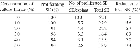
Sublethal Level of S. rolfsii Culture Filtrates. Addition of S. rolfsii culture filtrates did not affect the percentages of SE proliferation of embryogenic tissues of peanut cv. Kelinci. Addition of up to 50% of culture filtrates resulted in 96% of SE proliferation (only 4% reduction than that on P16 medium without addition of culture filtrates) (Table 1).
On the other hand, S. rolfsii culture filtrates were significantly reduced the average number of SE/explant and the total number of SE. On selective P16 medium containing 10% of S. rolfsii culture filtrates, number of SE/explant and total number of SE were only 5.7 and 229 SE, a reduction of approximately 56% as compared to that of P16 without culture filtrates (Table 1). Addition of up to 50% of culture filtrates into P16 medium further reduced number of SE/explant and total number of SE (up to 73% reduction) (Table 1). However, the reduction rate tended to level off at selective P16 medium containing 30, 40, and 50% of S. rolfsii culture filtrates (Table 1).
Single passage culture (one month) on selective P16 medium containing 30% of S. rolfsii culture filtrates did not result in sublethal level. Neither was addition of up to 50% of culture filtrates. Therefore, culturing peanut embryogenic tissues for more than one passages on selective P16 medium containing 30% of culture filtrates was necessary.
Responses against 30% of S. rolfsii Culture Filtrates.
Table 2 presented the results of culturing embryogenic tissues of peanut cv. Kelinci for up to three consecutive passages (3 months) on P16 medium with or without 30% of S. rolfsii
culture filtrates. All of the embryogenic tissues grown on P16 medium without S. rolfsii culture filtrate proliferated SE with an average of 14.2 SE/explant after the passage 1 and 14.1 SE/ explant after the passage 2. Total number of SE on P16 medium without S. rolfsii culture filtrates 3550 and 7050 SE, respectively (Tabel 2).
After one month on P16 medium with 30% culture filtrates (passage 1), SE proliferation from embryogenic tissues of peanut cv. Kelinci was 90% (Table 2). After two and three months on the same selective medium (passage 2 and passage 3); however, the percentages of SE proliferation were only 79 and 49%, respectively (Table 2). The average numbers of proliferated SE from embryogenic tissues at passage 1, passage 2, and passage 3 were 3.3 SE, 2.1 SE, and 1.1 SE per explant, respectively (Table 2).
The reduction of total SE after one month on P16 medium with 30% culture filtrates (passage 1) was 79%; while after two months (passage 2) and three months (passage 3) on the same selective medium were 95 and 98%, respectively (Table 2). Representative samples of SE proliferated from embryogenic tissues of peanut cv. Kelinci on P16 medium without culture filtrates, with 30% culture filtrates at passage 1 and passage 2 were presented in Figure 1a-d.
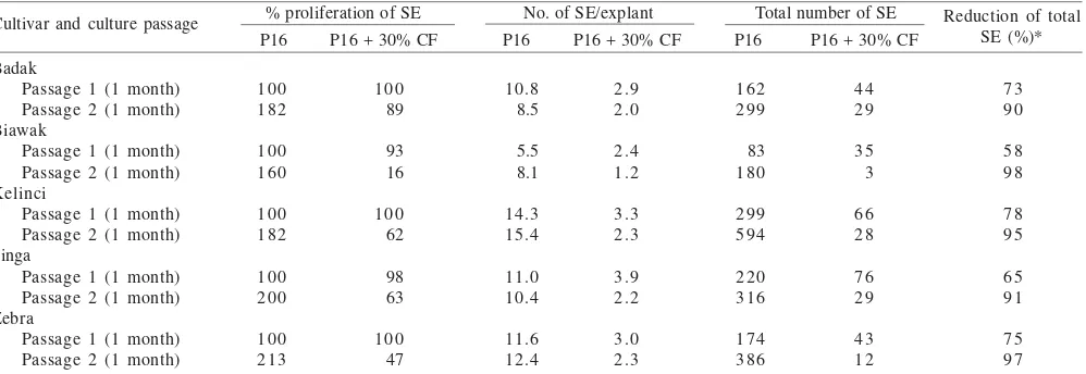
Responses of Peanut Cultivars against S. rolfsii Culture Filtrates. After the passage 1, percentage of SE proliferation on selective P16 medium containing 30% of S. rolfsii
culture filtrates was similar to P16 medium without culture filtrate for all peanut cultivars (Table 3). After the passage 2, however, the percentage of SE proliferation was almost twice as many as the passage 1 on P16 without culture filtrates. On P16 medium with 30% of culture filtrates after the passage 2, percentage of SE proliferation of peanut cv. Badak (89%) was only slightly decreased than that of without culture filtrates. While percentage of SE proliferation of peanut cv. Biawak,
Table 1. Effects of various concentrations of S. rolfsii culture filtrates in P16 medium for somatic embryo (SE) induction on proliferation of SE of peanut cv. Kelinci
No. of proliferated SE medium, without culture filtrates (0%), while Xt was one on the selective
media containing various concentration of S. rolfsii culture filtrates, respectively
Table 2. Effects of 30% of S. rolfsii culture filtrates in the P16 medium for somatic embryo (SE) induction on proliferation of SE of peanut cv. Kelinci after three consecutive passages of subculture (1 month/passage)
Culture passages (1 month/passage) Passage 1 Passage 2 Passage 3
Responses and medium for SE proliferation
Proliferating embryogenic tissues (%) P 1 6
P16 + 30% culture filtrate Number of SE/explant
P 1 6
P16 + 30% culture filtrate Total number of SE
P 1 6
P16 + 30% culture filtrate Reduction of total SE (%)*
100
medium, without culture filtrates (0%), while Xt was one on the selective
Zebra, Kelinci, and Singa were 16, 47, 62, and 63%, respectively (Table 3).
The average number of SE/explant of all peanut cultivars on P16 medium with 30% of S. rolfsii culture filtrates were less than that of P16 without culture filtrates either after the passage 1 or the passage 2. However, the average number of SE/explant on P16 medium without 30% of culture filtrates for all peanut cultivars was similar, while that on P16 medium with 30% of culture filtrates was decreased after the passage 1 and the passage 2 (Table 3). Total numbers of SE after the passage 1 and the passage 2 for peanut cv. Biawak were decreased by up to 58% and 98%, Singa - 65% and 91%, Badak - 73% and 90%, Zebra - 75% and 97%, and Kelinci - 78% and 95% (Table 3).
Culture Filtrate-Insensitive SE and Plantlet Regeneration. Most of 1500 clumps of embryogenic tissue of peanut cv. Kelinci were died after three consecutive passages of in vitro selection on selective medium containing 30% of S. rolfsii culture filtrates. However, 300 of them survived and produced an average of 1 to 2 SE. Browning of embryogenic tissues and proliferation of culture filtrate-insensitive SE after three passages of in vitro selection was presented in Figure 1e-f.
To obtain SE germination, the culture filtrate-insensitive SE was subjected to two consecutive passages of subculture on culture filtrates free P16 medium prior to germination. By adding these recovery processes, most of the culture filtrate-insensitive SE germinated successfully and plantlets were regenerated. Representative results of germination of culture filtrate-insensitive SE, regeneration of plantlets, and their acclimatization were presented in Figure 2a-d. Using the described procedures, up to fifty R0 peanut plants were successfully regenerated from surviving culture filtrate-insensitive SE of peanut cv. Kelinci. These plants were grown in the plastic-house to produce seeds for further evaluation. Various abnormal morphological characters were observed among R0 lines derived from culture filtrate-insensitive SE, such as: excessive branching, pentafoliate, hexafoliate, and
octafoliate leaves, variegated leaves, and reduced pod yield per plant (Figure 3a-f).
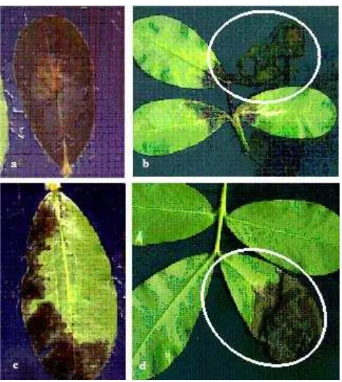
Identification of S. rolfsii Resistance of Peanut Somaclonal Variants. Results of detached leaf test on 26 R0 lines of peanut for resistance against S. rolfsii indicated that 16 lines were susceptible against direct infection of S. rolfsii. Most of the tested leaf tissues from these R0 plants were necroses and killed due to fungal infection six days after inoculation (Figure 4a-b). The inoculated leaflets and leaves of seven R0 lines of peanut showed variegation between necroses and healthy tissue (Figure 4c-d). These data indicated although most of the R0 peanut lines were regenerated from culture filtrates-insensitive SE, they were not resistance against direct infection of S. rolfsii on the leaf tissues.
DISCUSSIONS
An attempt to induce somaclonal variation among tissue culture derived planting materials, especially through long term culture of embryogenic tissues, in combination with in vitro selection have been used to evaluate the possibility of regenerating novel peanut lines with resistance against
S. rolfsii. Strategy to achieve such objectives has been evaluated in this experiment.
The success of in vitro selection depend on the availability of an established in vitro regeneration system and an effective selective agent (Hammerschlag 1992). Effective procedures for inducing SE from peanut explants have been developed previously (Edy 1998; Sulichantini 1998). This standard procedure proved to be effective for SE induction from 14 Indonesian peanut cultivars (Susilawati 2003) and peanut cv. Kelinci was reported to be the most responsive for SE regeneration. Peanut cv. Kelinci has also been reported as susceptible against infection of S. rolfsii (Yusnita & Sudarsono 2004). Therefore, peanut cv. Kelinci was selected as a model for the development of in vitro selection methods for resistance against S. rolfsii.
Table 3. Effects of addition of 30% of S. rolfsii culture filtrates (+30% CF) in the P16 medium for somatic embryo (SE) induction on SE proliferation of five peanut cultivars after two consecutive passages of subcultures (two months) on selective medium
% proliferation of SE No. of SE/explant Total number of SE P16 P16 + 30% CF P16 P16 + 30% CF P16 P16 + 30% CF
Certain pathogenic fungi secreted toxic metabolite onto culture filtrates and these filtrates have often been used as selective agents for the development of disease resistance somaclonal variant lines through in vitro selection (Hammerschlag 1992; Ahmed et al. 1996; Jin et al. 1996; Remotti 1997; Jayasankar & Litz 1998; Jayasankar et al. 2000). In this report, S. rolfsii culture filtrates presumably containing toxic metabolites has been added into medium for inducing peanut SE and used as selective agent.
Sclerotium cepiforum produced 1.8 mg oxalic acid/ml medium after 16 days under in vitro condition (Stone & Armentrout 1985). Oxalic acid is also the major phytotoxin secreted by S. rolfsii when infecting plants (Punja 1985). The amount of oxalic acid produced under in vitro condition was
not known nor was measured in this experiment. However, under the condition of this experiment, addition of S. rolfsii
culture filtrates into P16 medium drastically inhibited growth and SE proliferation.
Figure 1. Proliferation of somatic embryo (SE) of peanut cv. Kelinci on P16 medium with and without S. rolfsii culture filtrates (CF). (a) Many somatic embryos were developed from embryogenic tissues grown on P16 medium without CF; (b) After the first passage on P16 medium with 30% of CF, less SE were formed and part of embryogenic tissues necrosed; (c) Most of embryogenic tissues died on P16 with 30% of CF after the third passage of subculture. Close up pictures of (d) Proliferating SE of peanut cv. Kelinci on P16 medium without CF; (e) The browning of embryogenic tissues; and (f) Proliferation of CF-insensitive SE from mostly dead tissues.
Figure 2. Plantlet regeneration from S. rolfsii culture filtrates (CF)-insensitive somatic embryo (SE) of peanut cv. Kelinci. (a) SE germination on MS medium containing 2 g/l of activated charcoal (MSAC); (b) Well developed shoots on MSAC media ready for root induction; (c) Peanut plantlets after acclimatization and ready for transfer to the plastic-house; (d) The R0 peanut line grown in the plastic-house for seed production.
Figure 3. Representative of morphological variants among R0 lines of peanut cv. Kelinci regenerated from S. rolfsii culture filtrates (CF)-insensitive somatic embryo. (a) In vitro selection-derived (left) and seed-selection-derived (right) of peanut plants grown in the plastic house; (b) Normal branching of seed-derived peanut plant; (c) Abnormal branching of in vitro derived R0 peanut plant; (d) R0 plant with leaf variegation; (e) Leaf of R0 plant with abnormal eight leaflets (octafoliate); and (f) Leaf with normal leaflets from seed derived peanut plant.
Sublethal level was achieved by adding 30% S. rolfsii
culture filtrates into P16 medium and by culturing embryogenic tissues for three consecutive passages of selection period. Such methods of in vitro selection resulted in more than 95% reduction in SE proliferation. This suggests culture filtrates of S. rolfsii is effective as selective agent for in vitro selection. The use of fungal toxic metabolites as selective agent for in vitro selection has successfully been demonstrated for
Fusarium sp. (Arcioni et al. 1987; Ahmed et al. 1996; Jin et al. 1996) and Septoria glycines (Song et al. 1994).
Responses of five peanut cultivars against 30% of S. rolfsii
culture filtrates were also tested in this experiment. The results indicated that response against culture filtrates of S. rolfsii is genotype dependent. Previous evaluation under the controlled condition indicated peanut cv. Badak was highly susceptible against S. rolfsii infection while peanut cv. Kelinci and Singa were both susceptible (Yusnita & Sudarsono 2004). Response of peanut cv. Biawak and Zebra against S. rolfsii
have not been documented. Peanut cv. Badak (highly susceptible) was the least sensitive against culture filtrates than the other four peanut cultivars.
It is possible that the response of peanut cultivar against
S. rolfsii infection may not be directly correlated with their response against S. rolfsii culture filtrates. In this case, heat resistance toxic metabolites that were presumably present in the S. rolfsii culture filtrates may not be the only factor affecting response of peanut plants against S. rolfsii infection (Punja 1985).
Out of 1500 clumps of initial embryogenic tissues of peanut cv. Kelinci cultured on selective P16 medium containing 30% of S. rolfsii culture filtrates, 300 proliferated 1-2 culture filtrate-insensitive SE per clump. Therefore, the calculated frequency of recovering culture filtrate-insensitive SE using the developed in vitro selection procedure in this experiment was approximately 1%. However, this number did not reflect the frequent mutation of culture filtrate-insensitive phenotypes since the embryogenic tissues have been proliferated for at least one year prior to in vitro selection. The real frequency for this type of mutation was expected to be less.
Insensitive somatic embryos were able to proliferate on selective P16 medium containing 30% of S. rolfsii culture filtrates. Such ability may probably be due to acquisition of certain detoxification mechanisms. Related results previously reported that the ability of grapevine SE to survive on selective medium containing Elsinoe ampelina culture filtrates was due to induction of detoxifying enzymes during in vitro selection, leading to the systemic resistance character in the selected lines. Such mechanism may also operate among culture filtrate-insensitive SE of peanut identified in this study.
Not all of the culture filtrate-insensitive SE were able to grow and germinate normally on germination medium after recovery period. Abnormal form of germinated SE were often observed among culture filtrate-insensitive SE of peanut. Similar results of abnormal SE development, however, was also observed among germinated SE of peanut that have not been exposed to in vitro selection (Susilawati 2003).
In this experiment, 50 R0 lines of peanut cv. Kelinci have successfully been transferred to soil and grown to maturity in the plastic-house. Most of the R0 plants showed some degree of morphological variation, such as: excessive branching, abnormal number of leaflets, leaf variegation, male sterility, unable to produce flower nor pods, and yielded less pods/ plant. Soybean plants derived from SE have also been reported to exhibit various morphological variation (Barwale & Widholm 1987; Widoretno et al. 2003).
Although preliminary evaluation of the R0 lines against
S. rolfsii has been conducted and indication of the existence of S. rolfsii resistance peanut tissues was observed, it was too early to conclude the success of developing S. rolfsii
resistance peanut lines. Further evaluation using subsequent generation of peanut (R1 and R2) plants need to be conducted once seeds derived from the R0 lines were available.
ACKNOWLEDGMENT
Part of this research was supported by Proyek Peningkatan Penelitian Pendidikan Tinggi, Directorate General of Higher Education, Department of National Education - Graduate Team Research Grant (HPTP) – Batch I: Genetic Engineering and In Vitro Selection for the Development of Peanut Germplasm with Novel Characters –Drought Tolerance and Sclerotium Stem Rot Resistance. Contract No. 340/P4T/DPPM/IV/2003, on April 25, 2003. Yusnita was also supported by TMPD Scholarship for PhD program from the Department of National Education, Republic of Indonesia.
REFERENCES
Ahmed KZ, Mesterhazy A, Bartok T, Sagi F. 1996. In vitro techniques for selecting wheat (Triticum aestivum L.) for Fusarium-resistance. II. Culture filtrate technique and inheritance of Fusarium -resistance in the somaclones. Euphytica 91:341-349.
Arcioni S, Pezzotti M, Damoni F. 1987. In vitro selection of alfalfa plants resistant to Fusariumoxysporum f.sp. medicaganis. Theor Appl Genet 74:700-705.
Backman PA. 1984. Stem-rot. In: Porter DM, Smith DH, Rodriguez-Kabana R (ed). Compedium of Peanut Disease. St Paul: American Phytopathological Society Pr. p 15-16.
Barwale UB, Widholm JM. 1987. Somaclonal variation in plants regenerated from cultures of soybean. Plant Cell Rep 6:365-368. Dennis ES et al. 1984. Molecular analysis of the alcohol dehydrogenase
(adhI) gene of maize. Nuc Acid Res 12:3983-4000.
Edy A. 1998. Induksi embrio somatic dari eksplan poros embryo pada beberapa kultivar kacang tanah (Arachis hypogaea L.) secara in vitro [Tesis]. Bogor: Program Studi Agronomi, Institut Pertanian Bogor.
Gamborg OL, Miller RA, Ojima K. 1968. Nutrient requirement of suspension cultures of soybean root cells. Exp Cell Res 50:151-158.
Hammerschlag FA. 1992. Somaclonal variation. In: Hammerschlag FA, Litz RE (ed). Biotechnology of Perennial Fruit Crops. Walingford: CAB International. p 35-55.
Jayasankar S, Li Z, Gray DJ. 2000. In vitro selection of Vitis vinifera
‘Chardonay’ with Elsinoe ampelina culture filtrate is accompanied by fungal resistance and enhanced secretion of chitinase. Planta
211:200-208.
Jayasankar S, Litz RE. 1998. Characterization of embryogenic mango cultures selected for resistance to Colletotrichum gloeosporoides
Jin H, Hartman GL, Huang YH, Nickell CD, Widholm JM. 1996. Regeneration of soybean plants from embryogenic suspension cultures treated with toxic culture filtrate of Fusarium solani and screening of regeneraants for resistance. Phytopathology 86:714-718.
Karp A, Nelson RS, Thomas E, Bright SWJ. 1982. Chromosome variation in protoplast-derived potato plants. Theor Appl Gen
63:265-272.
Kuksova VB, Piven NM, Gleba YY. 1997. Somaclonal variation and in vitro induced mutagenesis in grapevine. Plant Cell Tiss Org Cult
49:17-227.
Kumar PS, Mathur VL. 2004. Chromosomal instability in callus culture of Pisum sativum. Plant Cell Tiss Org Cult 78:267-271. Larkin PJ, Scowcroft WR. 1981. Somaclonal variation - a novel source
of variability from cell cultures for plant improvement. Theor Appl Gen 60:197-214.
Matsumoto K, Barbosa ML, Souza LAC, Teixeira JB. 1995. Race 1
Fusarium wilt tolerance on banana plants selected by fusaric acid.
Euphytica 84:67-71.
Murashige T, Skoog F. 1962. A revised medium for rapid growth and bioassays with tobacco tissue cultures. Physiol Plant 15:473-497. Orton TJ. 1983. Experimental approaches to the study of somaclonal
variation. Plant Mol Biol Rep 1:67-76.
Phillips RL, Kaeppler SM, Peschkke VM. 1990. Do we understand somaclonal variation? In: Nijkamp HJJ, van Der Plas LHW, van Aartrijk J (ed). Progress in Plant Molecular Biology. Dordrecht: Kluwer Academic Publ. p 131-141.
Porter DM, Smith DH, Rodriguez-Kabana R. 1982. Peanut plant disease. In: Patee HT, Young CT (ed). Peanut Science and Technology. Yoakum, TX: American Peanut Research and Education Society. p 348-354.
Pratt RG. 1996. Screening for resistance to Sclerotinia trifoliarum in alfalfa by inoculation of excised leaf tissue. Phytopathology
86:923-928.
Punja ZK. 1985. The biology, ecology and control of S. rolfsii. Ann Rev Phytopathol 23:124-135
Ramos-Leal M et al. 1996. Somaclonal variation as a source of resistance to eyespot disease of sugarcane. Plant Breeding 115:37-42.
Remotti PC, Loffler HJM, Loten-Doting LV. 1997. Selection of cell-lines and regeneration of plants resistant to fusaric acid from
Gladiolus x grandiflorus cv. ‘Peter Pear’. Euphytica 96:237-245. Scowcroft WR, Ryan SA, Brettle RIS, Larkin PJ. 1985. Somaclonal variation in crop improvement. Proc Inter-Center Seminar on International Agricultural Research Center (IARCs) and Biotechnology: Biotechnology in International Agricultural Research. Los Banos, Manila, 23-27 Apr 1984. p 99-109. Shepard JF. 1981. Protoplast as sources of disease resistance in plants.
Ann Rev Phytopathol 19:145-166.
Skirvin RM, McPheeters KD, Norton M. 1994. Sources and frequency of somaclonal variation. Hort Science 29:1232-11237. Skirvin RM, Janick J. 1976. Tissue culture-induced variation in scented
Pellargonium spp. J Amer Soc Hort Sci 101:282-290.
Stone HE, Armentrout VN. 1985. Production of oxalic acid by
Sclerotium cepivorum during infection of onion. Ecologia 11:526-530.
Sulichantini EDS. 1998. Induksi embrio dari eksplan poros embrio dan embryonic leaflet pada beberapa kacang tanah (Arachis hypogaea
L.) komersial di Indonesia [Tesis]. Bogor: Program Studi Agronomi, Institut Pertanian Bogor.
Susilawati PN. 2003. Respons 14 kultivar kacang tanah terhadap stres kekeringan yang disimulasikan dengan penyiraman PEG dan kemampuan membentuk embrio somatiknya [Tesis]. Bogor: Program Studi Agronomi, Institut Pertanian Bogor.
Song HS, Lim SM, Widholm JM. 1994. Selection and regeneration of soybean resistant to patho-toxic culture filtrate of Septoria glycines. Phytopathology 84:948-951.
Widoretno W, Harran S, Sudarsono. 2003. Variation in qualitative and quantitative characters among somaclones of soybean derived from
in vitro selected somatic embryos. Hayati 10:110-117.