Vol. 17 No. 4, p 173-178 EISSN: 2086-4094
http://journal.ipb.ac.id/index.php/hayati DOI: 10.4308/hjb.17.4.173
_________________ ∗
∗∗
∗∗Corresponding author. Phone/Fax: +62-251-8622833, E-mail: [email protected]
INTRODUCTION
Enzyme inhibitors have been pay attention as useful tools, not only for the study of enzyme structures and reaction mechanisms but also for potential utilization in pharmacology (Bode & Huber 1992) and agriculture (Terashita et al. 1980). It is useful in the diagnosis and therapy of inflammation and pancreatitis (Delston et al. 2003). It is also useful for fruit-body formation of mushroom cultivation (Astuti et al. 2002). Specific and selective protease inhibitors are potentially powerful tools for inactivating target proteases in the pathogenic processes of human diseases such as emphysema, arthritis, pancreatitis, thrombosis, high blood pressure, muscular dystrophy, cancer, and AIDS (Demuth 1990).
For the last 10 years, attention on protease as the drugs target has increased slightly due to their role on illness mechanism of virus, such as influenza and HIV, malaria, cancer, and even degenerative illnesses, such as Alzheimer. Although many drugs that can inhibit protease mechanisms are already available, researches has being done to find natural protease inhibitor from many kinds of source, such as virus, bacteria, fungi, and also other organisms, such as sponges and tunicate (Imada 2004). Mayer and Lehmann (2000) reported that among sea organisms, sponge was the most potential producer of bioactive agents including enzyme inhibitor components
(Lee et al. 2001). Sponge-associated bacteria are also produce bioactive components (Webster et al. 2001). Bacterial and cyanobacterial symbions of sponge, especially Aplysina aerophobia, could reach up to 40% of total sponge biomass (Ahn et al. 2003). The symbiotic association with the sponge can be either intranucleus symbiotic, intracellular symbiotic, or extracellularr exosymbiotic. This symbiotic can happen due to sponge as a filter feeder organism. Lee et al. (2001) reported that bioactive components of the sponge originally come from biosynthetic products of its symbions. And bacteria was found the most in sponge, it was more than 3x10⁴ CFU/g (Nurhayati et al. 2006).
Protease inhibitors can keep a protease from working and splitting a protein into peptides, and It can blocked protease enzyme activity. However, there is a few number protease inhibitor had been found from marine environment, especially from sponge-associated bacteria. Therefore, this study was to screen bacteria producing protease inhibitor isolated from sponge Jaspis sp.and to analyze inhibitory activity against subtilisin, thermolysin, and crude extract from pathogenic bacteria.
MATERIALS AND METHODS
Bacterial Strains, Protease Substrates, and Growth Media. 138 bacteria isolated from Jaspis sp. were used in this study. Pathogenic bacteria, such as Enteropathogenic Escherichia coli (EPEC) K.1.1 (obtained from Dr. Sri Budiarti), Pseudomonas aeruginosa, and Staphylococcus
Screening and
Characterization of Protease Inhibitors from Marine
Bacteria Associated with Sponge Jaspis sp.
ARIS TRI WAHYUDI∗∗∗∗∗, QATRUNNADA, NISA RACHMANIA MUBARIK
Microbiology Division, Department of Biology, Faculty of Mathematics and Natural Sciences, Bogor Agricultural University, Darmaga Campus, Bogor 16680, Indonesia
Received April 22, 2010/Accepted November 11, 2010
Three isolates among 138 sponge-associated bacteria were isolated from Waigeo Island, Raja Ampat West Papua Province, Indonesia, have been shown protease inhibitory activity against subtilisin (serine protease), thermolysin (metalloprotease), and crude extract from pathogenic bacteria (Eschericia coli enteropathogenic/ EPEC K.1.1, Staphylococcus aureus, and Pseudomonas aeruginosa). Those three isolates were designated as sponge associated bacteria SAB S-12, SAB S-21, and SAB S-17. A simple casein and Sea Water Complete (SWC) double layer agar method was used to screen the bacteria against pathogenic bacteria producing protease, i.e. EPEC K.1.1, S. aureus, and P. aeruginosa. Among them, SAB S-12 isolate showed no inhibitory zone indicated. The isolate had the highest inhibitory activity against subtilisin and crude extract enzyme of pathogenic bacteria, the inhibitory activity was 91.6 and 98.9%, respectively. In addition, the SAB S-21 isolate had the highest inhibitory activity against thermolysin, it was 70.4%. The optimum pH and temperature for protease inhibition of the three isolates was at pH 7.0-8.0 and 40-50 oC respectively. Based on 16S rRNA gene sequence, the closest related with
SAB S-12, SAB-17, and SAB-21 isolates was Providencia sp. (92% identity), Paracoccus sp. (86% identity), and Bacillus sp. (100% identity), respectively.
Key words: sponge-associated bacteria, protease inhibitor, pathogenic bacteria, screening, 16S rRNA
aureus (obtained from PPSHB, IPB) were also used throughout this research, and routinely maintained in Luria Broth (LB; Oxoid). The following proteases were used in this research, Subtilisin from Bacillus licheniformis (SIGMA) and Thermolysin from Bacillus thermoproteolyticus rokko (SIGMA).
Screening of Sponge-Associated Bacteria Producing Protease Inhibitor. As many as 138 bacteria isolated from Jaspis sp. were determined their inhibitory protease activity using double layer medium of Imada’s (1985a) method. This method used sea water complete (SWC) as the lower layer medium and mixture of NA and SM medium as the upper layer medium, and the bacterial isolates were applied on the lower layer of medium and incubated for 24 hours at 37 oC. After 24 hours of incubation, bacterial colony was removed, then the medium was filled up with the upper layer medium. The pathogenic bacteria was subsequently inoculated on the upper layer medium and incubated for 24 hours at 37 oC. Inhibitory protease activity was indicated by no or reducing of proteolytic zone around the bacterial colony.
Protease Activity Assay. Three of pathogenic bacterial strains i.e. Enteropathogenic Escherichia coli (EPEC) K.1.1, P. aeruginosa, and S. aureus were inoculated on LB medium and pre-cultured at 37 oC on a 100 rpm rotary shaker until the optical density (OD) of 0.8 was achieved. Then, 10% (v/v) of the pre-culture was inoculated in 150 ml of the same medium and incubated for 56 hours at 37 oC. The culture was harvested by centrifugation at 3000 rpm and the supernatant was used for the assay of inhibitory activity. Protease activity was measured by Walter’s method (1984) with slight modification.
Protease Inhibition Assay. Inhibition activity was measured using method according to Imada et al. (1985b) with slight modification. Twenty-five µl of subtilisin, thermolysin, and crude extract of pathogenic bacteria (EPEC K.1.1, S. aureus, and P. aeruginosa) in 0.5 mlof 30 mM Tris-HCl buffer pH 8.0, was pre-incubated with 0.5 ml of culture supernatant for 12 minutes at 30 oC. The substrate which used were subtilisin (91 mg protein/ml) as one of the serine protease group, thermolysin (42 units/mg solid; 64 units/mg protein) as one of the metalloprotease group, and EPEC K.1.1, P. aeruginosa, and S. aureus which were produced based on Baehaki (2004). EPEC K. 1.1 (0.013 U/ml) for isolate SAB S-12, S. aureus (0.04 U/ml) for isolate SAB S-21, and P. aeruginosa (0.25 U/ml) for isolate SAB S-17.
One ml of 1% (w/v) Hammarsten casein (MERCK) in the same buffer was added to pre-incubated mixture. After incubation of the mixture for 12 minutes at 30 oC, 2 ml of 5% (w/v) trichloroacetic acid (TCA) was added to terminate the enzyme reaction and mixture was kept for 20 minutes at 30 oC. After that, the solutions were centrifuged at 3000 rpm for 10 minutes. The optical density of filtrate was measured by turbidimetric measurement at 280 nm in a GeneQuant pro 81670 type spectrophotometer (Cambridge Ltd, England). Simultaneusly, the optical density of the blank and control (without inhibitor solution) were also determined. The percentage of inhibition was calculated as = (1-B/A) x 100%, wherein A was the
increased optical density without the inhibitor after 12 minutes reaction, and B was the increased optical density with the inhibitor after 12 minutes. One unit of inhibitor activity was defined as the amount by which enzyme activity was reduced by 50% under the above condition. Characterization of Inhibitor Protease on Various Temperatures and pH. The culture supernatant solution containing protease inhibitor was adjusted to various temperatures from 10 to 90 oC for 10 minutes and the residual inhibitory activity was measured. The solution containing culture supernatant was adjusted to various pH levels from 3 to 9 for 10 minutes and incubated for 10 minutes at each optimum temperature. After incubation, pH was readjusted to assay pH (8.0) and the residual inhibitory activity was determined. Citrate buffer and Tris-(hydroxymethyl)-aminomethane (Tris) buffer were used as the buffer solutions (0.03 M).
Characterization and Identification of Bacteria Producing Protease Inhibitor. Characterization morphologically for potential bacteria which were extracted from Jaspis sp. producing protease inhibitor was done by Gram staining (Cappucino & Sherman 1983). The shape of the bacteria was observed by microscope (Olympus Type CH20BIMF200) in 1000 magnification. Molecular identification of three strains of bacteria producing inhibitor protease were identified based on 16S rRNA gene sequence, by three steps, (i) isolation of the DNA (Sambrook & Rusell 2001), (ii) amplification of 16S rRNA gene by PCR using amplification machine (Gene Amp PCR system 2400 Perkin Elmer), and (iii) sequencing of the 16S rRNA gene. The primer used to amplify the 16S rRNA were forward primer 63f (5’-CAG GCC TAA CAC ATG CAA GTC) and 1387r (5’-GGG CGG WGT GTA CAA GGC) (Marchesi et al. 1998) using method described by Bahri et al. (2009). The DNA fragments were sequenced by PT Charoen Pokphan Indonesia Jakarta. A comparison between the 16S rRNA gene sequence of sponge-associated bacteria producing protease inhibitor to that of 16S rRNA gene data found in the GenBank database was done by connecting to the BLASTN (basic local alignment search tool for nucleotide) program at http:// www.ncbi.nlm.nih.gov. By that software, the sequencing of the DNA would be gotten in high homology with the DNA sequence of SAB S-12. All DNA sequences alignment was arranged in FASTA3 format and then similarity analysis was done using ClustalW program from the website www.ebi.ec.uk/clustalW. The result was saved on TreeConW software, and by using this software, the phylogenetic tree was made in Phylip format with bootstrap replication 100X. In the making of phylogenetic tree, Alteromonas (FN661963.1), Passiflora rubra (AY261639.1), P. piscicida (X82215.1), Idiomarina zobellii
(GQ131629.1), Pseudoalteromonas bacteriolytica
(AF173962.1), and Algicola sagamiensis (AB063324.1) were used as the reference strains.
RESULTS
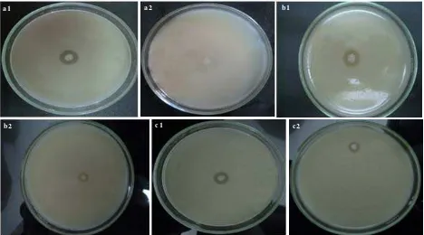
were isolated from marine sponge Jaspis sp., three strains produced protease inhibitor, they were designated as SAB S-12, SAB S-21, and SAB S-17, respectively. Those isolates were able to reduce or inhibit production of protease produced by pathogenic bacteria (EPEC K.1.1, P. aeruginosa, and S. aureus) indicated by no proteolytic zone or reducing of proteolytic zone surrounding bacterial colony. SAB S-12 showed the highest capability to produce protease inhibitor, as there was no proteolytic zone around it’s growth zone, while both SAB S-21 and SAB S-17 showed capability to produce protease inhibitor, but their capability was less than SAB S-12 (Figure 1).
Protease Activity. The period of the optimum protease production of EPEC K.1.1, S. aureus, and P. aeruginosa were gained after 24, 16, and 40 hours of incubation, respectively. The highest protease activity of EPEC K.1.1 was 0.01 U/ml at 16 hours. Its lag phase was within 8 hours, followed by an exponential phase up to 32 hours with maximum OD of 1.86. This result indicated that EPEC K.1.1 had optimum protease production period at 24 hours, where the protease activity on this time was the highest (0.01 U/ml), therefore the harvest time of EPEC K.1.1 was 24 hours. Protease was produced along with the cell growth and it reached the highest protease activity on its nearly stationer phase. The highest protease activity of S. aureus was 0.04 U/ml at 16 hours. The lag phase of S. aureus was within 8 hours, followed by an exponential phase up to 40 hours within maximum OD600 of 1.97. The highest protease activity of P. aeruginosa was 0.25 U/ml at 32 hours. Its lag phase was within 16 hours, followed by an exponential phase up to 32 hours with maximum OD600 of 1.98.
Protease Inhibition Activity. The highest inhibitory activity of isolates SAB S-12, SAB S-21, and SAB S-17 in subtilisin substrate were 62.06% at 28 hours, 27.58% at 28 hours, and 20.68% at 28 hours, respectively. In addition, the highest inhibitory activity of isolate SAB S-12, SAB S-21, and SAB S-17 in thermolysin substrate were 6.25% at 8 hours, 14.58% at 28 hours, and 12.50% at 28 hours, respectively. This result means that the optimum protease inhibitor activity of SAB S-12, SAB S-21, and SAB S-17 in subtilisin substrate were at 28 hours of cultivation, while SAB S-12 was at 8 hours of cultivation in thermolysin substrate. The highest inhibitory activity of isolate SAB S-12 in EPEC K.1.1’s enzyme extract substrate was 98.84% at 24 hours cultivation. The highest inhibitory activity of isolate SAB S-21 in S. aureus’s enzyme extract substrate was 51.29% at 24 hours cultivation. The highest inhibitory activity of isolate SAB S-17 in P. aeruginosa’s enzyme extract was 23.52% at 20 hours cultivation. The results showed that SAB S-12, SAB S-21, and SAB S-17 needed different time to reach protease inhibition capability. Each isolates reach its optimum condition after that their protease inhibition decreased slightly.
Characteristics of Inhibitor Protease. The highest inhibitory activity of isolate SAB S-12, SAB S-21, and SAB S-17 in subtilisin substrate were 88.45% at 40 oC, 44.67% at 50 oC, and 21.53% at 40 oC, respectively. The highest inhibitory activity of isolate SAB S-12, SAB S-21, and SAB S-17 in thermolysin substrate were 59.47% at 40 oC, 47.52% at 40 oC, and 50.52% at 40 oC, respectively. This result indicated that the optimum protease inhibitor activity of SAB S-12, SAB S-21, and SAB S-17 in subtilisin substrate were 40, 50, and 40 oC, while for the protease
a 1 a 2 b1
b2 c1 c2
Table 1. Homologous search analysis of the 16S rRNA gene sequence of three bacterial isolates producing protease inhibitor isolated from sponge Jaspis sp.
Isolate code Homology Identity (%) Access No. SAB S-12
SAB S-17 SAB S-21
Providencia sp.
Paracoccus sp.
Bacillus sp.
9 2 8 6 1 0 0
AY870456.1 GU338411.1 FN556567.1 inhibitor activity of the three of isolates mentioned above
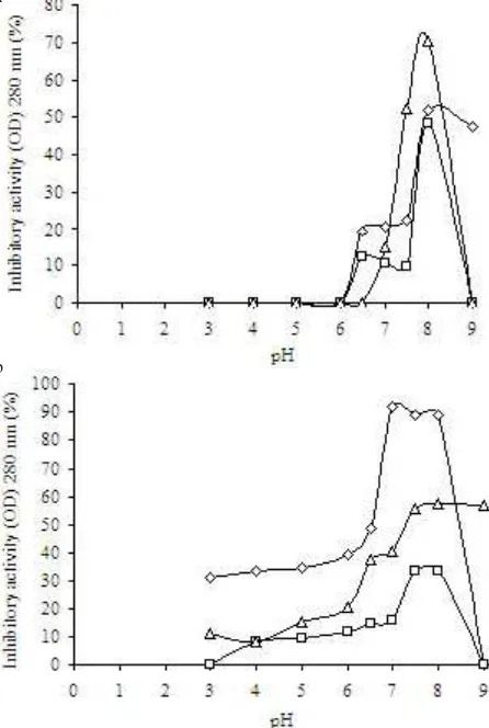
in thermolysin substrate were 40 oC (Figure 2). Furthermore, the highest inhibitory activity of isolate SAB S-12, SAB S-21, and SAB S-17 in thermolysin substrate was 50.43% at pH 8.0, 70.37% at pH 8.0, and 49.89% at pH 8.0 condition, respectively. The highest inhibitory activity of isolate SAB S-12, SAB S-21, and SAB S-17 in subtilisin substrate was 91.57% at pH 7.0, 57.23% at pH 8.0 condition, and 30.78% at pH 7.5, respectively (Figure 3).
Identification of Bacteria Producing Protease Inhibitors. The result of PCR of 16S rRNA genes of isolates SAB S-12, SAB S-17, and SAB S-21 were successfully amplified, i.e.1300 bp in size (data not shown). Sequence analysis of the 16S rRNA gene sequence of three bacterial isolates producing protease inhibitor are shown in Table 1. SAB S-12 was identified as Providencia sp. (92% identity), SAB S-17 was identified as Paracoccus (86% identity), and SAB S-21 was identified as Bacillus sp. (100% identity). The phylogenetic tree of of three isolates, SAB S-12, SAB S-21, and SAB S-17 are shown in Figure 4.
Temperature (oC)
Temperature (oC)
a
b
Figure 2. Inhibitory activity of SAB 12, SAB 21, and SAB S-17 against various temperature in (a) thermolysin substrate, (b) subtilisin substrate. : isolate SAB S-12,
: isolate SAB S-21, : isolate SAB S-17.
a
b
Figure 3. Inhibitory activity of SAB 12, SAB 21, and SAB S-17 against various pH levels in (a) thermolysin substrate, (b) subtilisin substrate. : isolate SAB S-12, : isolate SAB S-21, : isolate SAB S-17.
SAB_S_17 SAB_S_21
SAB_S_12
Alteromona P. rubra
P. piscicid I. zobellii P. bacterio A. sagamien
1 0 0 0.1
9 1
8 7
5 0 9 6 1 0 0
1 0 0
DISCUSSION
Three bacterial isolates of SAB S-12, SAB S-17, and SAB S-21 were isolated from marine sponge Jaspis sp. showed protease inhibitors activity. Protease inhibitor was produced by SAB S-12 could inhibit protease larger than SAB S-21 and SAB S-17. The preliminary protease inhibitor activity was determined from the disappearance of proteolytic zone of the isolates on the screening medium. Based on screening test results, SAB S-12 showed no inhibitory zone, while SAB S-21 and SAB S-17 showed proteolytic zone, which were 0.1 and 0.1 cm, respectively. The three strains mentioned above also inhibit the serine protease (subtilisin), metalloprotease (thermolysin), and crude extract from pathogenic bacteria (EPEC K.1.1, S. aureus, and P. aeruginosa). Isolate SAB S-12 had the highest activity in inhibiting subtilisin (91.57%) and crude extract from the pathogenic bacteria (98.84%). Meanwhile, isolate SAB S-21 has the metalloprotease (thermolysin) inhibitory activity at roughly 70.370%.
The similar study was conducted by Nurhayati et al. (2006) reported that a bacterium designed as isolate 6A3 (identified as Chromohalobacter sp.) isolated from sponge X. testudinaria from Kepulauan Seribu, produced protease inhibitor activity (93.5% activity) against protease produce by P. aeruginosa). However, they did not classify a protease inhibitor molecule based on their study. Their finding was really different with our results revealing that isolate SAB S-12 has the largest capability to produce serine protease inhibitor molecule or compound. It indicated that this molecule/compound produced by isolate SAB S-12 inhibited protease production of pathogenic bacteria (EPEC K.1.1). In the assay of its activity, casein from skim milk was used as the substrate for the growing of pathogenic bacteria. Normally, pathogenic bacteria produce protease enzyme which digests casein in the skim milk agar medium as the kinds of protein, therefore the pathogenic bacteria would made proteolytic zone around its colony.
Each bacterial isolate of SAB S-12, SAB S-21, and SAB S-17 had maximum protease inhibitor production in each optimum time of cultivation. It indicated that the optical density of them would be higher along with the time of cultivation. The optical density described the density of amount of bacteria in the solution. Thus, the protease inhibitor production would growing along with the growing of optical density until it faced its optimum time of cultivation and then the optical density would decrease and so did its production of protease inhibitor enzyme. The protease inhibitory activity of each bacterial isolates showed different percentage for different substrate. It was indicates that each isolates had specification to different kind of protease (Figure 2).
Isolate SAB S-12 had higher inhibition capability to serine protease (subtilisin) while, SAB S-21 had higher inhibition capability to metalloprotease (thermolysin). pH and temperature also influenced the inhibitory activity produced by each bacterial isolates. Those components
were also could enhanced the protease inhibitory activity of SAB S-12, SAB-21, and SAB-17, respectively (Figure 2). Hames and Hooper (2000) reported that temperature influences the reaction mechanism of protease inhibitor (protein) in 2 ways. The first way is the temperature will increase the mechanism of reaction until it reaches its optimum temperature, meanwhile in further higher temperature, changes in enzyme conformation will be happen. Therefore the enzyme can not enter the active site of inhibitor enzyme. After that, the activity of enzyme inhibitor will decrease. The second way is increasing of molecule thermal energy which develop structure of enzyme inhibitor protein will be affected in destroy of non-covalent interaction so that enzyme inhibitor will denature and its activity will decrease.
Based on 16S rRNA sequence, SAB S-12 homolog with Providencia. This bacterium is a Gram negative, motile bacterium of the family Enterobacteriaceae. This genus can be found commonly in soil, water, and sewage (Tumbarello 2004). This result is different from Imada (1985b) who successfully isolated marine bacteria producing protease inhibitor, Alteromonas sp. This isolate , produced protease inhibitor of marinostatin. It is also different from Kobayashi et al. (2003) who found Pseudomonas sagamiensis as the marine bacteria producing protease inhibitor. Based on the phylogenetic tree, it could be known that SAB S-12 (homolog with Providencia sp.), SAB 17 (homolog with Paracoccus sp.), and SAB S-21 (homolog with Bacillus sp.) were in the same groups (Figure 4 & Table 1). They were relatively closed each other than to bacterial reference strains. The others bacteria, such as Alteromonas, P. piscicida, P. rubra, I. zobelii, and A. sagamiensis were used as the comparator bacteria. They were definitively classified as marine bacteria producing protease inhibitor (Kobayashi et al. 2003).
REFERENCES
Ahn Y-B, Rhee S-K, Fennell DE, Kerkhof LJ, Hentschel U, Haggblom MM. 2003. Reductive dehalogenation of brominated phenolic compounds by microorganisms associated with the marine sponge Aplysina aerophoba. Appl Environ Microbiol
69:4159-4166.
Astuti P, Pratiwi SUT, Hertiani T, Alam G, Tahir A, Wahyuono S. 2002. Marine sponge Jaspis sp, a potential bioactive natural source against linfectious diseases. Berkala Ilmu Kedok 34:135-140.
Baehaki A. 2004. Karakterisasi protease beberapa bakteri patogen [Thesis]. Bogor: Postgraduate Program, Bogor Agriculture University.
Bahri S, Wahyudi AT, Mubarik NR. 2009. Genetic diversity of plant growth promoting rhizobacteria of Bacillus sp. based on 16S rRNA sequence and amplified rDNA restriction analysis.
Microbiol Indones 3:1978-3477.
Bode W, Huber R. 1992. Natural protein proteinase inhibitors and their interaction with proteinases. Eur J Biochem 204:433– 451.
Cappucino JG, Sherman N. 1983. Microbiology: A Laboratory
Manual. New York: Addison-Wesley Publ Comp.
Demuth HU. 1990. Recent developments in inhibiting cysteineand serine proteases. J Enz Inhibit 3:249-278.
Hames BD, Hooper NM. 2000. Biochemistry: The Instant Notes. 2nd Ed. Hongkong: Springer-Verlag.
Imada C. 2004. Enzyme inhibitors of marine microbial origin with pharmaceutical importance. Mar Biotechnol 6:193-198. Imada C, Simidu U, Taga N. 1985a. Isolation and characterization of marine bacteria producing alkaline protease inhibitor. Bull
Jap Soc Sci Fish 51:799-803.
Imada C, Taga N, Maeda N. 1985b. Cultivation conditions for subtilisin inhibitor-producing bacterium and general properties of the inhibitor “marinostatin”. Bull Jap Soc Sci Fish 51:805-810.
Kobayashi T, Imada C, Hiraishi A, Tsujibo H, Miyamo K. 2003.
Pseudoalteromonas sagamiensis sp. nov., a marine bacterium
that produces protease inhibitors. Int J Syst Evol Microbiol
53:1807-1811.
Lee YK, Lee JH, Lee HK. 2001. Microbial symbiosis in marine sponges. J Microbiol 30:254-264.
Marchesi JR, Sato T,Weightman AJ,Martin TA,Fry JC,Hiom SJ, Wade WG. 1998. Design and evaluation of useful bacterium-specific PCR primers that amplify genes coding for bacterial 16S rRNA. Appl Environ Microbiol 64:795-799.
Mayer AMS, Lehmann VKB. 2000. Marine pharmacology.
Pharmacology 42:62-69.
Nurhayati T, Suhartono MT, Nuraida L, Poerwanto SB. 2006. Karakterisasi awal inhibitor protease dari bakteri yang berasosiasi dengan spons asal Pulau Panggang, Kepulauan Seribu. Hayati 13:58-64.
Sambrook J, Russel DW. 2001. Molecular Cloning: A Laboratory
Manual. Vol 1, 3rd Ed. New York: Cold Spring Harbor
Laboratory Pr.
Terashita T, Kono M, Murao S. 1980. Promoting effect of S-PI on fruiting of Lentinus edodes. Trans Mycol Soc Jpn 21:137-140.
Tumbarello M, Citton R, Spanu T, Sanguinetti M, Romano L, Fadda G, Cauda R. 2004. ESBL-producing multidrug-resistant
Providencia stuartii infections in a university hospital. J Antimicrob Chemother 53:277-282.
Walter P, Gilmorer R, Blobel G. 1984. Protein translocation across the endoplasmic reticulum. Cell 38:5-8.
Webster NS, Wilson KJ, Blackall LL, Hill RT. 2001. Phylogenetic diversity of bacteria associated with the marine sponge