Majalah Kedokteran Nusantara Volume 41 y No. 3 y September 2008 219
Ewing’s Sarcoma
Soekimin, Reno Keumalazia Kamarlis
Departemen Patologi Anatomi, Fakultas Kedokteran, Universitas Sumatera Utara, Medan
Abstract: It was reported a teenager, aged 16 years old with adnexal tumor, with a chief complain of pelvic cavity enlargement and pain. Diagnose was made by histopathologic examination. Histopathologic features showed proliferation of small-cell, round, and some spheroidal and uniform cells. The nuclei are round or slightly oval, have finely dispersed chromatin, some of them with vesicular nuclei and one or two distinct nucleoli. The tumor cells have clear cytoplasm without defined cytoplasm. Clusters of tumor cells separated by fibrous septa. Mitotic activity is variable There are proliferation of blood vessel without angioinvasion. Keywords: ewing’s sarcoma, small-cell, round, and some spheroidal and uniform cells, fibrous septa
Abstrak: Dilaporkan satu kasus, seorang gadis, usia 16 tahun, dengan diagnosa tumor adneksa, dengan keluhan utama adanya massa pada rongga pelvik dan nyeri. Berdasarkan pemeriksaan histopatologi didapatkan gambaran proliferasi sel-sel berukuran kecil, bentuk bulat dan sferoid, uniform. Inti bulat – oval, kromatin tersebar merata, beberapa diantaranya dengan inti vesikuler dengan satu dua anak inti, sitoplasma jernih dengan batas tidak jelas. Kelompokan sel-sel tumor dibatasi oleh sekat-sekat jaringan ikat. Gambaran mitosis dapat dijumpai. Tampak proliferasi pembuluh darah dan tidak dijumpai angioinvasi.
Kata kunci: ewing’s sarcoma, sel-sel bentuk bulat, sferoid dan uniform, septa jaringan ikat
INTRODUCTION
Ewing’s sarcoma is a small-cell malignant neoplasm of bone which develops in the diaphysis or metaphysis of long bones, most often the femur, tibia and humerus, as well as in the pelvis (pelvic girdle, and illium). In this case, the patient suffered with enlarge abdominal and pain for two month. She consulted to the gynecologist and from USG examination the was big mass in pelvic cavity. Intra operation finding the was a mass at the pelvic bone.1
From the biopsy, the specimen shows a Ewing’s sarcoma with proliferation of small-cell, round, and some spheroidal and uniform cells. . The tumor cells have clear cytoplasm with-out well-defined cytoplasm and island of tumor cells separated by fibrous septa. There are proliferation of blood vessel and interstitial bleeding features. Mitotic activity is variable.1,2,3
CASE REPORT
A teenager, aged 16 years old with adnexal tumor, with a chief complain of pelvic cavity enlargement and pain. Diagnose was made by histopathologic examination.
From gross finding specimen from mass pelvic biopsy, fragmented, volume about 5 cc, pale yellow colored and elastic.
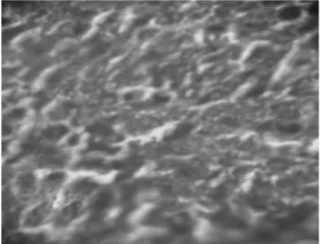
Microscopic features, the preparation contained of proliferation of small-cell, round, and some spheroidal and uniform cells. The nuclei are round or slightly oval, have finely dispersed chromatin, some of them with vesicular nuclei and one or two indistinct nucleoli. The tumor cells have clear cytoplasm without defined cytoplasm. Clusters of tumor cells separated by fibrous septa. Mitotic activity is variable There are proliferation of blood vessel without angioinvasion.
Laporan Kasus
Majalah Kedokteran Nusantara Volume 41 y No. 3 y September 2008 220
Figure 1. Clusters of small-cell, round and uniform, separated by fibrous septa
Figure 2. The nuclei are round and oval, dispered chromatin, clear and indistinct cytoplasm
DISCUSSION
Ewing’s sarcoma classically presents as pain, often accompanied by local inflammation. Fever is fairly common and may initially suggest the possibility of inflammatory lesion.1,2
There is definite male predilection. Children and adolescents are usually affected, a younger population than with any other primary bone tumor. About 75 percent of the patients with Ewing’s sarcoma are in the first two decades of life. The youngest patients was 5 months old and about 9 percent were in the first 5 years of life.2,3
Patients present with pain, with or without swelling. Pathologic fracture is rare. Some patients present with fever associated with laboratory findings of anemia, leucocytyosis, and increased erythrocyte sedimentation rate, all of which suggest infection.1,2,3,4,5
Gross finding, Ewing’s sarcoma tumor is a soft, white (fish-flesh) mass of almost liquid consistency. When tumor is sectioned, the contents may run like pus, and this may lead to a mistaken diagnosis of osteomyelitis. Geographic or punctate areas of necrosis may
be prominent. The cortex is often thickened, and fleshy tumor is seen between layers of thickened periosteum. In the modern era, almost all patients receive preoperative chemotherapy, radiotherapy, or both before surgical resection. In a resected specimen, the area of the tumor may be fibrotic or scarred. Occationally, the area is cystic, containing liquid necrotic debris.2,4
From cytologic findings, fine-needle aspiration biopsy (FNAB) specimens of primary Ewing’s sarcoma/PNET of bone are typically highly cellular. Two main types can be recognized in classics cases of Ewing’s sarcoma. The predominant population consists of relatively uniform cells with sparse, poorly defined cytoplasm and rounded, uniform nucleoli containing finely dispered chromatin and inconspicuous nucleoli. Less frequent are scattered, smaller cells, with hyperchromatic, dense nuclei. In addition, occasional cells with abundant glycogen and finely vacuolated cytoplasm are seen.
Atypical variants of Ewing’s sarcoma/PNET have a uniform population of somewhat larger cells with more-developed cytoplasm, enlarged nuclei with more irregular chromatin, and clearly discernable nucleoli. In these cases, mitotic figures are often seen. Although rossette-like arrangements of tumor cells have been described in aspirates of PNET, its distinction from classic Ewing’s sarcoma is usually impossible with FNA cytology.3,5,6,7
Histopathology findings, the essential microscopic finding is proliferation of small-cell, round, uniform cells and no matrix production. The Intergroup Ewing’s Sarcoma Study divided the growth pattern of this tumor into three types based on histologic findings: 1) diffuse: broad fields of tumor without topographic features; 2) lobular: island of tumor cells separated by fibrous septa; 3) filagree: delicate bicellular anastomosing strands separated by fibrovascular stroma.
Soekimin dkk. Ewing’s Sarcoma...
Majalah Kedokteran Nusantara Volume 41 y No. 3 y September 2008 221 peppered with almost uniformly placed
nuclei. The cytoplasm contains a moderate to large amount of glycogen granules.8
The nuclei have finely dispersed chromatin and one or two indistinct nucleoli. Mitotic activity is variable.
Necrosis may be slight or extensive; viable tumor may be arranged perivascular pattern. Nuclear dust may encrust vessels, producing the Azzopardi phenomenon that, best known with small cell carcinoma of the lung.
Since the original description by Ewing, it has been recognized that the tumor may contain rossettes, which is a feature considered typical of primitive neural tumors. Indeed, a small subset of Ewing’s sarcoma has a lobulated growth pattern, prominent rossettes, and even a fibrillary background, features that strongly suggest neural differentiation. The term primitive neuronectodermal tumor (PNET) has been applied to this lesion. Immunohistochemical and cytogenetic studies have confirmed that Ewing’s sarcoma may derived from neuroectoderm and that classic Ewing’s sarcoma, large cell Ewing’s sarcoma, and PNET (including the so-called Askin’s tumor) all belong to the same family but have different degrees of formation.7
Histologic grading is not practical because of the uniform appearance from tumor to tumor. All small celll sarcomas are high-grade tumors (grade 4 of 4).
Open biopsies are rarely necessary to confirm the diagnosis of Ewing’s sarcoma. If the findings on fine-needle aspiration cytology are not diagnostic, open biopsy performed and frozen section must be examined to confirm the presence of viable tumor and to allocate material for all special studies.1,2,4,5,9
CONCLUSION
We report a rare case, young female, with enlarge abdominal and from biopsy result diagnosed as Ewing’s sarcoma. From microscopic findings shows proliferation of small-cell, round, and some spheroidal and uniform cells. The nuclei are round or slightly oval, have finely dispersed chromatin, some of them with vesicular nuclei and one or two indistinct nucleoli. The tumor cells have clear
cytoplasm without defined cytoplasm. Clusters of tumor cells separated by fibrous septa. Mitotic activity is variable There are proliferation of blood vessel without angioinvasion.
Unfortunately, we didn’t find result of laboratory examination to confirm clinical features. To make sure, it’s better to make special stain (Periodic Acid Schiff and reticulin) to differ with malignant lymphoma and immunohistochemistry (CD99, vimentin, NSE, and S-100 protein).
The microscopic differential diagnosis of Ewing’s sarcoma includes osteomyelitis, eosinophilic granuloma, and the group of small-cell tumors that includes lymphoma, leukemia, and metastatic neuroblastoma (and, in the case of soft-tissue Ewing’s sarcoma).
REFERENCES
1. Bullough Peter, Orthopedic Pathology, fourth edition, Mosby, 2004, p. 471-4. 2. Robbins et all, Basic Pathology, 7th
edition, Saunders, 2003, p. 770-2.
3. Mills Stacey et all, Stenberg’s Diagnostic Surgical Pathology, volume IA, 4th edition, Lippincott Williams & Wilkins, 2004, p. 272-3.
4. Krishnan, et all, Tumors of The Bones and Joints 2, AFIP Atlas of Tumor Pathology series 4, ARP, 2005, p. 209-22.
5. Rosai, Rosai and Ackerman’s Surgical Pathology, volume two, ninth edition, Mosby, 2004, p.2172-7.
6. Orel, Svante R et all, Fine Needle Aspiration Cytology, fourth edition, Elsevier, 2005, p. 442-3.
7. Fine-needle Aspiration Cytology of Extraskeletal Ewing’s sarcoma, available at
http:///www.javascript:AL_get(this,’jour’ Diagn Cytopathol.’)
8. Kissane, John M, Anderson’s Pathology, volume II, 9th
edition, Mosby, 1990, p. 2045-47.