Effect of extracorporeal irradiation on segmental bone
autograft incorporation in Sprague-Dawley rats
Abstrak
Latar belakang: Autograft tulang digunakan pada operasi rekonstruksi kasus tumor tulang. Extracorporeal irradiation (ECI) pada tulang digunakan untuk membunuh sel tumor. Namun radiasi juga menimbulkan efek negatif terhadap osteoinduksi dan osteogenensis untuk inkorporasi autograft tulang. Penelitian ini bertujuan mempelajari pengaruh iradiasi dengan dosis 50-300 Gy terhadap kemampuan inkorporasi graft tulang.
Metode: Pada 24 ekor tikus Sprague-Dawley dilakukan reseksi en bloc 7 mm pada diaisis tibia. Tikus dibagi menjadi 4 kelompok: kelompok 1 tidak mendapat radiasi; kelompok 2, 3 dan 4 berturut-turut mendapat iradiasi 50, 150 dan 300 Gy pada tulang yang direseksi, selanjutnya diimplantasi kembali. Inkorporasi tulang dievaluasi pada minggu ke-6 dan -8 secara radiologi dan pada minggu ke-8 dilakukan evaluasi histopatologis, hitung osteoblas dan pengukuran ekspresi BMP-2. Data dianalisis menggunakan uji Anova dan Kruskal-Wallis.
Hasil: Pada minggu ke-6, skor radiologis kelompok iradiasi 150 dan 300 Gy lebih rendah dibandingkan kontrol (4 vs 6 dan 4 vs 6; p = 0,01). Demikian juga pada minggu ke-8 (5,40 vs 7,14; p = 0,009 pada kelompok 150 Gy dan 5,60 vs 7,14; p = 0,018 pada kelompok 300 Gy). Pemeriksaan histopatologis memperlihatkan bahwa median skor histologis kelompok iradiasi 50, 150 dan 300 Gy lebih rendah secara bermakna dibanding kontrol (6 vs 7 , p = 0,017; 4 vs 7, p = 0,005; 6 vs 7, p = 0,013). Hitung osteoblas dan ekspresi BMP-2 tidak menunjukkan perbedaan bermakna pada tiap kelompok.
Kesimpulan: ECI dengan dosis 50 sampai 300 Gy terhadap autograft tulang menyebabkan keterlambatan dalam inkorporasi autograft tulang, namun autograft tersebut masih memperlihatkan kemampuan osteoinduktif dan osteogenesis.
Abstract
Background: Bone graft has been widely used in bone tumor reconstructive surgery. Extracorporeal irradiation (ECI) is commonly used to eliminate malignant cells before bone autograft. However, it may have negative effects on autograft incorporation. This study aimed to evaluate the ability of bone autograft incorporation after extra corporeal irradiation.
Methods: 24 Sprague-Dawley rats underwent 7-mm en bloc resection of tibial diaphysis, and were divided into 4 groups. The irst group did not receive irradiation; the 2nd, 3rd, and 4th groups received 50, 150 and 300 Gy
bone irradiation respectively, and then reimplanted. Radiologic score were evaluated at week-6 and -8, while histopathology, osteoblast count and BMP-2 expression were examined at week-8. Data were analyzed with ANOVA or Kruskal-Wallis tests.
Results: At week-6, radiologic scores in group 150 and 300 Gy were signiicantly lower compared to control group (4 vs 6 dan 4 vs 6; p = 0.011; p = 0.01). The same results were also obtained at week-8 (5.40 vs 7.14; p = 0.009 in the group 150 Gy and 5.60 vs 7.14; p = 0.018 in the group 300 Gy. Histopathological scores of the groups receiving 50, 150 and 300 Gy were signiicantly lower compared to the control group (6 vs 7, p = 0.017; 4 vs 7, p = 0.005; 6 vs 7, p = 0.013). Osteoblast count and BMP-2 expression were not signiicantly different among all groups.
Conclusion: ECI with the dose of 50 to 300 Gy is associated with delayed bone autograft incorporation. However, the osteoinductive and osteogenesis capacity for autograft incorporation were maintained.
Keywords: autograft incorporation, BMP-2, extracorporeal irradiation (ECI), osteoblast, osteoinductive, osteogenesis
pISSN: 0853-1773 • eISSN: 2252-8083 • http://dx.doi.org/10.13181/mji.v23i3.1082 • Med J Indones. 2014;23:147-53
Correspondence author: Muhammad Wahyudi, [email protected]
B a s i c M e d i c a l R e s e a r c h
Copyright @ 2014 Authors. This is an open access article distributed under the terms of the Creative Commons Attribution-NonCommercial-ShareAlike 4.0 International License (http://creativecommons.org/licenses/by-nc-sa/4.0/), which permits unrestricted non-commercial use, distribution, and reproduction in any medium, provided the original author and source are properly cited.
Muhammad Wahyudi,1 Achmad F. Kamal,1 Nurjati C. Siregar,2 Marcel Prasetyo3
Nowadays, bone graft is widely used in orthopedic surgery, either for fracture healing or bone tumor reconstructive surgery. Reconstruction of the bone tumor needs a massive bone graft, either by autograft or allograft as a substitute of prostheses, depends on the case and graft availability. Bone allograft, which are common in the countries that have tissue banks, has some disadvantages including the risk of viral transmission and the possibility of rejection due to recipient’s immune system. In the absence of tissue bank facility, bone autograft become an option. This autograft brings additional advantages such as a much lower risk of immune reaction, as well as a lower cost for the patients. However, the possibility of residual bone tumor in the autografted segment represents
a signiicant drawback. An attempt to overcome
this problem is by extracorporeal irradiation (ECI) of bone segment to be implanted. In this method, the bone undergoes en bloc resection, irradiation and re-implantation. Irradiation dose of 50-300 Gy is usually used and it has been known to be quite effective to kill tumor cells. But, radiation may also have negative effects on cells of the bone graft.1,2
The bone components, such as the osteogenic and bone marrow cells are radiosensitive, while the inorganic matrix, the major components of the bone is relatively radio resistant.
Osteogenic cells are precursor cells which play roles in the formation of the new bone. A lot of studies have been conducted with the aim to identify the effect of irradiation on the bones, bone healing and bone graft incorporation.3,4
Besides osteogenic cells, the bone matrix also contains growth factors which are essential for bone healing and bone graft incorporation. One of the growth factors is bone morphogenic protein-2 (BMP-2), a derivate of transforming growth
factor-β (TGF-β). Study that has been conducted
by Tarmo et al, showed that a irradiation up to 25 kGy on BMP-2 extracted in gelatin capsules did not affect BMP-2 function which acts as osteoinductive factor.5
The effect of different doses of ECI on bone autograft incorporation and BMP-2 expression in the osteotomy healing area has not been clearly
identiied. This study was aimed to evaluate the inluence of extracorporeal irradiation on the
incorporation ability of bone autograft. In addition,
its inluence on histological features and expression
of BMP-2 were also evaluated.
METHODS
This was an experimental study using post-test only control group design conducted at the Laboratory of Experimental Animal and Toxicology, Research and Development Unit, Ministry of Health, Indonesia. Extra corporeal irradiation was performed at National Nuclear Energy Agency of Indonesia
(BATAN), Jakarta; while histopathological and
immunohistochemical studies were conducted at Department of Pathological Anatomy, Faculty of Medicine, Universitas Indonesia.
The study was conducted on 24 white Sprague-Dawley rats, 8-12 weeks, 250-350 g BW, which were bred at the Laboratory of Experimental Animal and Toxicology, Research and Development Unit, Ministry of Health, Indonesia, between December 2010 and February 2011. The rats were devided into four groups, the control group without irradiation and three groups receiving 50 Gy, 150 Gy and 300 Gy irradiation, respectively. The protocol of this study has been approved by Ethical Committee of the Faculty of Medicine, Universitas Indonesia (472/PT02.FK/ETIK/2010).
Surgical procedure
The rat was placed in supine position, under aseptic condition an anterior incision (1.5 cm) was made on the left tibia. En bloc resection of the left tibial diaphysis was made about 7 mm in size. The bone was temporarily
ixed with 0.8 mm Kirschner wire, soft tissues and skin was stitched using 4.0 vycril ilament. The resected bone
segments were put into transportation device equipped with dry ice. The bone for irradiation was taken to BATAN and received gamma irradiation of 50 Gy, 150 Gy and 300 Gy emitted by a calibrated radiotherapy Cobalt-60 source. After irradiation, the bone segments
were reimplanted, ixated with a 0.8 mm K-wire and
put into the medullary canal in a retrograde fashion toward proximal tibia and subsequently inserted to distal. After reimplantation, the experimental animal was kept at animal laboratory.
Evaluation procedure
At the 6th and 8th week after bone autograft, implantation
anterio-posterior (AP) and lateral projections of tibial radiograph were performed. At the 8th all experimental animals were sacriiced, the tibial bone was resected and sent for histologic evaluation. Parafin-embedded block
were stained with hematoxylin eosin (HE) and some were left unstained for immunohistochemical analysis. The HE stained slides were evaluated for radiological
score using modiied Salkeld histological score and
for osteoblast count. Immunohistochemical analysis was performed using antibody antiBMP-2 reagent to evaluate BMP-2 expression. These parts of study were conducted at the Department of Pathological Anatomy, Faculty of Medicine, Universitas Indonesia.
Radiographic assessment
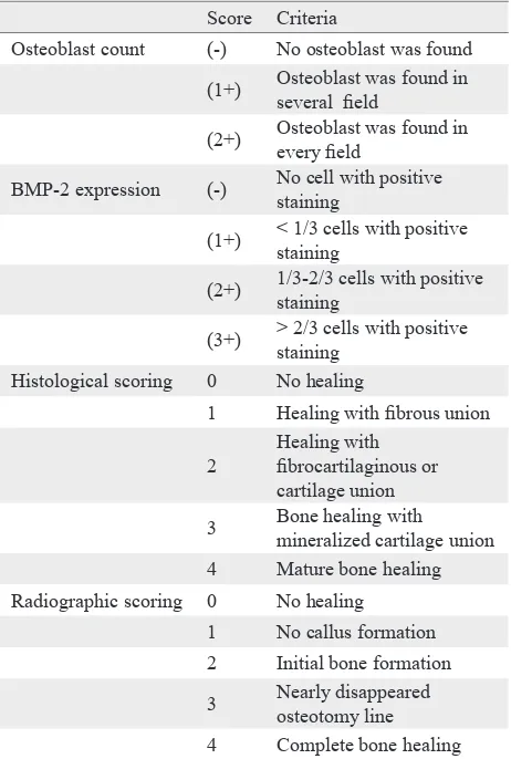
The radiographic evaluation of bone graft incorpora-tion was made based on radiographic grading score as described by Lane and Sandhu.6 Score are shown
in table 1. The assessment was performed by observ-ing the proximal and distal osteotomy line on AP and lateral projection of radiograph at sixth and eighth week. The assessment was carried out using X-ray instrument by Philips bulky diagnosis, 46 kV 5.00 mAs. The distance between the light source and the
ilm was 90 cm. The total score of complete radio -graphic assessment between proximal and distal dis-tance was 8. The assessment was carried out by an expert in musculoskeletal radiology.
Osteoblast count
On microscopic examination (40X magniication, HE
stain), osteoblast count was evaluated by examining the proximal to distal osteotomy area and autograft cortex. The evaluation was performed by an expert in pathological anatomy. The evaluation criteria are shown in table 1.
BMP-2 assessment
Immunohistochemical analysis was performed using the BMP-2 (P275) pAb reagent kit (Bioworld Technology, USA) with product category number of BS3473 to detect BMP-2 expression in rat bones. The evaluation was performed by using a
microscope at 40x magniication and analyzing three ields of view in intramedullary area of the bone
autograft. The number of cells expressing BMP-2 was counted.7,8 The semi-quantitative assessments of
BMP-2 expression are shown in table 1.
Histopathological assessment
The assessment of autograft incorporation was evaluated histologically after performing the
hematoxilyn eosin (HE) staining based on modiied
Score Criteria
Osteoblast count (-) No osteoblast was found (1+) Osteoblast was found in
several ield
(2+) Osteoblast was found in every ield
BMP-2 expression (-) No cell with positive staining
(1+) < 1/3 cells with positive staining
(2+) 1/3-2/3 cells with positive staining
(3+) > 2/3 cells with positive staining
Histological scoring 0 No healing
1 Healing with ibrous union
2
Healing with ibrocartilaginous or cartilage union 3 Bone healing with
mineralized cartilage union 4 Mature bone healing Radiographic scoring 0 No healing
1 No callus formation 2 Initial bone formation 3 Nearly disappeared
osteotomy line 4 Complete bone healing
Table 1. Evaluation criteria of osteoblast count, BMP-2 expression, histological and radiographic score
Salkeld fracture healing scoring. The proximal and distal area of osteotomy was evaluated. The histology scoring was the total score of proximal and distal osteotomy area. The range of score was between 0 and 8.9
Data analysis
Data are presented descriptively to indicate the characteristics of the tested bones and their similarity to control group. ANOVA test and Kruskal-Wallis test were used to analyze the correlation between independent and dependent variables.
RESULTS
Radiographic assessment of autograft incorporation
At the 6th week, the post-hoc analysis revealed p =
group 6.0 (5.0-7.0). Following 300 Gy irradiation, the median score of bone graft incorporation rate at 6th week was 5.0 (4.0-6.0) which was signiicantly lower compared control group, which is 6.0 (5.0;
7.0). There was no signiicant difference among the irradiation groups.
At the 8th week, the mean score of bone autograft
incorporation rate following 150 Gy irradiation is 5.40 ± 1.14 which was signiicantly lower compared to the control group (7.14 ± 0.69, p < 0.05). In the group receiving 300 Gy irradiation, the score was 5.60 ± 1.14 (p < 0.05 vs control group). No signiicant difference among the irradiation groups (Table 2).
Comparison of histopathological score
Table 3 shows comparison of histopathological, osteoblast count and BMP-2 expression. The median value of bone histopathological score following 50, 150, and 300 Gy irradiation are 6.0 (5.0-6.0), 4.0 (4.0-6.0), and 6.0 (4.0-6.0). All of these values were
signiicantly different from control group [7.0
(6.0-8.0), p < 0.05)]. There was also signiicant different between the group of 150 Gy and 50 Gy (p = 0.028).
DISCUSSION
This study used radiographic grading score as described by Lane and Sandhu6 to assess fracture
union. By using this score, the quality of healing in proximal and distal osteotomy can be evaluated. The timing of X-ray assessment at the 6th week was
based on the fact that the X-ray assessment for tibial
fracture union showed no signiicant changes after
Score of incorporation
Control
(n = 7) 50 Gy irradiation(n = 5)
150 Gy irradiation (n = 5)
300 Gy irradiation
(n = 5) Statistical test p Median at 6th week 6.0 (5.0 ; 7.0) 6.0 (4.0 ; 7.0) 4.0 (3.0 ; 5.0) 4.0 (4.0 ; 6.0) Kruskal-Wallis 0.012
Mean at 8th week 7.14 ± 0.69 6.60 ± 1.14 5.40 ± 1.14 5.60 ± 1.14 One-Way ANOVA 0.027
Table 2. Comparison of radiographic scoring for bone graft incorporation at 6th and 8th week after extracorporeal irradiation
Variable Control
(n = 7)
50 Gy irradiation
(n = 5)
150 Gy irradiation
(n = 5)
300 Gy irradiation
(n = 5)
Statistical test p
Median of histopathological score 7.0 (6.0 ; 8.0) 6.0 (5.0 ; 6.0) 4.0 (4.0 ; 6.0) 6.0 (2.0 ; 6.0) Kruskal-Wallis 0.004 Median of osteoblast count constant 2.0 (1.0 ; 2.0) 1.0 (1.0 ; 2.0) 2.0 (1.0 ; 2.0) Kruskal-Wallis 0.16 Median of BMP-2 expression 2.0 (2.0;3.0) 2.0 (1.0 ; 3.0) 1.0 (1.0 ; 3.0) 2.0 (1.0 ; 3.0) Kruskal-Wallis 0.77
Table 3. Comparison of histopathological, osteoblast count and BMP-2 expression
Control
50
150 Gy irradiation
30
150 Gy irradiation
30
50 Gy irradiation
300 Gy irradiation
300 Gy irradiation
Control 50 Gy irradiation
150 Gy irradiation 300 Gy irradiation
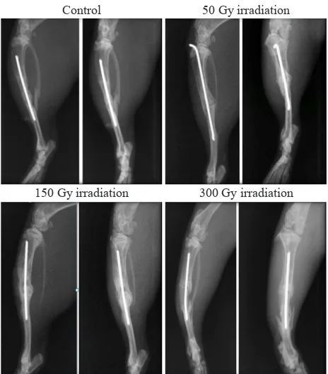
Figure 1. Tibial bone X-Ray at 8th week
6 weeks.10 In other word, on the sixth week, normal
tibial bone fracture had undergone radiographical healing. We expected to see delayed bone healing by performing the X-ray at the 8th week.
At the 6th week, animals in the control group and those
receiving 50 Gy irradiation showed radiographic healing, or at least had undergone bone formation. In fact, there were segments which showed complete bone healing according to Lane and Sandhu score.
This inding is consistent with the results of the study
on the 6th week, the bone autograft, which was
not exposed to irradiation had been healed with bone formation, and immature bone (woven bone) formation can be shown histologically.Meanwhile, most of bones which are exposed to 150 Gy and 300 Gy irradiations, were still in callus form and
just began to have bone formation; although, there
were 2 cases with complete bone healing. Thus, it can be assumed that 150-300 Gy irradiations
may cause delayed bone autograft incorporation;
while 50 Gy irradiation group showed similar incorporation rate with control group at 6th week
observation.
After the 8th week, radiographic assessment in
the control group revealed that there were 2 rats experiencing complete bone healing on both
segments; while the other still demonstrated
osteotomy line though the line has almost been disappeared. Some rats in the groups receiving irradiation also demonstrated complete bone healing at 8th week, which showed diminished osteotomy line. After post-hoc analysis was performed, a signiicant
difference was found regarding the average rate of bone graft incorporation between control group and those with 150 Gy and 300 Gy irradiations.
A study conducted by Voggenreiter, et al6
demonstrated that at the 9th week, the bone autograft
without irradiation had experience bone healing
by bone formation; moreover, in some cases the
osteotomy line had disappreared and the osteotomy area was hardly recognized. It can be said that the radiographic results of the control group at 8th week in
our study are similar with the results of Voggenreiter at 9th week. Higher dose of irradiation (1kGy) has shown signiicant difference between the irradiated
non irradiated autograft.
It can be concluded from the radiographic grading score that 150-300 Gy irradiation causes delayed incorporation of bone autograft. However, the irradiated bone still has the capacity for incorporation, as indicated by the increased score between the 6th and 8th week. Radiographically, the
irradiation with 50 Gy does not give any signs of delayed incorporation.
The union process between autograft and recipient bones is similar to fracture healing process. However, the autograft transplantation of cortex bone may be accompanied by a process of creeping substitution, which includes revascularization at the beginning
of healing process, and subsequently followed by resorption of bone graft and formation of the new bones.11
In this study, the union process of proximal and distal segment of bone autograft against recipient
bone was evaluated histologically using the modiied
Salkeld score, which assess the quality of the union.10 We used K-wire during implantation for ixation and
after implantation the rats directly went through
weight bearing on their operated extremities;
therefore, the healing model would be through the
process of endochondral ossiication.
Endochondral ossiication involves recruitment,
proliferation and differentiation of undifferentiated
mesenchymal cells into calciied cartilage
that eventually will be replaced by bone. The characteristic of such healing process includes some
stages: hematoma formation, inlammatory phase, cartilage formation, calciication of the cartilage,
cartilage resorption, bone formation and eventually bone remodeling.12
It has been known that irradiation may damage cells, both directly and indirectly. By direct mechanism, the irradiation ionizes cell DNA or other essential components of cell causing chemical changes in the cells or DNA. Bone graft irradiation may affect osteogenesis and osteoinductive components that play role in bone autograft incorporation.
The effect of irradiation on bone at initial stage includes vascular dilatation accompanied with injured myeloid cells and subsequently followed by bone resorption and cell lost in bone marrow. It can be due to macrophage disintegration in the bone marrow, which releases enzymes and causes myeloid aplasia. Occurrence of bone resorption is indicated by increased osteoclast lining of resorbed trabecula.13
Damage in bone marrow may affect autograft incorporation since the stroma of bone marrow is a major contributing factor in osteogenesis. However, it should be noted that after each damage, the repairing process will follow.14
Maeda, et al15 revealed that at initial stage of
radiation, there were decreases of bone cells and osteoid, resulting in lower osteoblastic and osteoclastic activity. After this period, there will be
regeneration of the dead cells; however, osteoblastic
Osteoblast is one of cell component of the bone which has essential function in collagen synthesis
to develop osteoid and calciication process of the
osteoid. Osteoblast is derived from osteoprogenitor cells of bone marrow stroma or from the adjacent soft tissues, and act as component of osteogenesis. Active osteoblast can change into bone lining cell and osteocyte.11,12,16
It has been known that the higher the levels of cell proliferation and differentiation, the more resistant the cells against radiation are. A study conducted by Jacobson et al on rat bones with irradiation dose of 40 Gy indicated that the osteocytes was still viable.17
A study conducted by Dare, et al18 demonstrated that
following 40 Gy irradiation in rats, there was reduced capacity of osteoblast differentiation. Moreover, it was also found that there was reduced alkaline phosphatase activity.
Sugimoto, et al13 found that during irradiation up to
50 Gy, the osteocytes stayed intact and only several empty lacuna were detected. Moreover, after a few
weeks of irradiation there was signiicant lower
capacity of developing new bone and reduced number of cells in bone marrow. However, when followed for a longer period, increased new bone formation, bone marrow cells and reduced porous bone could be observed.13 Since new bone formation is the function
of osteoblast, it can be concluded that immediately after irradiation, osteoblast damage occurs. However, since the bone marrow has a capacity for regeneration and it is one of osteoprogrenitor
cells sources, therefore our study did not ind any signiicant different between the control group and
irradiated groups. It is assumed that at 8th weeks
following the irradiation, the bone marrow cells have regenerated and produced osteoblast cells.
BMP-2 is part of transforming growth factor-β superfamily and it is one of bone growth factor components and has has chemotactic characteristic for osteoprogenitor cells in vitro.19 A study conducted
by Bostrom, et al 20 showed the expression of BMP-2
in fracture callus. Thus, they concluded that BMP-2 has essential role in fracture healing.
Bone autograft has osteoinductive properties. It has a capacity to recruit mesenchymal cells in the host and its surrounding to differentiate into osteoblast and chondroblast. The recruitment is induced by
growth factor inside the autograft, such as BMP-2.11 Anderson, et al21 has conducted a study to
identify BMP-2 expression in bone. By performing immunohistochemical analysis, the study found BMP-2 expression in osteoblast, chondrocytes, osteoprogenitor cells of medullary bone stroma and cells that have role in vascular formation.
In our study, there was no signiicant difference of BMP-2 expression at least in two groups. It is similar to the study conducted by Anderson, et al.21 There was no signiicant difference between
control and irradiated groups, because after 8 weeks, cell regeneration has occurred in bone
marrow; therefore, during the assessment, it has
been able to express BMP-2. Another rational for this includes BMP-2 denaturation has not been proven to be present following irradiation in a dose of 50-300 Gy. It is in accordance with the study conducted by Wintroub et al,22 which demonstrated
that BMP still has osteoinductive function at dose range of 50-70 kGy on demineralized allograft. Osteogenesis was evaluated by osteoblast count and osteoinductive factor was assessed by BMP-2 expression. Both factors have important role in autograft incorporation. The osteoblast count and BMP-2 expression at 8th week did not show signiicant difference between control group and
autograft that had received irradiation.
If we consider the histological and radiographical score, it can be said that at 8th week, there has been
regeneration of bone marrow cells. Thus, osteoblast
count and BMP-2 expression show non-signiicant
difference. This is consistent with a study conducted by Sugimoto, et al13 which indicated that following
irradiation, the bone marrow has a capacity for regeneration. The larger the dose of irradiation, the longer the time needed for regeneration.
A study by Tarmo, et al5 showed that BMP-2
extracts which received 4 kGy irradiation still has osteoinductive function. It can be also considered that bone autograft irradiation up to 300Gy does not cause denaturation of BMP-2.
It can be concluded from our study, that extracorporeal irradiation at the dose of 50-300 Gy is associated with delayed incorporation without impairing the capacity of bone autograft incorporation since it still retain osteoinduction and osteoconduction capabilities.
Acknowledgment
We would like to thank those who have facilitated the conduct of this study and writing of this manuscript. In particular, all staff at Department of Orthopaedic and Traumatology, Department of Radiology, and Department of Pathological Anatomy Faculty of Medicine, Universitas Indonesia/Cipto Mangunkusumo Hospital. Also we would like to thank Mrs. Febrida Anas for guidance and support the process of irradiation at BATAN.
Conlic of interest
All authors have nothing to disclose.
REFERENCES
1. El-Wahidi GF, Eldesoky I, Kotb S, Awad I, Thabet M, Thaleh Y. Neoadjuvant chemotherapy & low dose extracorporeal irradiation for treatment of osteosarcoma. J Clin Oncol. 2005;23(16S):9074.
2. DiCaprio MR, Friedlaender GE. Malignant bone tumors: limb sparing versus amputation. J Am Acad Orthop Surg. 2003;11(1):25-37.
3. Yusof N. Interaction of radiation with tissues. Singapore: World scientiic; 2007. p. 99-107.
4. Currey JD, Foreman J, Laketić I, Mitchell J, Pegg DE, Reilly GC. Effect of ionizing radiation on mechanical properties of human bone. J Orthop Res. 1997;15(1):111-7. 5. Pekkarinen T, Hietala O, Jämsä T, Jalovaara P. Effect
of gamma irradiation on the osteoinductivity of morphogenetic protein extract from reindeer bone. Acta Orthop. 2005;76(2):231-6.
6. Voggenreiter G, Ascherl R, Blümel G, Schmit-Neuerburg KP. Extracorporeal irradiation and incorporation of bone graft. Acta Orthop Scand. 1996;67(6):583-8.
7. Wang FS, Yang KD, Kuo YR, Wang CJ, Sheen-Chen SM, Huang HC, et al. Temporal and spatial expression of bone morphogenic protein in extracorporeal shock wave promoted healing of segmental defect. Bone. 2003;32(4): 387-96.
8. Rauch F, Lauzier D, Croteau S, Travers R, Glorieux FH, Hamdy R. Temporal and spatial expression of bone
morphogenetic protein-2, -4, and -7 during distraction osteogenesis in rabbits. Bone. 2000;27(3):453-9.
9. Oztürk A, Yetkin H, Memis L, Cila E, Bolukbasi S, Gemalmaz C. Demineralized bone matrix and hydroxyapatite/ tri-calcium phosphate mixture for bone healing in rat. Int Orthop. 2006;30(3):147-52.
10. Watanabe Y, Nishizawa Y, Takenaka N, Kobayashi M, Matsushita T. Ability and limitation of radiographic assessment of fracture healing in rats. Clin Orthop Relat Res. 2009;467(8):1981-5.
11. Khan SN, Cammisa FP Jr, Sandhu HS, Diwan AD, Girardi FP, Lane JM. The biology of bone grafting. J Am Acad Orthop Surg. 2005;13(1):77-86.
12. Dimitriou R, Tsiridis E, Giannoudis PV. Current concepts of molecular aspects of bone healing. Injury. 2005;36(12):1392-404.
13. Sugimoto M, Takahashi S, Toguchida J, Kotoura Y, Shibamoto Y, Yamamuro T. Changes in bone afer high doses irradiation. J Bone Joint Surg Br. 1991;73(3):492-7. 14. Furtsman LL. Effect of radiation on bone. J Dent Res.
1972;51(2):596-604.
15. Maeda M, Bryant MH, Yamagata M, Gi L, Earle JD, Chao EY. Effects of irradiation on cortical bone and their time-related changes. A biomechanical and histomorphological study. J Bone Joint Surg Am. 1988;70(3):392-9.
16. Salter RB. Normal structure and function of musculoskeletal tissues. In: Textbook of disorders and injuries of the musculoskeletal systems. 3rd ed. Philadelphia: Lippincott Williams & Wilkins; 1999. p. 7-16.
17. Jacobsson M, Kälebo P, Tjellström A, Turesson I. Bone cell viability after irradiation. An enzyme histochemical study. Acta Oncol. 1987;26(6):463-5.
18. Dare A, Hachisu R, Yamaguchi A, Yokose S, Yoshiki S, Okano T. Effects of ionizing radiation on proliferation and differentiation of osteoblast-like cells. J Dent Res. 1997;76(2):658-64.
19. Welch RD, Jones AL, Bucholz RW, Reinert CM, Tjia JS, Pierce WA, et al. Effect of recombinant human bone morphogenetic protein-2 on fracture healing in a goat tibial fracture model. J Bone Miner Res. 1998;13(9):1483-90.
20. Bostrom MP, Lane JM, Berberian WS, Missri AA, Tomin E, Weiland A, et al. Immunolocalization and expression of bone morphogenic proteins 2 and 4 in fracture healing. J Orthop Res. 1995;13(3):357-67.
21. Anderson HC, Hodges PT, Aguilera XM, Missana L, Moylan PE. Bone morphogenetic protein (BMP) localization in developing human and rat growth plate, metaphysis, epiphysis, and articular cartilage. J Histochem Cytochem. 2000;48(11):1493-502.