Tamaelg et al. Med
J
Univ IndonSimplifying Excretory Urography
in
Children
with
Urinary Tract
Infection
Lebrien
Agustine
Tamaela, I ahja Zacharia,Widhodho
Titiet
Karyoman ggolo
Abstrak
Infeksi saluran kemih (ISK) anak yang tidak ditanggulangi baik bisa berdampakfatal. Pencitraan uroradiologis, a.l. pielografi intravena (PIV), pemeriksaan terpilih untuk mengganbarkan saluran kenih dengan lengkap, harus dilakukan. Tetapi dosis radiasi PIV tinggi. Tujuan penelitian ini ialah menilai eftsiensi PIV tanpa kehilangan infornnsi diagnostik. Dilakuknn studi observasi (memperoleh urogram dan kombinasifoto minimal yang lengknp nrengganbarkan konponen saluran kenih) dan studi perbandingan kemampuan diagnostik Qnenilai kematnpuan pola PIV optimal, dibandingkan dengan PIV klasik). Tiapfoto
dari
100 seri PIV (foto polos perut, foto urograrn nenit ke 4, 8, 12 dan 3O) periode Januari 1986 -April
1990, dinilai 3 penilai independen dalam hal kejelasan dan kemanrpuan deteksi kelainan. Yang dibandingkan adalah indeks efficacy uji diagnostik (sensitivitas, spesifisitas, nilai prediksi positif, nilai prediksi negafif, akurasi, rasio kemungkinan positif, rasio kemungkinan negatifl. PIV klasik yang dinilai panel 2 pakar radiologi anak digunakan sebagai baku emas. Foto urogram menit ke 8 adalahfoto tunggal paling lengkap untuk netnvisualisasikan seluruh komponen PIV. Kombinasifoto
polos dengan urogratn menit ke 8 dengan atau tanpa urogran menit ke 4, adalah kombinasi PIV paling optimal. Kemampuan diagnostiknya ternyata setara dengan PIV klasik (penguranganfoto tidak nengurangi kenanpuan deteksinya). Kesitnpulan yang diperoleh ialah sebagian besar PIV dapat dihentiknn setelahfoto urogra,n nenit ke E. Pola PIV optinal adalah konbinasifoto polos dan urogra,n menit ke 8 dengan atau tanpa urogram menit ke 4. Ke 2 pola tersebut dapat digunakan sebagai alternatif pola petneriksaan PIV rutin untuk pasien ISK anak.Abstract
Failure to detect abnonnalities in children with urinary tract infections may end up fatal. Excretory urography (EU) has been the exanination of choice to visualize details of the urinary tract, but EU gives a relatively high radiation dose. The purpose of this study is to evaluate sitnplifying EU without loosing its diagnostic efficacy. Observational and diagnostik tests cot,rparison study has been done. Reevaluation of eachfilm of lOO pediatric EU series in proven UTI cases expntined front January 1986 -
April
1990, by 3 independent investigators, describing visualization clarity and the presetrce of abnonnality of the urinary tract. The 'gold standard' used was a consensus panel of 2 senior pediatric radiologists. The 8 ninute urogra,,$ were found to be the most cotnplete singlefihn.
As an exarnination, cotnbination of plain photo of the abdotuetr and the 8 ,,rinute urogra,il with or without the 4 minute urogra,n, were the optimal 2 and 3 filtns EU procedures. By reducing exposure there was insignificant diagttostic efficacy lost. It is concluded that in,nost pediatric UTI cases, EU examination could be stopped afier cottrplete visualizarion of the uri,rary tract : after plain photo ofthe abdotnen and 8 minute urograrn with or without the 4 minute urogran. These could be reconnended to be thenininal
routine procedure.Keywords : pediatric imaging, urinary tract infection, sitttplification, excretory urography, diagnostic tests,
fficacy
indexesINTRODUCTION
Urinary tract infection (UTI) in infants
andchildren is
a
common
andsignificant
childhood
morbidity;
many
childhood
UTI
cases have somestructural
and/orfunc-tional urinary tract abnormalities, predisposing to
recurrent
andpersistent UTI. Failure to
detectunder-lying urinary tract anomalies
andfurther
mismanage-ment of complicated
UTI. may
endin chronic
pyelo-nephritis
andend-stag"
r.nul fuilur".
t'2'3'o'sThe excretory urography (EU)
has beenthe
ex-amination
of choice
to visualize both the details
andthe overview
of
the urinary
tract.5'6Unfortunately
it
gives a relatively
high gonadal
radiation
dosewhich
could
increase the risk of
malignancy induction
andgenetic
durnug".8'9'loRoutine EU examination
startswith
aplain photo
of
the abdomenwith
akidney ureter
andbladder
over-view (KUB). This
film usually detects
radioopaquecalculi, skeletal anomaly, calcification, and foreign
Vol 3, No l, Junuury - March 1994
objects
andis
usedto
evaluate the
bowel
preparation
of the patient and as
a
control of the radiological
technique.
After IV injection of a
contrast agent, someexposures
are
done to
visualize the kidney,
pel-viocalyceal
system, the ureter
andthe bladder,
while
the agent
is being excreted.l0'l
I'12To
get acomplete
visualization
of
theseEU
com-ponents,
the experts have different ideas about
thenumber
andthe
sequenceof
the
exposures.6'10'l I'12'13Since
1966EU in
our
pediatric imaging
subdivision,
has beenperformed
in a
routine procedure:
1plain
AP
photo
of
the
abdomen and
4 AP
urograms,
eachex-posed
at
4
(restricted
to
renal area),
8,
12, and
30minutes.
These exposuresgive overlapping
visualiza-tion of
theEU
components, thus wasting
X-ray
radia-tion.
The main
purpose
of
the study
was
to find out
whether EU could be simplified, without loosing
its
diagnostic
efficacy.
Simplified EU
examinations
will
decreaseradiation
dosein
pediatric patients (reducing
pediatric radiation
exposure), and
on the other
hand increases thepossibility
toperform
moreEU
examina-Simplified EU
Excretory Urography in ChiUren with
IITI
3l
tions
for
children
needingthis examination, especially
in developing countries,
with limited
medical
funding,
man
power (radiographers)
andfilms.
MATERIAL
AND METHODS
The
study
was conducted toevaluate
I2Opediatric
EU
of UTI
cases
done in
the
Pediatric
Imaging
SubDivision
of the
Child
Health Department, Medical
School
University
of Indonesia/Cipto Mangunkusumo
General
Hospital,
Jakarta,
from
January
1986-
April
1990.Two
steps of the study consisted ofobservational
studies anddiagnostic
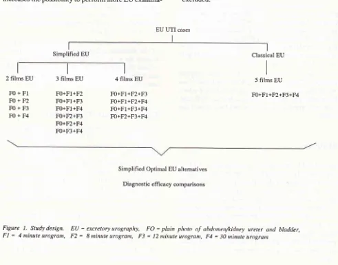
testscomparison study (Fig.1).
Eligible EU
series hadto
have aproven culture
of
themid-stream
urine
>
100.000
bacterial
colony/ml
(proven
UTI cases),
and consistedof5
photos:
1plain
AP photo
of the abdomen
(FO)
and
4 AP
urograms,
done at 4minutes, restricted
to the renal area(Fl),
andat
8(F2),
12(F3)
and 30minutes (F4),
giving
a com-pletevisualization of
each ofEU
components.Any EU
series withmissing
filrn
and EU ofumproven
UTI
were
excluded.Classical EU
I I I 5 films EU
Fo+Fl+F2+F3+F4 I
2 films EU
F0+Fl
F0+F2
F0+F3
F0+F4
I 3 films EU
Fo+Fl+F2
Fo+Fl+F3
Fo+Fl+F4
F0+F2+F3 F0+F2+F4 F0+F3+F4
I 4 films EU
F0+Fl+F2+F3 F0+Fl+F2+F4 FO+Fl+F3+F4 F0+F2+F3+F4
EU UTI cases
Simplified Optimal EU alternatives
[image:2.595.67.558.354.739.2]Diagnostic effi cacy comparisons
32
Tanaela et al.Data
collection was done
by reevaluation of
eachUE
component
(visualization
of
the kidney,
pel-viocalyceal
system,
ureter
and
the bladder)
on
eachfilm.
Eachfilm
was evaluated independently
with
theusual
sequence.Through
adetailed
criteria
which
had been discussed andtried
out to several EU seriesbefore
the study,
radiological
image
of each
component
wastransformed
tocategorical
data. Theevaluation
of eachcomponent
consistedof
thevisualization
quality
(clas-sified
as clear,unclear
and others) and theabnormality
shown (classified
asnormal, abnormal
and others).The first part
of
the study was to determinewhich
single andminimal
combination
ofEU
films
visualizes
every
EU
component completely.
And the second
part
was to determinewhether
therewould
be anyinforma-tion
lost
by
simplifying EU. This
part
was
done by
comparing diagnostic
efficacy
indexesl8
(sensitivity,
specificity, positive and
negative predictive value,
ac-curacy,
positive
and negative
likelihood ratio) of
simplified optimal EU
combinations
and thediagnos-tic yield of
acomplete
5 filmsEU.
The whole
data
collection was
done
by
3
inde-pendent investigators
(l
pediatric
resident and
2pediatric radiologists)
with
a blinded fashion twice
(with
> 6months
interval)
to
get an unbiasedobserva-tion
and
the
agreement
index
(kappa index).
The
evaluation
of thewhole
EU seriesby
a consensus panelof 2
senior pediatric radiologists
was used as thegold
standard
(alternative diagnostic
standard).and analvsed
with
comouter
R)
and
Microsoft Excel(S, for
described as texts and tables.Diagnostic test efficacy indexes and
its
957o
con-fidence interval
for
proportion
and
ratio were
com-pared.
RESULTS
From
120EU
series
20 EU
were excluded,
5 EU of
unproven
UTI
and
15EU
uncomplete
sets or with abad
visualization
of
the EU
components.
Twelve
of
these 15 serieswere
from
young
children (< 5 years).
On
13
of these
series, there was much bowel
gasobscuring
visualization of EU
components
that
grew
worse
in
later
urograms/exposures.
Two
series
werenot clear because
of
inadequate radiographical
tech-nique.
Most
eligible EU series
(58%) were
from <
5years
old children (Table
I),
as
52,8%
of UTI
cases(ICD No. 599.0) in our
outpatient
clinic
were young
children.
Med
J
Univ IndonTable
l.
Age and sex distribution of pediatric UTI cases with satisfactory EU exami nationsAg"
(yr)Sex Total
Male Female
0-
I
l-
55-10
l0-15
15-6 20
l9
70
6 26
l3
30
t2
46 32
t0
0
Total
52
48
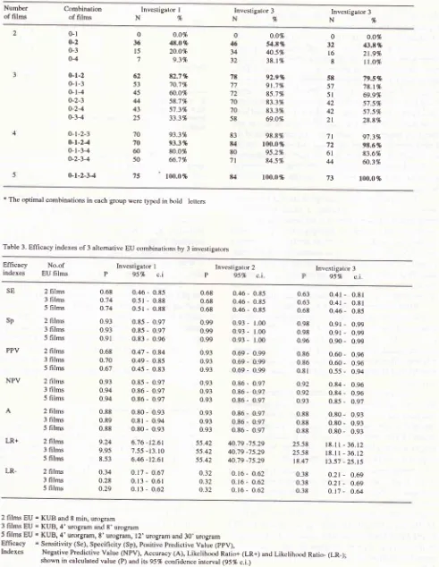
100Optimal Simplified EU
The
first
stepin the
study
was to identifywhich single
film
and whichoptimal
simplified
combination
would
give
complete
visualization
of
EU
components.
Thestudy showed
that 8 minute
urograms
(F2), followed
by
12and 30
minute
urograms (F3
andF4)
were
the bestsingle
films with a
complete
visualization
of the
urinary
tract
components.As an
examination, combinations
of plain
photo
of
the
abdomen
and the
8
minute urogram
with
or
without
the 4 minute urogram (2
films EU:
F0+F2 or
3 films EU: FO+Fl+F2),
arethe
optimal 2 films EU
and 3 films EU procedureswith
acomplete
visualiza-tion of EU
components (Table
2).Diagnostic
Efficacy of Simplified EU
The presence
of
abnormalities found
by
the experts
panel
in
the
5 films EU
(classical
EU)
was used to
calculate
the
diagnostic test
efficacy
indexes. To
simplify
the results, evaluation was
only
done
to
2optimal simplified
combinations,
i.e.
2
films
EU
(F0+F2)
and
3 films EU (F0+F1+F2),
and
compared to the complete 5films
EU
(F0+Fl+F2+F3+F4):
Diag-nostic
efficacy
of
thesecombinations were
describedin
diagnostic
test efficacy
indexes and
its
957o
con-fidence
interval
(Table
3).In EU series
in which
the
simplified EU
gave
acomplete
visualization
of
the urinary tract, no
diag-nosis
or
abnormality
were missed
by
either
inves-tigator. The efficacy
indexes and
its 95%
confidence
interval could be
compared between those
3 EU
[image:3.595.309.543.92.311.2]Vrtl 3, No
l, Juttuurv
March 1994 Excretory Urography in Children withIITI
33
Table 2' Combinations of EU films of pediatric UTI patients with clear visualization of EU comJxrncnts according to 3 investigators.
Number of films
Combination of films
lnvestigalor I
N%
N%
Investigakrr 3N%
Investigator 30-l 0-2 o-3 o-4 o-r-2 o-l-3 o-l-4 o-2-3 o-2-4 0-3-4 o-t-2-3
o-t-24
0-l-3-4 o-2-3-4 o-t-2-34 o.o% 4t.o% 20.o% 9.3% 82.7% 70.1% û.o% s8.7% 57.3% 33.3% 93.3% 93.3% 80.o% 66.'t% 100.0% o.o%g.t%
40.5% 38.t% 92.9% 9t.7% 8s.7% 833% 83.3% 69.O% 98.8% 100.0% 95.2% 84.s% 100.0% o.o1i 43.t% 21.9% tt.o% 79.5% 78.t% 69.994 s7.5% s7.5% 28.8% g',t.3% 98.6% 83.6% û.3%100.0 %
0 36 l5 7 62 53 45 44 43 25 70 70 60 50 75 0 46 34 32 o 32 l6 8 5t 57 5l 42 42 2l '7t 72 6l 44 73 7t 77 72 70 70 58 83
u
80 7lu
* The optimal combinations in each group were typetl in txrltl
letters
Table 3. Efhcacy indexes of 3 altemative EU combinations by 3 investigators
Efficacy indexes
No.of
EU films
Investigator I
P
95%
c.iInvestigalor 2
P
95%
c.iInvestigator 3
95%
c.i. SESp
PPV
2 films 3 films 5 films
2 films 3 films
5 films
2 films 3 films 5 films
2 films
3 films
5 films
2 films 3 films
5 films 2 films 3 films 5 films
2 films 3 films
5 films
0.68 o.74 o.74 0.93 0.93 0.91 0.68 0.70 o.67 0.93 o.94 0.94 0.88 0.89 0.88 9.24 9.95 8.53 0.34 0.28 o.29
0.46
-
0.850.5r
-
0.88 0.51-
0.88 0.85-
0.97 0.85-
0.97 0.83-
0.96 0.47-
o.84 0.49-
0.85 0.45-
0.83 0.85-
0.97 0.86-
0.97 0.86-
0.97 0.80-
0.93 0.81-
0.94 0.80-
0.93 6.76 -12.617.55 -13. l0
6.46 -t2.6t o.t'l
-
0.67 0.13-
0.610.13
-
0.620.63 0.63 0.68 0.98 0.98 0.96 0.86 0.86 0.81 o.92 o.92 0.93 0.88 0.88 0.88 25.58 25.58 18.4'l 0.38 0.38 0.38
0.68
0.46-
0.850.68
0.46-
0-850.68
0.46-
0.850.99
0.93-0.99
0.93-0.99
0.93-0.93
0.69-0.93
0.69-0.93
0.69-1.00 1.00 1.00 0.99 0.99 0.99
0.93
0.86-
0.970.93
0.86-
0.970.93
0.86-
0.970.93
0.86-
0.970.93
0.86-
0.970.93
0.86-
0.9755.42
40.'19 -75.2955.42
40.79 -75.2955.42
40.79 -75.29o.32
0. 16-
0.62o.32
0. t6-
0.62o.32
0.t6-
0.620-41
-
0.810.41
-
o.8l0.46
-
0.85 0.91-
0.99 0.91-
0.99 0.90-
0.99 0.60-
0.96 0.60-
0.96 0.55-
0.94 0.84-
0.96 0.84-
0.96 0.85-
0.97 0.80-
0.93 0.80-
0.93 0.80-
0.93l8.ll
-36.12l8.ll
- 36.1213.57 - 25.15
0.2r
-
0.69 0.21-
0.69o. l7
-
0.64 NPVA
LR+
LR-2 films EU = KUB and 8 min, urogram
3 films EU = KUB,4'urogram and 8'urogram
5 films EU = KUB, 4' urorgram, 8'urogram, l2'urogram and 30'urogram
Efficacy
= Sensitivity (Se), Specificity (Sp), pmitive predictive Value (ppV), [image:4.595.53.544.106.740.2]34
Tamaela et al.DISCCUSSIONS
Although
in
the literature
UTI in
children
is
often
most common infection in
till
suggested
in
every child
in
+4
U2
yearsstudy period
there
were
only
120 EU series found withthat
indica-tion. The explanation might be the fact that
UTI
in
infants
andchildren
areoften asymptomatic
andunder-diagnosed,
not only clinically but
also
microbiologi-cally and
radiographically
due to financialrestrictions.
And for
this reason, we usedall EU
series that had been doneduring
those4
Il2
yearc.We
haveto
acceptthat
the
study
sample was not thevery
representative of thetarget
population,
and
we could
not perform 'on
thespot evaluation'
of
each
film
like in
prospective
studies.
However we designed
the study
asnatural
aspossible,
by
reading every
film
in
the
usual 'clinical
sequence'.The data
collection,
though
from old EU
series,were done through
a
detailed,
2
times and
blinded
reevaluation
of
every
film,
not
influenced
by
thepre-vious/excisting
expertise
(primary
data).
To increase
theprecision
and accuracyof the
study,
we designed a standardizedstrict evaluation criteria of
eachEU
com-ponent,
to
transform
radiological image
to
nominal
data. This criteria was tried
out
and discussedprior
to
the study to ensure
visual perception
agreement,which
is very
crucial in
diagnostic
radiological
studies.More
than oneinvestigator
was meantto
get the
idea that resultsof this
simplifying
procedure werenot
influenced
by
the investigator's knowledge and
ex-perience. The
multi
observers' diagnostic capabilities
were
not
meant to be compared,but to show
theinsig-nificant
differences between
the efficacyindexes
of 2
simplified EU
and the classical
EU by
oneparticular
investigator.
Some
previous investigators
onthis
matter select-edcertain
film(q) and
evaluate its
efficacy
against thecomplete set.t9'2o'21'22
In this
study
acertain urogram
was
not chosen,
weither a
proposed hypothesis
madeto
avoid
film
selection bias.
The evaluation
of
theurogram
was also
stressed
to
be done
in
the
usual sequence,with no
backwards evaluation,
Transformation of
radiological image,
ablending
of black-grey-white
spectrum, to categorigal
datawill
certainly be
affected by
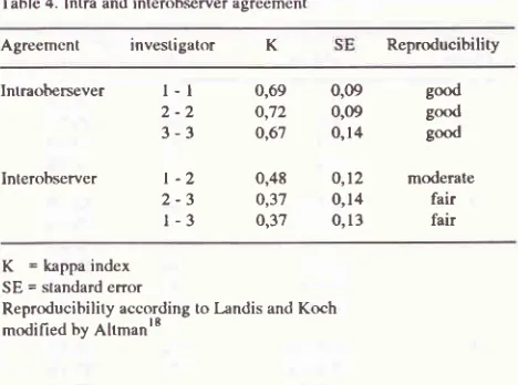
theintraobserver
andinterob-server perception.
To measure
this factor,
evaluation
was done twice
to
get the reproducibilityindex (Table
4). The
interevaluation period
was more than 6months
to
get
areal
blinded second
evaluation, that
was usedin
the
analysis.
The result
showed
that
intraobserver
reproducibility
was
good,ls
showing
thereliability of
further analysis.
Table 4. Intra and intenrbserver agreement
Med
J
Univ IndonAgreemcnt investigator
K ReproducibilityIntraobersever
Interobserver
0,09
good0,O9
good0,14
goodO,l2
moderate0,14
fair0,13
fair0,69 o,72 o,67
0,48 o,37 0,37
K
= kappa index SE = standard errorReproducibility according to l,andis and Koch
modified by Altmanls
To
avoid
'measurement
bias', no clinical
datawere
disclosed,
like
fever
as
an important
symptom
related
to the
presenceof urinary
tract
abnormality
or
ballotement
in
hydronephrosis
cases. Theevaluation
by
the 2pediatric radiologists experts
panelto
reach aconsensus
was also done
last. The
results
of
this
evaluation were
considered as
the
truth
(expert
validity),
functioning as
the
alternative
diagnostic
standard.
In the
ideal
utopic
setting, the gold
standardof
urinary tract
abnormality
should
be theresult
of
a
complete
uroradiological investigation,23 which
should not
beinfluenceà
by the resuli of EU
itself.18To
make
a simple2
x 2
diagnostic test table, we
simplify
theevaluation
of abnormality
to
a dichotomy(normal
and abnormal)
by classifying both the
"nor-mal"
and"others"
classification as "normal".
This
was considered as onecategory
becauseboth
donot
imply
special treatment.
And
disregarding the
EU
result,
most
UTI
in
young
children
still
needother
meansof
uroradiological investigations as indicated,
like
theultrasound examination and/or
mictiocystourethrog-r^phy.4's't2'23Optimal urogram and EU combinations
In EU
serieswith a
complete
visualization
of
the
uri-nary
tract
components, the
combination
of
plain
film
+ 8
minute urogram (2
film
EU) and
the combination
of plain
film
+ 4minute urogram
+
8minute urogram
'already have
shown
43.8-54.8% and
79.5-92.9%
respectively
of
complete
visualization
of
the
com-ponents
(Table
2).The
diagnostic
ability
of
simplified optimal EU
procedures
(2
and
3 films
EU,
those
with
minimal
number
of films but with
agood
percentageof
com-plete
visualization of
thecomponents)
compared to theclassical
EU, showed
that
thedifferences
between theefficacy indexes
were
insignificant
(Table
3). Inother
words
adding
further exposures
we
would
not
give
l-t
2-2
3-3
t
-2
[image:5.595.326.560.99.273.2]Vol 3, No
l,
Jattuary - March 1994any extra diagnostic
information. This
result
con_formed the
statement
of
some experts,
that after
the;".::::.ldP'','îdr,,H,l?s'"'
no
rurther
exposure is
Simplified
2
film
EU
procedure
(plain
film
+
8minute urogram)
needed
further
exposures
if
the
gminute urogram did not show
the
urinary tract
com-ponentscompletely.
In this study there wasI
abnormal
caseundetected
by
investigator
I
on the
2
film
EU
becausethe
8
minute
urogram was
blurred
(the
child
moved during
exposure), and another
caseby
inves-tigator 3
becausethe pelviocalyceal
system was not
clear
yet.
So
'diagnostic information loss, would
not
have happened,had the
simplified EU
proceduresal-ready visualized the urinary tract completely.
This
findings
also conformed previous reports. Lâhde et al. reported that plainfilm
and a 10minute uro-gram
were enoughin pediatric
UTI
cases. There were no manage_ment
changesby
simplifying the
EU.20 Schey
et
al.reported that one
8minute urogram either detected or
excluded abnormality in
most
""".r.22
The accuracy
(83.27o), sensitivity
(56%)
and
specificity
(91.3%)
of
one
film (5
minute
urogram)
wasalso
reported
by
Leonidas
et
al.2l
Only
in
doubtful
cases
or
im-perfect
films,^^
they
suggested
to
make further
urograms."'"",tt
Thesereports
reemphasizedthe
im_portance
of
a radiologists
presencein
the simplified
procedure.to-determine the
necessity
of
any further
"*porur",lo'l
I
Although the principle
of
minimal
exposure in
pediatric imaging is
stressedby many centers, there is
no one
globally
acceptedfilm
sequencing. Some
con_servative experts
still
defended
the
multi
exposure(Table 5). McClennan
statedthat reducing the
proce_dure had
not proven efficacious,
and these shortened
urograms did not
provide
an acceptablelevel
of
diag_nostic confidence.la
But
f,om this study, the
abovereports,
andthe
recommendation
of
the
WHO
expert committee.14But from this study,
the above reports,
and the recommendation
of
the
WHO
expert
com_mitte,24 we should consider the idea of
simpiifying EU
as a
rational
approach
in uroradiologic
investigations
and-that
we
should proceed
with
a prospective studyperforming
these2 or
3
films
EU
procedure
for UTI
cases
in
children.
If
in
the
clinical
setting
EU
can beperformed in
the 2
film
EU procedure.If
the 2film
EU hàs not shownthe
lower urinary tract, we
still
can make
further
ex-posure(s).
In
case the 8minute
urogram
is too
late to
catch
a clear
nephrogram
(+
pelviocalyceal system),
we
still
can give
another
contrast
injéctionltle
andmake an
earlier urogram,
e.g. the
4 minute
urogram.
Rut
becauseany added
contrast agent
injection
iscon-sidered adding
child's discomfort
andrisk
plus
someExcretory Urography in Children with
IJTI
35extra
expenses,
the
3 film
EU
procedure could
beconsidered
to
be theoptimal simplified
EU
too.Tahlc
5.
Minimal routine exJxrsure in pctliatric EU according to some ped iatric radiology centers/experts.No. of
film
Exposure Experts2 films
3 films
4 films
7 fihns
KUB 6'AP urogram
KUB 6'AP urogram
20'AP urogram
KUB
3'-5'renal urogram
l0'AP urogram
KUB
l'renal urogram
lO'AP urogram
KUB
30" renal urogram
l0'AP urogram
KUB
4'-5'renal urogram
l5'-20'AP urogratn
KUB
2'rcnal urogram
7'AP urogram
KUB
3' rcnal urograrn
l2'rcnal urograln
120'AP urogram
KUB
4'renal urogram
8' AP urogram 12' AP urogram
30' AP urogram
KUB
0'renal urogram
3'-5' rcnal urogram
15'AP urogram
l5' lateral urogram
KUB
I'renal urogram
5' AP urogram
l0' AP urogram
l0' right oblique urogram
l0'
lcl't ohliquc urogramKUB
Dunbar & Nogardy, 1972
Emmett, 1964
Hartman & Hattery, 1976
Kirks, 1984
Hilton, 1984
Gordon, 1988
Dunhar, l99O
Smellie & Normand, 1976
Tamaela et al, 1989
(since 1966)
Aarorison & Cremin, 1984
Poznanski, 1976
McClcnnan, 1988
I'
rcnal urograrn4'-5'ronal urogram
8'- l0' cornprcssed AP urogram lO'- 15' right ohliquc urogram
lO'-15' lcft obliquc urogram
Jxrst compression AP urogram
KUB = kidncy uretcr bladdcr overvicw; Ap = anterior-posterior;
36
Tatnaela et al.Psychological approach
in EU examination
for
pediatric patients
In
13of
15EU
seriesexcluded from
this study, much
air in the bowel obscured the visualization of urinary
tract
components,
especially
in
later urograms. An
adequate
bowel preparation can
be
ruined
by
this
aerophagia,
very possibly
causedby
excessivecrying.
And
becausefor children excretory urography
is
con-sidered as oneof
the mostdisliked examinations
(start-ing with
aninjection in our department
the proceduretakes
at least 30 minutes), the approach
before
andwhile
undergoing
this procedure should
beimproved.
To
uncooperative young
children,
immobilization
restraint
is also needed.Failure to
do sowill
end upin
improper/uninterpretable and
blurred
EU
series.
Simplified shorter
EU procedure
reducesthe child's
discomfort
andhopefully
will
reduce thecrying
andits
hindering impact on the visualization of urinary
tract
components.
The impacts of EU simplification
Simplification
of EU
procedure
(2
or 3 films
EU
reduces the radiation
dose, especially
to
the
gonads,and
theoritically
reducesthe examination cost.
If
theaverage gonadal
radiation
dosefor
boys is 42 mrad andfor girls
420 mrad per examination2s undif
at least 50 %EU
examinations
could be ended after
3
exposures,like in this study, there would
be an average gonadalreduction
of
+
60 mrad/patient. The imaginary
radia-tion reducradia-tion calcularadia-tions
for
thewhole population
in
this study
are asfollows: 58 (boys)
x 50% (more
thanhalf
could
besimplified) x
6O%(3
films
comparedto
5
films) x 42
rnradplus 42 (girls) x
5O%x 60%
x
42Omrad = 6,022.8 mrad/ 100 patients.
If
the costof
3 films
EU is 60% of
the costof classical EU for
athird
classpatient (Rp. 37,50O,-),
with similar
calculations,
therewould
be animaginary reduction of
costof
Rp.7,500,-/examination
(5O%x 40% x Rp. 37,500,-).
Simplification
of
this
examination
means
alsocutting
the time spent, thus increasing the possibility
to
improve
the
psychological
approach
to
word
children by
theradiographers,
aswell
assaving
somelife
time of the
radiologic
equipment.
With
this
Med J Univ Indon
simplified procedure more examinations could be
ar-ranged
in
the same length
of
time,
with
the
samediagnostic
facility,
manpower
andfilm
stock.CONCLUSIONS
In most pediatric
UTI
cases,EU examination could
bestopped
after complete visualization
of
the urinary
tract,
i.e.,
after
plain
photo
of
the
abdomen
and
8minute urogram with
orwithout
the 4minute urogram.
The diagnostic
efficacy
indexes
of
these simplified
EU
procedures
were
not
increased
by
adding
other
urogram(s). These
2
or
3
films
EU could be
recom-mended
to
be
the
minimal routine
procedure
in
pediatric patients
suffering from UTL
ACKNOWLEDGEMENTS
We are
grateful
to Drs. BambangMadiyono
andSudig-do
Sastroasmorofor
their contributing
advice
on
thestatistical
aspects andto Mr. Heri Sukisno for
prepar-ing appropriate computer software.
APPENDIX
C onfid enc e inte rva
I
c a lculations
Confidence
intervals
of
proportion
diagnostic
radiol-ogy
efficacy
indexes(sensitivity,
specificity,
accuracy,positive
and negativepredictive value)
werecalculated
byt
2p+Q2+g
c,l. =
2+zg2
c.i.
= confidence intervalp
= proportion= Z'tz
I
,/{
Zop=
normal standard deviation for two-tailed a;for 95% confidence level in this study, Za
= l'96
N
= sampel sizeConfidence intervals
for
the likelihood ratios were
calculated
by2p (1-p)
I
""-
CC. A tutorial on confidence intervals for proportions in diagnostic radiology. Am J Roentg 1990; 154: 477-480.Vol 3, No I, Jurtuary - Morc'h 1994
c.i. =
ew-eX
e
= exponential function for antilog, transformationW
= log, LR-Zd
x SE log, LRX
= log" LR +Za x
SE log, LRloge = normal logaritm of the basic constant number
e (e = 2,718281)
For likelihood râtio + (LR+) SE log" LR+ =
tl
ll
b
b+d
Excretory Urography in Children with
IJTI
3710. Emmett
JL, editor. Methods
in
urographic diagnosis. [n:Clinical urography. 2no ed. Philadelphia: Saunders, 1964;
l-36.
ll.
Hartman GW, Hattery RR. Uroradiology: procedures andanatomy. [n: Kelalis, King, Belman, Clinical pediatric
urol-ogy. Philadelphia: Saunder, 1964; t5-37.
12. Hilton S. The child with a urinary tract infection. In Hilton,
Edwards,
Hilton,
editors, Practical pediatric radiology,Philadelphia: Saunders, 1964: 15-37.
13. Aaronson
IA,
Cremin BI, editors. Imaging techniques. In:Clinical
paediatric uroradiology. Edinburgh: Churchill Livingstone, 1984; 18-22.14. McClennan BL. Urography-anatomy and technique. In:
pol-lack, editor,
Clinical urography. Philadelphia:
Saunders,1988;
ll66-
7.15. Dunbar JS. Excretory urography in infants and children. In:
Pollack, editor, Clinical urography. Philadelphia: Saunders,
l99O;2O7.
16. Gordon I. Urinary tract in paediatrics: the role ofdiagnostic
imaging. Br I Radiol 1990; 63: 507-l
l.
17. Kirks DR, editor. Genitourinary tract. In: Practical pediatric
imaging. Diagnostic radiology of infants and children.
Bos-ton/Toronto: Little Brown, 1984; 67 6-7 43.
18. Altman DG, editor. Some common problems
in
medicalresearch. In: Practical statistics for medical research.
Lon-don: Chapman and
Hall, l99l:396-439.
19. Hilman B, Abrams HL, Hessel SJ, Herbert S, Benazzi RB,
Gerson
DE.
Simplifying radiological examinations. Theurogram as a model. Lancet 1979;
i:
1068-71.20. Lâhde
S,
Standerskjôd-NordenstamC-G,
Suoranta H,Phytinen
L
Two-picture urographyin urinary tract
infec-tions. J Urol
l98l;
125: 820-1.21. Leonidas JC, Schwartz R, Schwartz AM, Mc Cauley RGK,
Darling DB. The one-film urogram in urinary tract infection
in children. Am J Roentg 1983;14l:61-4.
22. Schey WL, Slrkolnick A, White H, Finder C. Eighrminute
excretory urographic
film: once is enough.
Urologyl98l;
l8:
5 l5-8.23. Nogardy MB. Diagnostic imaging of the urinary system in
infants and children. In: Behrman, Vaughan, Nelson, editors,
Textbook
of
Pediatrics.l2th
ed. Philadelphia: Saunders,1983;
l3l4-8.
24. WHO Study Group. Rational use of diagnostic imaging in
paediatrics. WHO Technical Report Series, 1987; 757:
49-51.
25. Troupin R, terjemahan. Pemaparan radiasi pada radiologi
diagnostik dalam klinik. Iakarta: ECG, 1989; 5-7.
a
a+c
For likelihood ratio - (LR-) SE log" LR- =
REFERENCES
l.
Smellie JM, Normand ICS. Urinary infections in children.Post- grad med J 1985;
6l:
895-905.2. Brouhard BH, Travis LB. Infections of the urinary tract. In:
Rudolph, Hoffman, Pediatrics. l8th ed. Norwalk: Appleron
& Lange, 1987;
ll97-2o2.
3. Tambunan
T.
Penanggulangan pielonefritiskronik
padaanak. Dalam
:
Pendidikan
TambahanBerkala Ilmu
Kesehatan Anak: Penanggulangan penyakit ginjal kronik
pada anak; Iakarta
I
luni
1983; 23-31.4. Alatas
H.
Penanggulangan infeksi traktus urinarius padaanak. Maj Kedoklndones 1983; 33:
lli-21.
5. Lebowitz RL, Mandell J. Urinary tract infection in children:
putting radiology in its place. Radiology 1987; 165: l-9.
6. Friedenberg RM, Dunbar JS. Excretory urography. In
pol-lack, editor,
Clinical urography. Philadelphia:
Saunders,1990;
l0l.
7. Badan Tenaga Atom Nasional. Pedoman proteksi radiasi di
rumah sakit dan tempat praktek umum lainnya. Jakarta:
BATAN,1985.
8. Pohlit W. Radiation risk in the pediatrc age group. Institut
fiir Biophysik
der Universitât FranKurt. In press.9. Walker JS. The controversy over radiation safety. A